It is an immensely challenging task to segment the tissue regions and cells in histological images of breast cancer with precision, but the results of this task are extremely significant for the field of computational pathology as a whole. To address this challenge, the integration of the Denoising Diffusion Probabilistic Model (DDPM) and the Generative Adversarial Network (GAN) has been explored. Specifically, we employ a conditional DDPM as the generator within the GAN framework, alongside a conditional adversarial network serving as the discriminator, to achieve segmentation of breast cancer histology images both at the regional and cellular levels. The forward process of the DDPM is first applied to the image mask. As the noise is added step by step, it is conditioned with the pathological image and estimated by a denoising network. To improve the estimated noise, the estimated noise is again conditioned with the pathological image and fed into the discriminator as part of the training process. As part of the test phase, a noise image conditioned with a pathological image is fed into a denoising model trained taking into consideration each time step, which segments the images into regions and cells in the reverse process. Three datasets were used for the experiments, one at a regional level and two at a cellular level. This method outperforms both GAN and diffusion models, as well as current state-of-the-art methods. Specifically, our method shows notable improvements in terms of Dice and IoU metrics over existing state-of-the-art methods.
Quantum computing introduces new computational capabilities that can support advanced cybersecurity solutions when combined with machine learning. In recent years, quantum machine learning (QML) has emerged as a promising approach for enhancing network intrusion detection systems (IDS), particularly for analyzing complex and high-dimensional network traffic. This paper presents a systematic survey of QML techniques applied to network intrusion detection. The survey reviews peer-reviewed studies published up to January 2026 that employ quantum, hybrid quantum–classical, and quantum-inspired learning models for IDS. The selected studies are analyzed with respect to the algorithms used, intrusion detection datasets, and evaluation metrics reported. The analysis shows that most current approaches rely on simulated quantum environments and legacy datasets, while evaluation practices remain inconsistent across studies. These findings highlight the early developmental stage of QML-based IDS and the need for standardized evaluation protocols and more realistic experimental settings. Finally, open challenges and future research directions are identified to support the development of reliable, scalable, and practically deployable QML-based intrusion detection systems.
Deep learning models often operate as black-boxes, raising trust concerns in sensitive applications such as mental health monitoring. Moreover, centralized training requires aggregating personal data on central servers, posing privacy risks. Existing lightweight models often struggle with cross-dataset generalization, while the evaluation of explainability methods remains largely qualitative, with limited quantitative validation. These challenges hinder the deployment of Facial Emotion Recognition (FER) systems in privacy-sensitive and resource-constrained environments. To address these issues, this work introduces FedXAI, a framework that integrates federated learning and explainability metrics to guide privacy-preserving model optimization for continuous mental health monitoring on edge devices. Lightweight architectures were evaluated on three in-the-wild datasets, FER2013, RAF-DB, and ExpW, using classification accuracy, cross-dataset generalization, and explainability quality as evaluation criteria. Explainability was assessed using Grad-CAM++, LIME, and SHAP with quantitative metrics aligned with three complementary domains of the Co-12 framework: Sharpness, Compactness, and Fidelity. The selected model, requiring only 0.42 GFLOPs, achieved a cross-dataset accuracy of 76.2% in centralized training and 74.3% under federated learning, outperforming existing lightweight methods and surpassing most large-scale architectures while achieving 17 frames per second on a Raspberry Pi 4, demonstrating its suitability for potential edge deployment. These improvements were validated through comparisons with strong cross-dataset baselines and were found to be statistically significant. Furthermore, SHAP emerged as the most reliable explainability method, and Fidelity analysis showed that the top 10% most important pixels corresponded to discriminative facial regions while introducing minimal noise. The results also revealed that models with similar classification accuracy can exhibit markedly different explainability performance, whereas higher class-level accuracy was associated with better explanation quality.
The COVID-19 pandemic has accelerated the adoption of contactless biometric modalities such as face, iris, voice, and periocular recognition, which offer a safer alternative to traditional methods by reducing the risk of disease transmission in public and private spaces. While face recognition technologies have shown robust performance even with partial facial occlusions, their accuracy significantly diminishes when individuals wear medical masks, highlighting the importance of periocular biometrics for reliable personal identification. To enhance security and accuracy, multi-biometric systems - combining multiple biometric traits - outperform single-modality approaches. We propose a multi-input convolutional neural network (MICNN) framework that fuses the left and right periocular traits from the same face image for enhanced biometric recognition. We evaluate our method on two challenging periocular datasets, achieving highly competitive correct recognition rates of 99.62% and 98.33%, respectively, outperforming recent benchmarks. These results underscore the efficacy of multi-sample periocular recognition using deep learning for contactless biometric identification.
The accuracy of breast cancer detection in histopathology images presents a critical challenge and remains a central focus in advancements in computational pathology. Scaling Convolutional Neural Networks (CNNs) can improve feature extraction, especially in multi-class problems that require varying levels of complexity to distinguish between different classes. However, selecting the optimal model complexity for each image remains a persistent challenge. Current approaches predominantly rely on fixed-complexity models regardless of individual image characteristics. This study introduces an adaptive method that intelligently matches model complexity to tumor characteristics by integrating a Denoising Diffusion Probabilistic Model (DDPM), a generative model that iteratively improves segmentation accuracy by progressively removing noise, for tumor region segmentation with a range of EfficientNet architectures for classification. The segmentation model, trained on a specialized dataset of breast cancer regions, identifies tumor regions in testing images from breast cancer detection datasets. Based on the size of the segmented tumor region, an appropriate EfficientNet model, ranging from B0 to B7, is dynamically selected for classification. The core insight is that smaller or undetected tumor regions require less computational complexity and can be accurately classified using efficient models like EfficientNet-B0, while larger tumor regions benefit from the enhanced feature extraction capabilities of deeper models like EfficientNet-B7 to capture more intricate features. This adaptive approach mitigates segmentation inaccuracies and ensures that the appropriate model complexity is applied based on tumor characteristics. Rigorously evaluated on three distinct datasets, the proposed approach demonstrates superior performance compared to leading techniques, particularly in terms of 95% confidence intervals, the area under the precision-recall curve (PR-AUC), and accuracy, achieving 93.86% accuracy with 95% confidence intervals of [93.15-95.31%] and PR-AUC of 0.986 on the BRACS dataset. This approach represents a notable advancement in breast cancer detection within histopathology images and holds significant potential as a tool in computational pathology.
Modern image restoration and super-resolution methods utilize deep learning due to its superior performance compared to traditional algorithms. However, deep learning typically requires large labeled training datasets, which are rarely available in astrophotography. Deep Image Prior (DIP) bypasses this constraint by performing unsupervised optimization on a single image without training data; however, DIP often suffers from overfitting, artifact generation, and instability. This work proposes DIPLI - a framework designed specifically for resolved, high-contrast astronomical targets that shifts from single-frame to multi-frame processing using the Back Projection technique, combined with dense optical flow estimation via the TVNet model, and replaces deterministic predictions with Monte Carlo estimation obtained through Stochastic Gradient Langevin Dynamics (SGLD). A comprehensive evaluation compares the method against the original DIP, the transformer-based model RVRT, and the diffusion-based model DiffIR2VR-Zero on synthetic data with ground truth, while comparing qualitatively against Lucky Imaging on real astronomical data. On synthetic datasets, DIPLI achieves the best perceptual fidelity scores (LPIPS in 12/12 and DISTS in 10/12 scenarios), while the diffusion-based DiffIR2VR-Zero achieves the best pixel-level distortion scores (PSNR in 9/12 and SSIM in 8/12 scenarios), consistent with the well-known perceptual-distortion trade-off in image restoration (Blau and Michaeli In Proceedings of the IEEE Conference on Computer Visionand Pattern Recognition 6228-6237 2018). Compared to classical Lucky Imaging, the model requires far fewer input frames (7-13 versus thousands) and avoids the need for early stopping that limits standard DIP. Qualitative evaluation on real-world data of resolved solar-system objects, where ground truth is unavailable and domain shifts typically hinder generalization, suggests that the method appears to preserve fine detail while suppressing noise and artifacts.
Deepfake videos have become increasingly realistic which poses serious threats to digital security and media integrity. The current detection methods face significant challenges at generalizing the new manipulation techniques and they often require access to camera reference patterns for forensic analysis. We propose a hybrid deepfake detection framework which combines the analysis of statistical Photo Response Non-Uniformity (PRNU) with the learning of Convolutional Neural Network (CNN). Traditional PRNU methods need a database of known camera fingerprints to work. Our approach is different; we extract directly statistical features from the wavelet noise without needing any reference patterns. This means we can detect deepfakes even when we do not know which camera was used. The proposed system fuses the features of PRNU with the predictions of CNN through a weighted combination where the achieved optimal performance is with 30% PRNU influence and 70% CNN influence. The experiments on the WildDeepfake dataset reveal that our hybrid approach achieves 82% accuracy which outperforms PRNU-only (65%) and CNN-only (77%) methods. We evaluate the framework on binary classification to distinguish authentic videos from deepfakes. The modular design separates feature extraction from classification which enables future extension to source attribution tasks without modifying the feature extraction pipeline.
Accurate molecular subtyping of breast cancer into luminal versus basal/HER2 subtypes remains difficult due to limited integration of histopathology and genomic data. Existing multimodal fusion strategies often underexploit cross-modal interactions and tend to overfit small cohorts, resulting in suboptimal classification. To address this, we propose a hypernetwork-based fusion framework where gene expression profiles dynamically modulate CNN parameters for histopathology analysis. Results on 5-fold cross-validation show that unimodal genomic models perform strongly, whereas image-only models perform poorly, and conventional early fusion provides no significant improvement. In contrast, the proposed hypernetwork-mediated fusion achieves 0.96 accuracy, with consistently high F1-score and AUC, clearly outperforming both unimodal and standard multimodal approaches on the same cohort. This establishes hypernetwork fusion with cross-modal augmentation as an effective and generalizable strategy for robust breast cancer subtyping, offering potential for precision oncology applications.
The early and reliable diagnosis of skin cancer is critical to mitigating its metastatic potential and progression. However, even highly accurate artificial intelligence (AI) classifiers tend to produce overconfident predictions on ambiguous lesions, increasing the risk of misdiagnosis. Uncertainty quantification (UQ) addresses this challenge by evaluating model confidence and distinguishing definitive classifications from cases requiring clinician review—a cornerstone for safely integrating AI into clinical practice. We propose GraphSkinUQ, a hybrid framework that integrates convolutional neural network (CNN)-based feature extraction and graph-based relational reasoning to model lesion contextual relationships, together with Bayesian Monte Carlo dropout, to simultaneously quantify epistemic uncertainty (model limitations) and aleatoric uncertainty (data noise). Through iterative sampling of the predictive posterior distribution, GraphSkinUQ generates well-calibrated confidence scores, evaluated using average predictive entropy (0.10 bits), Brier score (0.1382), and expected calibration error (ECE). Experimental results show that GraphSkinUQ achieves a Brier score of 0.1382, Receiver Operating Characteristic (ROC) area under the curve (AUC) of 0.954, and average predictive entropy of 0.10, outperforming conventional classifiers in calibration. The framework effectively identifies high-uncertainty cases, allowing clinicians to prioritize ambiguous lesions for additional scrutiny. By bridging AI-driven diagnostics with clinically interpretable confidence metrics, GraphSkinUQ enhances the safety and transparency of automated skin cancer screening, fostering trustworthy and robust human–AI collaboration in clinical decision-making, and aligning machine-driven insights with the nuanced demands of real-world oncology practice.
Accurate breast cancer subtyping guides treatment selection, yet
histopathology captures morphology without molecular state, while genomic
profiling captures molecular signatures without spatial context. Existing
fusion methods rely on concatenation, or on attention applied only after each
modality is encoded independently. This work identifies a scale-dependent
asymmetry in the direction of cross-modal conditioning: the direction that
performs best under limited samples is not the one that holds at scale, and
the reversal is traced to the capacity of the modulation pathway rather than
to the fusion principle. The comparison is carried out within a
hypernetwork-guided framework in which an auxiliary network maps one
modality to conditioning parameters that modulate the other's feature
representation, shaping features at the parametric level rather than the
decision stage; modulation is patient-specific rather than patch-specific.
Both directions are instantiated --- gene-to-image (HyperG2I) and
image-to-gene (HyperI2G) --- and trained under a label-aware MixUp strategy
that interpolates within-class samples across both modalities, preserving the
hard binary labels clinical decisions require. The framework is evaluated on
two paired TCGA-BRCA cohorts --- one limited-sample, one independently
assembled at scale --- under a single protocol spanning two whole-slide
representations, multiple visual backbones, and both conditioning directions.
On the limited-sample cohort, gene-to-image conditioning at its optimal
augmentation setting exceeds early fusion and both unimodal baselines, giving
the highest recall on the aggressive Basal/HER2 class of any configuration
evaluated, and an ablation favours intra-class over inter-class mixing. At
scale this ordering does not hold: image-to-gene conditioning sustains its
performance whereas gene-to-image does not, recovering only partially under
the full tissue bag and isolating the capacity of the modulation pathway as
the binding constraint. Direction and capacity of cross-modal conditioning,
rather than fusion depth alone, therefore govern how such frameworks scale.
Carotid artery stenosis risk stratification is one of the most sought-after methods for diagnosing the chances of stroke. There is an inherent requirement to identify the risk before its onset through techniques such as ultrasound imaging. The carotid artery intima-media thickness, a marker for stenosis, can be identified, marked, and assessed. Typically performed by a trained operator, now automated approaches have been introduced that can automatically segment and classify the status of the carotid artery intima-media, aiding in the diagnosis of the chances of stroke. In this paper, a new framework based on two components is presented to segment the intima-media layer of the carotid artery to aid in diagnosis of the status. Firstly, the segmentation model is based on an enhanced Unet using multi-scale squeeze and excite operations. Secondly, a novel patch-wise dice loss function is introduced to optimize the normal dice loss function. The obtained results using augmentation on two combined datasets indicate an improvement in different metrics with respect to the state of the art. Notably, 89.4% dice coefficient index and 80.85% IoU, with data augmentation. The source code for the functions discussed in this paper will be available at https://github.com/Vlabgit/MSEUnet.git.
Retinal vascular segmentation, a widely researched topic in biomedical image processing, aims to reduce the workload of ophthalmologists in treating and detecting retinal disorders. Segmenting retinal vessels presents unique challenges; previous techniques often failed to effectively segment branches and microvascular structures. Recent neural network approaches struggle to balance local and global properties and frequently miss tiny end vessels, hindering the achievement of desired results. To address these issues in retinal vessel segmentation, we propose a comprehensive micro-vessel extraction mechanism based on an encoder- decoder neural network architecture. This network includes residual, encoder booster, bottleneck enhancement, squeeze, and excitation building blocks. These components synergistically enhance feature extraction and improve the prediction accuracy of the segmentation map. Our solution has been evaluated using the DRIVE, CHASE-DB1, and STARE datasets, yielding competitive results compared to previous studies. The AUC and accuracy on the DRIVE dataset are 0.9884 and 0.9702, respectively. For the CHASE-DB1 dataset, these scores are 0.9903 and 0.9755, respectively, and for the STARE dataset, they are 0.9916 and 0.9750. Given its accurate and robust performance, the proposed approach is a solid candidate for being implemented in real-life diagnostic centers and aiding ophthalmologists.
With the rapid evolution of digital identification techniques, ensuring the security of personal identifiers, particularly in facial recognition systems, is crucial for preserving privacy and safeguarding sensitive data. This paper investigates the use of Homomorphic Encryption (HE) to enable secure facial recognition, driven by the growing demand for privacy-preserving biometric solutions. Specifically, Partially Homomorphic Encryption (PHE) based on the Paillier algorithm is employed to perform computations directly on encrypted data, ensuring data confidentiality and integrity throughout the recognition process. The Histogram of Oriented Gradients (HOG) is utilized for feature extraction, enhancing robustness against variations in lighting and noise. To further optimize the encryption and decryption efficiency, Principal Component Analysis (PCA) is applied to reduce feature dimensionality. The study utilizes two publicly available datasets and evaluates performance based on geometric mean (GM). The results demonstrate the feasibility of this approach for secure facial recognition, highlighting its efficiency and improved processing speed compared to conventional methods while maintaining privacy protection.
Over the last decade, biometrics has witnessed significant advancements in various forensic and security applications for human identification and authentication, with growing interest in effective and discriminative traits such as palmprints. However, practical applications still face challenges, especially when palmprints are collected in portions, such as at crime scenes, or partially acquired for authentication in uncontrolled environments. This paper presents a novel method that incorporates the local binary patterns (LBP) operator into the conventional scale-invariant feature transform (SIFT) algorithm to detect and extract robust keypoint features. While SIFT employs a Gaussian filter to detect keypoints on the palmprint, the proposed method leverages the multi-scale LBP operator to detect stable points prior to computing the corresponding descriptors. Furthermore, an efficient method for filtering keypoints, namely the Self-Geometric Relationship (SGR) filter, is introduced to eliminate potential false matches. The proposed palmprint recognition system, LBPSIFT-SGR, demonstrates competitive performance on full palmprints compared to state-of-the-art techniques and exhibits clear superiority on partial palmprint images, where competing systems fail, across different datasets.
AI-based segmentation enables longitudinal tracking of tumor growth and changes over time. This helps clinicians monitor disease progression, assess treatment efficacy, and make informed decisions regarding patient management. The combination of fuzzy C-means (FCM) clustering and short-term memory (LSTM) networks enhances the robustness of brain tumor segmentation in MR images by leveraging the complementary strengths of both techniques. FCM clustering provides initial segmentation based on intensity distributions, while LSTM networks refine the segmentation by incorporating contextual information from neighboring regions, making the segmentation more robust to noise and artifacts in the images. To evaluate our segmentation system, we conducted experiments on the BraTS benchmark using different distance metrics. The True positive rate and True negative rate are highest on Mahalanobis, moreover, false positive rate and false negative rate are highest on Euclidean distance, and these results show that the Mahalanobis distance is more suitable for the proposed hybrid approach compared to the Euclidean distance. The experimental findings demonstrate that our own approach can yield comparable results to the ground truth segmentation of brain tumors, the proposed computer-aided diagnosis (CAD) system achieved promising results in the detection and segmentation of brain tumors on MR images, and demonstrated a Dice similarity coefficient of 91.8
The ability to predict breast cancer metastases is essential for making effective clinical decisions and managing patients. Traditional models predominantly rely on structured clinical data, which often lacks essential contextual details, limiting their predictive accuracy. In order to address this limitation, a multi-modal approach is introduced in which structured data is transformed into unstructured text, while contextual richness is preserved. Using this text, synthetic images are generated across three key diagnostic modalities, histopathology, mammography, and ultrasound, to enhance predictive capabilities. Based on converted text data, a pre-trained diffusion model was used to generate synthetic medical images in histopathology, mammography, and ultrasound modalities. The impact of a variety of text description variants on image quality and metastasis prediction was assessed. Comprehensive tumor descriptions or a combination of histological type and differentiation status were the most effective generation strategies. A comparison was conducted between three prediction approaches: a unimodal approach, an early fusion approach based on concatenation, and the Multi Co-Guided Attention (MCGA) approach. Through mutual attention, MCGA enhances feature alignment by addressing inter- and intra-modal heterogeneity and capturing complex relationships. Unimodal and multi-modal methods were evaluated with the application of SMOTE to mitigate the impact of data imbalance. Multi-modal fusion significantly outperforms unimodal methods, especially when class imbalances are mitigated by using SMOTE. When SMOTE was applied, Ultrasound+BERT achieved the highest level of accuracy (0.90), followed by Histopathology+BERT (0.88), and Mammogram+BERT (0.88). As compared to early fusion, MCGA demonstrated better class balance and improved minority class detection. Incorporating unstructured text with synthetic imaging modalities improves the accuracy of metastasis prediction by preserving contextual information. A MCGA fusion is particularly effective in ensuring balanced class performance, particularly for rectifying class imbalances. Through this approach, the complementary strengths of textual and visual data are leveraged to overcome limitations in multi-modality integration. These results demonstrate the potential for advancing the prediction of breast cancer metastasis, offering a more robust and context-aware framework for clinical decision-making.
Traditional machine learning assumes that training and test sets are derived from the same distribution; however, this assumption does not always hold in practical applications. This distribution disparity can lead to severe performance drops when the trained model is used in new data sets. Domain adaptation (DA) is a machine learning technique that aims to address this problem by reducing the differences between domains. This paper presents simulation-based algorithms of recent DA techniques, mainly related to unsupervised domain adaptation (UDA), where labels are available only in the source domain. Our study compares these techniques with public data sets and diverse characteristics, highlighting their respective strengths and drawbacks. For example, Safe Self-Refinement for Transformer-based DA (SSRT) achieved the highest accuracy (91.6%) in the office-31 data set during our simulations, however, the accuracy dropped to 72.4% in the Office-Home data set when using limited batch sizes. In addition to improving the reader’s comprehension of recent techniques in DA, our study also highlights challenges and upcoming directions for research in this domain. The codes are available at https://github.com/AIPMLab/Domain_Adaptation.
The precise detection of breast cancer in histopathological images remains a critical challenge in computational pathology, where accurate tissue segmentation significantly enhances diagnostic accuracy. This study introduces a novel approach leveraging a Conditional Denoising Diffusion Probabilistic Model (DDPM) to improve breast cancer detection through advanced segmentation and feature fusion. The method employs a conditional channel within the DDPM framework, first trained on a breast cancer histopathology dataset and extended to additional datasets to achieve regional-level segmentation of tumor areas and other tissue regions. These segmented regions, combined with predicted noise from the diffusion model and original images, are processed through an EfficientNet-B0 network to extract enhanced features. A transformer decoder then fuses these features to generate final detection results. Extensive experiments optimizing the network architecture and fusion strategies were conducted, and the proposed method was evaluated across four distinct datasets, achieving a peak accuracy of 92.86% on the BRACS dataset, 100% on the BreCaHAD dataset, 96.66% the ICIAR2018 dataset. This approach represents a significant advancement in computational pathology, offering a robust tool for breast cancer detection with potential applications in broader medical imaging contexts.
Stock market is pivotal to economical systems. It is a platform where listed stocks of companies can be bought/sold by market participants to gain profit. In 2020, the total capitalization of all markets reached 95 trillion. Within such profiting platforms, malicious attacks like stock price/market manipulation are conducted. Stock price manipulation attack refers to the strategic act of trading either a stock, a future (derivative financial contracts), or a security (financial instrument) to increase or decrease their prices. Hence, making significant profits. Manipulation types vary by sources and/or exploited methods. To detect and deter such attacks/practices, extensive research have been conducted. To further assist the future studies, in this paper, we conduct a systematic literature review to extract research articles relevant to stock price/market manipulation attacks and their state-of-art detection systems/solutions. To the best of our knowledge, this is the first systematic literature review that is based on a predefined search methodology and systematically collects the relevant articles that were published between 2011 and 2021. After applying the systematic search methodology, we extracted and surveyed 66 articles and classified them into four main detection approaches. Namely, statistical-based, Machine Learning based, system-based/artefacts, and visualization-based/observation-based. We provide a thorough comparison among the detection systems in terms of utilized datasets, leveraged features, employed optimization techniques, and applied performance measures. Our survey also identifies and discusses a set of existing research gaps such as generality of detection approaches, near/real-time detection capability, validity of real/synthesised datasets, etc. We concluded our survey by proposing several research topics and recommendations to be investigated and addressed by future works.
Accurate breast cancer survival prediction using multi-modal data is vital for enhancing clinical decisions. This study evaluates deep learning based fusion strategies, early, intermediate, late, and a hybrid approach, to integrate histopathology images and genomic data for one year survival prediction. We developed a robust evaluation framework, employing tailored deep learning architectures and metrics including accuracy, precision, recall, F1 score, and AUC. Model performance was validated using Kaplan–Meier curves and log-rank tests, with SHAP-based feature importance analysis enhancing interpretability. Results highlight the strengths and limitations of each fusion strategy, offering insights into optimal multi-modal learning approaches for breast cancer prognosis. Our findings underscore the importance of selecting task specific fusion methods, providing a reproducible, interpretable framework to advance survival prediction. All code and configurations are publicly available.
 Somaya Al-Maadeed合作论文数Department of Computer Science and Engineering, College of Engineering, Qatar University88
 Fouad Khelifi合作论文数Queen's University Belfast56Fatih Kurugollu合作论文数Queen's University, Belfast
School of Computer Science49Salim Chitroub合作论文数Signal and Image Processing Laboratory;Electronics and Computer Science Faculty;Signal and Image Processing Laboratory|Electronics and Computer Science Faculty41
Fouad Khelifi合作论文数Queen's University Belfast56Fatih Kurugollu合作论文数Queen's University, Belfast
School of Computer Science49Salim Chitroub合作论文数Signal and Image Processing Laboratory;Electronics and Computer Science Faculty;Signal and Image Processing Laboratory|Electronics and Computer Science Faculty41 Richard Jiang合作论文数Department of Computer Science and Digital Technologies, Northumbria University39
 Abdallah Meraoumia合作论文数USTHB Signal and Image Processing Laboratory, Electronics and Computer Science Faculty P.O. Box 32 El Alia, Bab Ezzouar 16111 Algiers Algeria34
 Danny Crookes合作论文数School of Computer Science, The Queen’s University of Belfast32
 Younes Akbari合作论文数22
 Omar Nibouche合作论文数21
 Camel Tanougast合作论文数20
 Larbi Boubchir合作论文数LIASD Res Lab, Univ Paris 0820
 Lahouari Ghouti合作论文数Dept Informat & Comp Sci, King Fahd Univ Petr & Minerals20
 Noor Almaadeed合作论文数Department of Computer Engineering, Brunel University19Muhammad Atif Tahir合作论文数18
 Mokhtar Nibouche合作论文数Department of Engineering Design and Mathematics, University of the West of England, Bristol17
 Rifat Hamoudi合作论文数Sharjah Institute of Medical Research, College of Medicine, University of Sharjah13
 Omar Elharrouss合作论文数Qatar Univ, Dept Comp Sci & Engn, Doha, Qatar13Peter Milligan合作论文数12
 Ashref Lawgaly合作论文数12
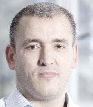 Ammar Belatreche合作论文数Department Computer and Information Sciences, Faculty of Engineering and Environment, Northumbria University12
 Ahmad Chaddad合作论文数11Said Boussakta合作论文数University of Newcastle upon Tyne10
 Abbes Amira合作论文数College of Computing and Informatics, University of Sharjah10
 A. Beghdadi合作论文数Institut Galilée, Université Sorbonne Paris Nord9
 Imad Rida合作论文数9
 Asim Baig合作论文数8
 Kashif Saghar合作论文数8
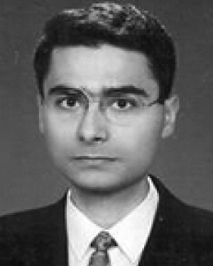 Huseyin Seker合作论文数8
 David Kendall合作论文数Dept Comp & Informat Sci, Northumbria Univ7Suchithra Kunhoth合作论文数7
 Duaa Hamed AlSaeed合作论文数Sch. of Comput. Sci. & Digital Technol., Northumbria Univ.;c;Sch. of Comput. Sci. & Digital Technol., Northumbria Univ.7
 Chang-Tsun Li合作论文数Faculty of Science Engineering and Built Environment, Deakin University;School of Information Technology, Deakin University7
 Ali El-Zaart合作论文数Department of Computer Science;College of Computer and Information Sciences;King Saud University;College of Computer and Information Sciences, King Saud University7M. Gueham合作论文数7Noureddine Mehallegue合作论文数University of Belfast7Zohir Dibi合作论文数7
 Faraz Khan合作论文数School of Biosciences, Faculty of Science, The University of Nottingham6
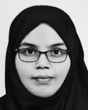 Najmath Ottakath合作论文数6
 Nandhini Subramanian合作论文数Qatar Univ, Dept Comp Sci & Engn, Doha, Qatar6
 Ismahane Cheheb合作论文数Northumbria University6Ferdi Sarac合作论文数6
 Mohamed Salah Azzaz合作论文数Lab. Syst. de Commun., Ecole Militaire Polytech.;c;Lab. Syst. de Commun., Ecole Militaire Polytech.6
 Lahoulou Atidel合作论文数Ecole Nat. Polytech.;c;Ecole Nat. Polytech.6
 Dina Shehada合作论文数College of Engineering & IT, University of Dubai6
 Gang Qu合作论文数Institute for Systems Research, University of Maryland;Department of Electrical and Computer Engineering, A. James Clark School of Engineering, University of Maryland;Maryland Embedded System and Hardware Security Lab, University of Maryland;Wireless Sensor Laboratory, University of Maryland6
 Muhammad Ali Tahir合作论文数Computer Science, Queens University of Belfast6Makram Nabti合作论文数6Moussadek Laadjel合作论文数6Khalil Zebbiche合作论文数Institute of Electronics, Communications and Information Technology, Queen's University Belfast;Algerian National Centre for Research and Development Algeria6




