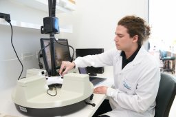
INTRODUCTION:Peripheral intravenous catheter (PIVC) failure prior to therapy completion is a major issue. Technology to identify more suitable veins for cannulation is essential to improve successful insertion and reduce post-insertion complications and avoidable costs. OBJECTIVE:The experimental study was designed to compare vessel selection for cannulation when using a novel ultrasound-based vein visualization prototype versus traditional landmark technique. METHODS:Three clinicians competent in cannulation (CCC) used landmark technique to select and mark a vein indicated for short peripheral catheter (SPC) placement on each arm of the 12 patients recruited (24 arms). This process was repeated using the vein visualization prototype. A blinded Vascular Access Specialist (VAS) evaluated each marked site using ultrasound (GE Logiq S7). Vessel cannulation suitability was rated on a 5-point Likert (⩾3 considered suitable). Ethics approval was obtained (SMHS HREC RGS0000006930). RESULTS:Landmark-selected veins were deemed suitable less often (52.2% vs 78.3%; p = 0.08) than prototype-selected veins and had lower average suitability scores (2.57 vs 3.22; p = 0.03). Landmark technique selected veins from various sites: hand (4.3%), wrist (17.4%), mid-forearm (73.9%), and antecubital fossa (4.3%), whereas the prototype predominantly selected mid-forearm veins (95.7%). Landmark-selected veins were significantly shallower (1.83 mm vs 2.61 mm; p = 0.03) than prototype-selected veins, however the diameter of these veins were not significantly different between the two techniques (2.20 mm vs 2.72 mm; p = 0.05). Both techniques detected veins with 95.8% sensitivity. CONCLUSION:The prototype outperformed the landmark technique by visualizing non-palpable, non-visible vessels. The veins identified were larger and optimally located in the mid-forearm, both attributes linked to fewer complications and improved first-time insertion success. Future prototype refinement will include ergonomic improvements and clinical data to enhance performance.
First-pass peripheral intravenous catheter (PIVC) insertion failure rates are up to 40
BACKGROUND:Both coronary atherosclerotic plaque activity and low endothelial shear stress (ESS) are predictive of adverse cardiovascular events. We aimed to investigate their association and relationship with high-risk plaque features. METHODS:Coronary computed tomography angiography (CCTA) based flow simulations were used to compute ESS in patients presenting with acute coronary syndrome proceeding percutaneous coronary intervention. Associations between ESS, CCTA plaque features and coronary plaque activity, measured by 18F-sodium fluoride (18F-NaF) positron emission tomography (PET), were investigated at the coronary segment and vessel level. RESULTS:ESS and coronary plaque activity were both analyzed in 330 coronary segments and 123 vessels. The area of low ESS (<0.4 Pa), termed low shear area (LSA), was larger in 18F-NaF positive regions increasing from median 11.7 mm2 (IQR: 4.6-27.4) to 29.0 mm2 (IQR: 14.1-55.2) at the segment level (P < 0.0001) and from median 27.3 mm2 (IQR: 8.6-65.3) to 57.8 mm2 (26.6-108.2) at the vessel level (P = 0.0049). The maximum tissue-to-background ratio of 18F-NaF activity positively correlated with LSA at the segment level (rs = 0.27; P < 0.0001) and at the vessel level (rs = 0.38; P < 0.0001). LSA was associated with spotty calcification at both the segment (P <0.0001) and vessel level (P = 0.0042) and positive remodeling at the vessel level (P = 0.025). CONCLUSIONS:In patients with acute coronary syndrome, LSA is associated with increased coronary atherosclerotic plaque activity, as measured by 18F-NaF PET.
Objective: Inserting needles into veins is fundamental to medical care with up to 90% of inpatients requiring a peripheral intravenous catheter/cannula (PIVC) during their stay. Yet 40%-50% of PIVC insertions fail on the first attempt. Here, we present an easy-to-use novel vein visualizing ultrasound prototype device and data from in vitro and in vivo performance. Methods: Locational accuracy was determined through phantom simulated forearm veins, across variations of vein diameter (3-5 mm), depth (10-20 mm), and velocity (10-100 mm/s). Usability studies were conducted on nine clinicians to establish effectiveness and ease of use of the proposed prototype assisted cannulation workflow. Sensitivity of the prototype was demonstrated by scanning 80 forearm veins across 40 healthy volunteers. Results: Our prototype's locational accuracy in simulated forearm veins is 0.21 mm +/- 1.71 mm (s.d.) (97.7% agreement to the ground truth, p < .001). Usability studies found that 100% of users were able to handle the prototype in a sterile manner with minimal assistance. The sensitivity was excellent at finding veins (94%). In comparison, sensitivity of vein finding using landmark technique with torniquet (visible 46% and palpable 74%) were far inferior. Conclusion: Initial performance verification and validation studies presented suggest that the proposed ultrasound visualization method can simply and reliably help clinicians detect well-perfused veins at depth and visualize in the coronal view onboard the probe in alignment with the transducers. With improved ergonomics, the device has the potential to be an easy to use device for clinicians performing vascular access.
BACKGROUND:Peripheral intravenous catheters (PIVCs) are the most commonly used invasive medical device, yet despite best efforts by end-users, PIVCs experience unacceptably high early failure rates. We aimed to design a new PIVC that reduces the early failure rate of in-dwelling PIVCs and we conducted preliminary tests to assess its efficacy and safety in a porcine model of intravenous access.METHODS:We used computer-aided design and simulation to create a PIVC with a ramped tip geometry, which directs the infused fluid away from the vein wall; we called the design the FloRamp™. We created FloRamp prototypes (test device) and tested them against a market-leading device (BD Insyte™; control device) in a highly-controlled setting with five insertion sites per device in four pigs. We measured resistance to infusion and visual infusion phlebitis (VIP) every 6 h and terminated the experiment at 48 h. Veins were harvested for histology and seven pathological markers were assessed.RESULTS:Computer simulations showed that the optimum FloRamp tip reduced maximum endothelial shear stress by 60%, from 12.7 Pa to 5.1 Pa, compared to a typical PIVC tip and improved the infusion dynamics of saline in the blood stream. In the animal study, we found that 2/5 of the control devices were occluded after 24 h, whereas all test devices remained patent and functional. The FloRamp created less resistance to infusion (0.73 ± 0.81 vs 0.47 ± 0.50, p = 0.06) and lower VIP scores (0.60 ± 0.93 vs 0.31 ± 0.70, p = 0.09) than the control device, although neither findings were significantly different. Histopathology revealed that 5/7 of the assessed markers were lower in veins with the FloRamp.CONCLUSIONS:Herein we report preliminary assessment of a novel PIVC design, which could be advantageous in clinical settings through decreased device occlusion and reduced early failure rates.
The etiology of spaceflight-associated neuro-ocular syndrome (SANS) remains unclear. Recent murine studies indicate there may be a link between the space environment and retinal endothelial dysfunction.Post-fixed control (N = 4) and 14-day tail-suspended (TS) (N = 4) mice eye samples were stained and imaged for the vessel plexus and co-located regions of endothelial cell death. A custom workflow combined whole-mounted and tear reconstructed three-dimensional (3D) spherical retinal plexus models with computational fluid dynamics (CFD) simulation that accounted for the Fåhræus-Lindqvist effect and boundary conditions that accommodated TS fluid pressure measurements and deeper capillary layer blood flow distribution.TS samples exhibited reduced surface area (4.6 ± 0.5 mm2 vs. 3.5 ± 0.3 mm2, P = 0.010) and shorter lengths between branches in small vessels (<10 μm, 69.5 ± 0.6 μm vs. 60.4 ± 1.1 μm, P < 0.001). Wall shear stress (WSS) and pressure were higher in TS mice compared to controls, particularly in smaller vessels (<10 μm, WSS: 6.57 ± 1.08 Pa vs. 4.72 ± 0.67 Pa, P = 0.034, Pressure: 72.04 ± 3.14 mmHg vs. 50.64 ± 6.74 mmHg, P = 0.004). Rates of retinal endothelial cell death were variable in TS mice compared to controls. WSS and pressure were generally higher in cell death regions, both within and between cohorts, but significance was variable and limited to small to medium-sized vessels (<20 μm).These findings suggest a link may exist between emulated microgravity and retinal endothelial dysfunction that may have implications for SANS development. Future work with increased sample sizes of larger species or spaceflight cohorts should be considered.
Compression optical coherence elastography (OCE) has shown promise in detecting diseases, such as breast cancer, based on changes in the elasticity of tissue. While the mechanical models used in OCE assume a flat surface, tissue typically exhibits surface topography that can lead to inaccurate estimation of elasticity in OCE images (elastograms). The coupling of surface topography and heterogeneous mechanical properties in tissue make it challenging to study the relative impact of surface roughness on image formation. To address this, we developed mechanically homogeneous phantoms that accurately replicate the surface topography of human breast tissue. Surface topography of breast tissue specimens was captured in three-dimensional (3-D) optical coherence tomography (OCT) scans. Molds replicating the surface roughness of tissue were generated using 3-D modelling and 3-D printing. The phantoms were then fabricated in the molds using polydimethylsiloxane. OCT was used to validate the accuracy of the phantom surface topography and the phantoms were then used to determine the impact of surface roughness in compression OCE elastograms. Surface roughness phantoms were shown to accurately reflect the surface topography of six breast tissue specimens with a correlation of 0.96 +/- 0.03. When imaged with a variant of compression OCE called quantitative micro-elastography, surface roughness resulted in a slight decrease in elasticity accuracy with a mean percentage error of 8.2 % for a flat phantom compared to 11.7 % for the roughness phantom, and a larger decrease in elasticity precision, or sensitivity, with a mean relative standard deviation of 9.4 % for the flat phantom compared to 21.2 %, for the roughness phantom. Our results show that contrast in elastograms moderately correlated with the phantom surface profile (r = 0.59, p < 0.001) and that surface roughness introduces spatially-varying elasticity that should be considered in the analysis of elastograms in compression OCE.
INTRODUCTION:The complex arborization of the feto-placental vasculature is crucial for optimal fetal nutrition, waste exchange and ultimately, development. Ethical and experimental limitations constrain research into the human placenta, hence experimental animal models such as mice and rats, are crucial to understand placental function. It is unclear how well the mouse and rat feto-placental vascular structure emulates human. Moreover, the implications of differences in vascular structure, especially in arborization, for placental function remain unclear. METHODS:We use micro-computed tomography imaging, high frequency Doppler ultrasound and computational fluid dynamics to characterize feto-placental vasculature structure and haemodynamics in mice, rats, and human. RESULTS:Our data suggest that despite structural differences between rat and mouse placenta, haemodynamics are similar and that both hold applicability to investigating feto-placental structure and function. We show that human cotyledons demonstrate vascularity-dependent haemodynamic behaviour (including flow deceleration and oxygen exchange) similar to rodents and can be analysed in the same spectrum as rodents. Finally, we show strong structure-function relationships when interspecies datasets are combined; notably, we demonstrate that surrogate measures such as vascularity, can be used to estimate placental oxygen exchange function. DISCUSSION:Pre-clinical placental research utilising rat and mouse placentae to understand the impact of feto-placental arborization on placental function and fetal development can inform the human context.
Core temperature (TC) changes, alongside exercise, affect hemodynamic responses across different conduit and microvascular beds. This study investigated impacts of ecologically valid environmental heat and exercise exposures on cerebral, skin and retinal vascular responses by combining physiological assessments alongside computational fluid dynamics (CFD) modeling. Young, healthy participants (n = 12) were exposed to environmental passive heating (PH), and heated exercise (HE) (ergometer cycling), in climate-controlled conditions (50 mins, 40°C, 50% relative humidity) while maintaining upright posture. Blood flow responses in the common carotid (CCA), internal carotid (ICA) and central retinal (CRA) arteries were assessed using Duplex ultrasound, while forearm skin microvascular blood flow responses were measured using optical coherence tomography angiography. Three-dimensional retinal hemodynamics (flow and pressure) were calculated via CFD simulation, enabling assessment of wall shear stress (WSS). TC rose following PH (+0.2°C, p = 0.004) and HE (+1.4°C, p < 0.001). PH increased skin microvascular blood flow (p < 0.001), whereas microvascular CRA flow decreased (p = 0.038), despite unchanged ICA flow. HE exacerbated these differences, with increased CCA flow (p = 0.007), unchanging ICA flow and decreased CRA flow (p < 0.001), and interactions between vascular (CCA vs. ICA p = 0.018; CCA vs. CRA p = 0.004) and microvascular (skin vs. retinal arteriolar p < 0.001) territories. Simulations revealed patterns of WSS and lumen pressure that uniformly decreased following HE. Under ecologically valid thermal challenge, different responses occur in distinct conduit and microvascular territories, with blood flow distribution favoring systemic thermoregulation, while flow may redistribute within the brain.
3D printing technologies have the potential to revolutionize the manufacture of heart valves through the ability to create bespoke, complex constructs. In light of recent technological advances, we review the progress made towards 3D printing of heart valves, focusing on studies that have utilised these technologies beyond manufacturing patient-specific moulds. We first overview the key requirements of a heart valve to assess functionality. We then present the 3D printing technologies used to engineer heart valves. By referencing International Organisation for Standardisation (ISO) Standard 5840 (Cardiovascular implants - Cardiac valve prostheses), we provide insight into the achieved functionality of these valves. Overall, 3D printing promises to have a significant positive impact on the creation of artificial heart valves and potentially unlock full complex functionality.
We investigated variations in haemodynamics in response to simulated microgravity across a semi-subject-specific three-dimensional (3D) continuous arterial network connecting the heart to the eye using computational fluid dynamics (CFD) simulations. Using this model we simulated pulsatile blood flow in an upright Earth gravity case and a simulated microgravity case. Under simulated microgravity, regional time-averaged wall shear stress (TAWSS) increased and oscillatory shear index (OSI) decreased in upper body arteries, whilst the opposite was observed in the lower body. Between cases, uniform changes in TAWSS and OSI were found in the retina across diameters. This work demonstrates that 3D CFD simulations can be performed across continuously connected networks of small and large arteries. Simulated results exhibited similarities to low dimensional spaceflight simulations and measured data—specifically that blood flow and shear stress decrease towards the lower limbs and increase towards the cerebrovasculature and eyes in response to simulated microgravity, relative to an upright position in Earth gravity.
BACKGROUND:The stent-assisted balloon-induced intimal disruption and relamination (STABILISE) technique for treatment of type B dissection has shown promising clinical results at mid-term. Computational modeling is a way of noninvasively obtaining hemodynamic effects, such as pressure and wall shear stress, leading to a better understanding of potential benefits. Particular areas of interest are (1) the effect of intimal disruption and re-lamination and (2) the effect of the bare metal stent in the visceral aortic segment.METHODS:Single-center prospective case series. Data from 5 consecutive locally performed cases of STABILISE technique were analyzed. Included cases were type B aortic dissection with or without prior de-branching. The STABILISE procedure had to be performed without 30-day major complications. Preoperative and postoperative imaging data for each patient were transferred to the biomedical engineering team. Each case was reconstructed, meshed, and simulated with computational fluid dynamics using patient-specific data (heart rate, blood pressure, height, and weight). Hemodynamic parameters were then extracted from the simulations.RESULTS:In all cases, computational analysis showed for postoperative patients: (1) a drop in pressure difference between lumina and (2) lower wall shear stress effects, compared to their preoperative status. These observations were most pronounced in the visceral aortic segment.CONCLUSIONS:Computational modeling shows favourable changes in the flow dynamics of type B dissection treated using the STABILISE technique. This may suggest protective effects of this technique for long-term aortic healing and cicatrization.
Biopolymers play a critical role as scaffolds used in tendon and ligament (TL) regeneration. Although advanced biopolymer materials have been proposed with optimised mechanical properties, biocompatibility, degradation, and processability, it is still challenging to find the right balance between these properties. Here, we aim to develop novel hybrid biocomposites based on poly(p-dioxanone) (PDO), poly(lactide-co-caprolactone) (LCL) and silk to produce high-performance grafts suitable for TL tissue repair. Biocomposites containing 1-15% of silk were studied through a range of characterisation techniques. We then explored biocompatibility through in vitro and in vivo studies using a mouse model. We found that adding up to 5% silk increases the tensile properties, degradation rate and miscibility between PDO and LCL phases without agglomeration of silk inside the composites. Furthermore, addition of silk increases surface roughness and hydrophilicity. In vitro experiments show that the silk improved attachment of tendon-derived stem cells and proliferation over 72 h, while in vivo studies indicate that the silk can reduce the expression of pro-inflammatory cytokines after six weeks of implantation. Finally, we selected a promising biocomposite and created a prototype TL graft based on extruded fibres. We found that the tensile properties of both individual fibres and braided grafts could be suitable for anterior cruciate ligament (ACL) repair applications.
Fenelli et al1 recently presented results from a two-center retrospective and observational cohort study into outcomes from endovascular iliac branch device deployment in aortic aneurysms with iliac involvement (n = 256). Analysis is centered on three measures of iliac tortuosity: pelvic tortuosity index, external tortuosity index, and double iliac sign. Pelvic tortuosity index >1.4 was an independent predictor of endovascular complications and reinterventions, whereas external tortuosity index >1.7 predicted endoleak.
Pancreaticoduodenal artery aneurysms have been associated with coeliac artery stenosis or occlusion. These aneurysms are rare, and their size may not be predictive of rupture risk. Wall shear stress has a potential role in the initiation, development and rupture of aneurysms. We investigated the influence of coeliac artery stenosis on wall shear stress in developing PDA aneurysm and in both ruptured and intact aneurysms to determine if ruptured aneurysms experience low WSS compared to intact cases. Computed tomography imaging was retrospectively collected and reconstructed into three-dimensional geometries with which blood flow simulations were performed to compare aneurysm wall shear stress between ruptured vs intact groups. Aneurysm initiation was investigated by digitally augmenting a case to represent a healthy geometry, with incremental coeliac artery stenosis then virtually applied. Coeliac artery stenosis resulted in gastroduodenal artery flow reversal and increased wall shear stress in the inferior pancreaticoduodenal arteries, where wall shear stress reached reaching 7.55 Pa at 80% stenosis. Wall shear stress was lower in ruptured aneurysms compared to intact cases (0.23(0.25) Pa vs. 0.37(0.26) Pa). Aneurysm WSS was not correlated to diameter (rs = 0.196, p = .564). Our results suggest 70% to 80% CA stenosis as a threshold for flow reversal through the inferior PDA and GDA, with corresponding high levels of WSS in the IPDA which may be responsible for the aneurysm initiation.
Cerebrovascular haemodynamics are sensitive to multiple physiological stimuli that require synergistic response to maintain adequate perfusion. Understanding haemodynamic changes within cerebral arteries is important to inform how the brain regulates perfusion; however, methods for direct measurement of cerebral haemodynamics in these environments are challenging. The aim of this study was to assess velocity waveform metrics obtained using transcranial Doppler (TCD) with flow-conserving subject-specific three-dimensional (3D) simulations using computational fluid dynamics (CFD). Twelve healthy participants underwent head and neck imaging with 3 T magnetic resonance angiography. Velocity waveforms in the middle cerebral artery were measured with TCD ultrasound, while diameter and velocity were measured using duplex ultrasound in the internal carotid and vertebral arteries to calculate incoming cerebral flow at rest, during hypercapnia and exercise. CFD simulations were developed for each condition, with velocity waveform metrics extracted in the same insonation region as TCD. Exposure to stimuli induced significant changes in cardiorespiratory measures across all participants. Measured absolute TCD velocities were significantly higher than those calculated from CFD ( P range < 0.001–0.004), and these data were not correlated across conditions ( r range 0.030–0.377, P range 0.227–0.925). However, relative changes in systolic and time-averaged velocity from resting levels exhibited significant positive correlations when the distinct techniques were compared ( r range 0.577–0.770, P range 0.003–0.049). Our data indicate that while absolute measures of cerebral velocity differ between TCD and 3D CFD simulation, physiological changes from resting levels in systolic and time-averaged velocity are significantly correlated between techniques.
The interface tissue between bone and soft tissues, such as tendon and ligament (TL), is highly prone to injury. Although different biomaterials have been developed for TL regeneration, few address the challenges of the TL-bone interface. Here, we aim to develop novel hybrid nanocomposites based on poly(p-dioxanone) (PDO), poly(lactide-co-caprolactone) (LCL), and hydroxyapatite (HA) nanoparticles suitable for TL-bone interface repair. Nanocomposites, containing 3-10% of both unmodified and chemically modified hydroxyapatite (mHA) with a silane coupling agent. We then explored biocompatibility through in vitro and in vivo studies using a subcutaneous mouse model. Through different characterisation tests, we found that mHA increases tensile properties, creates rougher surfaces, and reduces crystallinity and hydrophilicity. Morphological observations indicate that mHA nanoparticles are attracted by PDO rather than LCL phase, resulting in a higher degradation rate for mHA group. We found that adding the 5% of nanoparticles gives a balance between the properties. In vitro experiments show that osteoblasts' activities are more affected by increasing the nanoparticle content compared with fibroblasts. Animal studies indicate that both HA and mHA nanoparticles (10%) can reduce the expression of pro-inflammatory cytokines after six weeks of implantation. In summary, this work highlights the potential of PDO/LCL/HA nanocomposites as an excellent biomaterial for TL-bone interface tissue engineering applications.
Tendon and ligament (TL) injuries affect millions of people annually. Biopolymers play a significant role in TL tissue repair, whether the treatment relies on tissue engineering strategies or using artificial tendon grafts. The biopolymer governs the mechanical properties, biocompatibility, degradation, and fabrication method of the TL scaffold. Many natural, synthetic and hybrid biopolymers have been studied in TL regeneration, often combined with therapeutic agents and minerals to engineer novel scaffold systems. However, most of the advanced biopolymers have not advanced to clinical use yet. Here, we aim to review recent biopolymers and discuss their features for TL tissue engineering. After introducing the properties of the native tissue, we discuss different types of natural, synthetic and hybrid biopolymers used in TL tissue engineering. Then, we review biopolymers used in commercial absorbable and non-absorbable TL grafts. Finally, we explain the challenges and future directions for the development of novel biopolymers in TL regenerative treatment.
Endovascular treatment of aortic disorders has gained wide acceptance due to reduced physiological burden to the patient compared to open surgery, and ongoing stent-graft evolution has made aortic repair an option for patients with more complex anatomies. To date, commercial stent-grafts are typically developed from established production techniques with simple design structures and limited material ranges. Despite the numerous updated versions of stent-grafts by manufacturers, the reoccurrence of device-related complications raises questions about whether the current manfacturing methods are technically able to eliminate these problems. The technology trend to produce efficient medical devices, including stent-grafts and all similar implants, should eventually change direction to advanced manufacturing techniques. It is expected that through recent advancements, especially the emergence of 4D-printing and smart materials, unprecedented features can be defined for cardiovascular medical implants, like shape change and remote battery-free self-monitoring. 4D-printing technology promises adaptive functionality, a highly desirable feature enabling printed cardiovascular implants to physically transform with time to perform a programmed task. This review provides a thorough assessment of the established technologies for existing stent-grafts and provides technical commentaries on known failure modes. They then discuss the future of advanced technologies and the efforts needed to produce next-generation endovascular implants.
Heart valve tissue engineering (HVTE) aims to provide living autologous heart valve implants endowed with regenerative capabilities and life‐long durability. However, fabrication of biomimetic scaffolds capable of providing the required functionality in terms of mechanical performance and tunable porosity to enable cellular infiltration remains a major challenge. Here, the additive manufacturing of bioinspired, spatially heterogeneous, tubular scaffolds enclosing the leaflets, inter‐leaflet triangles, and their interface for in situ HVTE using melt electrowriting (MEW) is demonstrated. The innovative platform enables the digital fabrication of scaffolds with ad hoc architecture (e.g., tunable location, specific fiber pattern, and orientation) and customizable geometry via a custom‐made control software. The user‐friendly interface allows for the definition of areas of the scaffold with specific patterns to obtain properties such as tunable J‐shaped stress–stain curve and anisotropy typical of the heart valve leaflet, compliant inter‐leaflet triangles, and reinforced curvilinear boundary between them. Heterogeneous, tubular, heart valve MEW scaffolds are then embedded with a microporous elastin‐like recombinamer (ELR) hydrogel to develop a soft‐network composite favoring cell infiltration and ensuring hemocompatibility. The acute systolic hemodynamic functionality of the MEW/ELR composite satisfies the ISO 5840 requirements, under aortic and pulmonary conditions.
 Tim M Mcgloughlin合作论文数40
 Lachlan J Kelsey合作论文数School of Engineering, The University of Western Australia38
 Karol Miller合作论文数School of Engineering, Mechanical Engineering, The University of Western Australia21
 Paul E Norman合作论文数21
 Nikhilesh Bappoo合作论文数Sch Engn, Univ Western Australia17
 Louis P Parker合作论文数Department of Engineering Mechanics, Royal Institute of Technology17
 Peter Hoskins合作论文数University of Dundee16
 Michael T. Walsh合作论文数and the Materials and Surface Science Institute, University of Limerick16
 Anthony Callanan合作论文数Mechanical Engineering, School of Engineering, The University of Edinburgh15
 David Newby合作论文数Centre for Cardiovascular Science, The University of Edinburgh12Adam Wittek合作论文数Intelligent Systems for Medicine Laboratory School of Mechanical Engineering;University of Western Australia;Intelligent Systems for Medicine Laboratory School of Mechanical Engineering, University of Western Australia12
 Grand Roman Joldes合作论文数School of Mechanical Engineering, The University of Western Australia12
 Shirley J Fearn合作论文数11
 Harrison Caddy合作论文数10P. A. Grace合作论文数9
 Yutthapong Tongpob合作论文数Fac Med Sci, Naresuan Univ9
 Behzad Shiroud Heidari合作论文数Harry Perkins Institute of Medical Research, The University of Western Australia9
 Caitlin Wyrwoll合作论文数School of Anatomy and Human Biology, The University of Western Australia9
 Siobhan O'Leary合作论文数Align Technol Inc8
 David Vorp合作论文数Department of Chemical & Petroleum Engineering, Swanson School of Engineering, University of Pittsburgh;Department of Mechanical Engineering & Materials Science, Swanson School of Engineering, University of Pittsburgh;McGowan Institute for Regenerative Medicine, University of Pittsburgh8Powell Janet T合作论文数8Eamon G. Kavanagh合作论文数8
 Bijit Munshi合作论文数Medical School, The University of Western Australia7
 De-Juan-Pardo Elena M合作论文数7
 Rodney J Dilley合作论文数Alfred Hospital, Baker Heart Research Institute7David S. Molony合作论文数6Rui Ruan合作论文数6
 Rachael O Forsythe合作论文数6
 Scott Semple合作论文数Centre for Cardiovascular Science, University of Edinburgh;Centre for Inflammation Research, University of Edinburgh6
 Minghao Zheng合作论文数School of Biomedical Sciences, The University of Western Australia;Oceans Institute, The University of Western Australia;Perron Institute for Neurological and Translational Science6
 Daniel Green合作论文数5Timothy Corbett合作论文数5Olivia M B McBride合作论文数5
 Peilin Chen合作论文数Perron Institute for Neurological and Translational Science5
 Peter J Carr合作论文数5
 Liam Morris合作论文数5Adrian Thomson合作论文数4
 Maaz Syed合作论文数4
 Carl J Schultz合作论文数Medical School, The University of Western Australia;Royal Perth Hospital4
 Seyed Mohammad Davachi合作论文数Texas A&M International University4
 Aidan J. Cloonan合作论文数4Elena De Juan Pardo合作论文数3Ryley A. Macrae合作论文数3h a chowdhury合作论文数3Georgia Khinsoe合作论文数3Michael Vernon合作论文数3
 Ebrahim Vahabli合作论文数Harry Perkins Institute of Medical Research, The University of Western Australia3
 Petra Mela合作论文数Department of Mechanical Engineering, Technische Universität München3
 Marc Dweck合作论文数Centre for Cardiovascular Science, University of Edinburgh3




