OBJECTIVES:SLE is a heterogeneous autoimmune disorder often complicated by vascular events, with or without antiphospholipid antibody syndrome (APS). This study aimed to explore subclinical venous involvement in SLE using biochemical and imaging modalities, focusing on vein wall thickness (VWT) and inflammation-related biomarkers. METHODS:In this cross-sectional study, 68 SLE patients were categorized based on antiphospholipid antibody (APA) status and clinical APS. Results were compared with 22 RA patients and 20 healthy controls. Serum levels of P-selectin, growth differentiation factor 15 (GDF15) and citrullinated histone 3 (CH3) were measured using ELISA. Ultrasonographic assessments evaluated VWT at bilateral jugular veins, femoral veins, portal vein and femoral artery. Correlations and predictors of vascular changes were analysed statistically. RESULTS:VWT was significantly increased in SLE patients compared with both the control groups (P < 0.001), regardless of APA and APS status. Serum P-selectin, GDF15 and CH3 levels were elevated in SLE-APS patients. GDF15 levels correlated positively with VWT, and increased portal VWT was independently associated with thrombosis. Biomarkers showed significant associations with APS, suggesting their role as indicators of prothrombotic states. No significant associations were found between vascular parameters and disease activity score. CONCLUSION:Subclinical venous involvement appears to be a novel vascular feature of SLE, reflected by increased VWT and its association with thrombosis and biomarkers. Increased portal vein thickness may indicate vascular risk. Whether these changes result from inflammation or vascular remodelling remains unclear, but their presence irrespective of disease activity highlights their clinical relevance.
PURPOSE:This study aimed to evaluate the diagnostic performance of vector flow imaging (VFI)-derived hemodynamic parameters and radiofrequency echo-tracking-derived arterial stiffness indices (RF-ASI) for detecting severe carotid artery stenosis. METHODS:This prospective, single-center observational study included 90 patients with carotid artery disease examined between May and July 2024. Stenosis severity was categorized as <50%, 50%-69%, and ≥70% according to the Society of Radiologists in Ultrasound (SRU) Doppler criteria. In patients with stenosis ≥50%, severity was confirmed using digital subtraction angiography, computed tomography angiography, or magnetic resonance angiography. At the plaque level, VFI-derived wall shear stress (WSS) and turbulence metrics, as well as RF-ASI, were measured. Correlation analyses, group comparisons, and receiver operating characteristic analysis were performed to evaluate each parameter's ability to detect ≥70% stenosis. RESULTS:Among the 90 patients (mean age, 70.4±8.7 years; 55.6% male), peak-segment mean wall shear stress (WSSmean) demonstrated the strongest correlation with stenosis severity (rₛ=0.87, P<0.001). WSSmean also showed the highest diagnostic performance for detecting ≥70% stenosis (area under the curve, 0.96; 95% confidence interval, 0.92 to 0.99; cutoff, 2.25 Pa), compared with other VFI-derived metrics and RF-ASI. Segmental analysis revealed that WSS values were highest at the plaque peak, whereas turbulence-related metrics were more pronounced in distal segments. In SRU-discordant cases (7 of 90), WSSmean agreed with anatomic imaging-based classification (≥70% vs. <70%) in six of seven lesions. CONCLUSION:VFI-derived WSSmean demonstrated the highest diagnostic performance for detecting severe (≥70%) carotid stenosis. In contrast, RF-ASI showed limited discriminatory ability and may primarily reflect global arterial stiffness rather than focal plaque severity.
Metabolic dysfunction-associated steatotic liver disease (MASLD) is a multisystem disorder characterized by hepatic lipid accumulation, low-grade inflammation, and metabolic dysregulation. Although the kynurenine pathway has been implicated in the pathogenesis of metabolic and inflammatory diseases, its contribution to MASLD remains unclear. This study aimed to evaluate tryptophan (TRP) metabolism and its relationship with inflammatory biomarkers and lipid parameters across different grades of steatosis. A total of 88 adults (62 MASLD, 26 healthy controls; aged 20-69 years) were enrolled in this study. Serum concentrations of inflammatory markers (CRP, IL-6, and TNF-α) were determined using immunoassay-based methods, and TRP metabolites (TRP, KYN, KYNA, 3-HK, 3-HAA, QA, PIC) were quantified using validated LC-MS/MS. Steatosis grades (0-3) were assessed via ultrasonography. Statistical analyses included the Mann-Whitney U test, the Kruskal-Wallis test, and the Spearman correlation test. MASLD subjects showed significantly higher BMI, triglycerides, AST, ALT, GGT, CRP, TNF-α, and KYNA levels compared with controls (all P < .05), while HDL-C levels were lower (P = .014). Across steatosis grades, BMI, TG, and CRP increased progressively (P < .001), and IL-6 showed a positive correlation with steatosis severity (r = .280, P < .05). KYNA levels were elevated in early steatosis (Grade 1, P = .008) and inversely correlated with TC and LDL-C (r = -.393 and r = -.384, respectively). The elevation of KYNA may reflect an early compensatory mechanism imitigating hepatic and metabolic stress. Integrating inflammatory and kynurenine pathway biomarkers could improve disease stratification and therapeutic targeting in MASLD.
Sodium-glucose cotransporter-2 (SGLT-2) inhibitors show promising hepatic benefits in patients with metabolic dysfunction-associated steatotic liver disease (MASLD); however, their effects on underlying metabolic mechanisms remain unclear. This study included 29 patients with type 2 diabetes mellitus (T2DM) and MASLD who initiated SGLT-2 inhibitor therapy. Hepatic fat content was assessed using advanced ultrasound techniques, including tissue attenuation imaging (TAI) and tissue scatter distribution imaging (TSI). Biochemical analyses, gas chromatography-mass spectrometry (GC-MS)-based metabolomics, and liquid chromatography-mass spectrometry (LC-MS)-based lipidomics were performed at baseline and after six months of treatment. Treatment resulted in significant reductions in hepatic fat fraction (17.0 +/- 5.5% to 13.7 +/- 5.9%, p = 0.002), glycated hemoglobin (HbA1c; p < 0.001), and body mass index (BMI; p < 0.001). Metabolomic analysis identified 17 significantly altered metabolites, predominantly amino acids (beta-alanine, glycine, valine, and isoleucine), with enrichment of aminoacyl-transfer RNA (tRNA) biosynthesis and adenosine triphosphate-binding cassette (ABC) transporter pathways. Lipidomic profiling revealed remodeling of 492 lipid species, particularly phosphatidylcholine, phosphatidylethanolamine, sphingomyelin, and triacylglycerol subclasses. SGLT-2 inhibitor therapy improved hepatic steatosis and induced metabolic remodeling in patients with T2DM and MASLD. Integrated metabolomic and lipidomic analyses suggest hepatic benefits beyond glycemic control, involving amino acid and lipid metabolic pathways.
BACKGROUND:Vitamin K deficiency impairs the activation of calcification inhibitors such as matrix Gla protein, promoting vascular calcification in CKD. Protein induced by vitamin K absence-II (PIVKA-II) is a filtration-independent marker of vitamin K status validated in hemodialysis, yet no data exist in peritoneal dialysis (PD). METHODS:Sixty prevalent PD patients at a single tertiary center were prospectively followed for 1 year. Serum PIVKA-II, dephosphorylated uncarboxylated matrix Gla protein (dp-ucMGP), and bone morphogenetic protein-2 (BMP-2) were measured at baseline and 12 months. Carotid plaque was assessed by ultrasonography. Independent predictors of plaque were identified by multivariable logistic regression and ROC analysis. RESULTS:Carotid plaque was present in 36 patients (60%). Vitamin K deficiency (PIVKA-II >40 mAU/mL) was identified in 43.3% and was more prevalent in plaque-positive patients (55.5% vs 25.0%; p = 0.019). All three biomarkers were significantly elevated in the plaque group. Age (OR 1.26; 95% CI 1.07-1.42; p = 0.004) and PIVKA-II (OR 1.71; 95% CI 1.09-2.70; p = 0.022) independently predicted plaque. The area under the ROC curve was 0.766 (p = 0.001) with an optimal cutoff of 35.09 mAU/mL (sensitivity 86.1%, specificity 50.0%). Over 12 months, plaque prevalence rose to 70%; baseline BMP-2 was the sole predictor of new plaque development (p = 0.022). CONCLUSION:PIVKA-II-assessed vitamin K deficiency is common in PD and independently associated with carotid plaque. A threshold of 35 mAU/mL may be more sensitive than the conventional 40 mAU/mL cutoff, supporting PIVKA-II screening and targeted supplementation trials in PD.
Ultrasonic speed of sound (SS) estimation is one of the techniques that can be used to assess hepatic fat content. Current literature lacks of information for ultrasonic SS estimation technique in terms of depth dependence. We aimed to evaluate the impact of measurement depth on SS measurements. A total of 90 patients who diagnosed with metabolic dysfunction-associated steatotic liver disease (MASLD) were included in the study. Upper edge of the region of interests (ROI) were positioned at 4 cm and 6 cm deep from the skin. Five measurements were obtained for both depths and the median values were noted for the analysis. Paired-t test was used to compare the measurements. The means of the SS measurements for 4 cm and 6 cm depths were 1514 ± 13.5 m/s and 1512.2 ± 15.1 m/s, respectively. There was a significant difference between the SS measurements obtained at 4 cm and 6 cm depths (P = 0.018). Subgroup analyses for BMI (< 30 kg/m2 vs. ≥ 30 kg/m2) and skin to liver capsule (SLC) distance (< 25 mm vs. ≥ 25 mm) revealed that there were significant differences in SS measurements between 4 cm and 6 cm depths in patients with high BMI (P = 0.047) and high SLC (P = 0.044), while there were no significant differences detected in patients with low BMI (P = 0.198) and low SLC (P = 0.140). Our results revealed that ultrasonic SS measurements are affected by the measurement depth in patients with high BMI and high SLC. These findings may be valuable for technical standardization of SS technique.
Respiratory complications significantly result in morbidity and mortality in patients with Systemic Lupus Erythematosus (SLE). However, the impact of SLE on respiratory muscle function, particularly diaphragm thickness and strength, has been the subject of limited investigation. The study aims to investigate the key physiological and respiratory parameters affecting diaphragm thickness in SLE patients and to assess its role as a predictive indicator of respiratory dysfunction. The cross-sectional study included 29 patients with SLE and 48 healthy individuals. Diaphragm muscle thickness in both the inspiration and expiration phases was evaluated by ultrasonography. Fatigue, self-reported physical activity, pulmonary function tests, and respiratory muscle strength were assessed in the study. Diaphragm thickness during inspiration and expiration phases, along with maximal inspiratory pressure (MIP) values, fatigue, and physical activity levels, were significantly lower in SLE patients compared to healthy controls (p < 0.05). Diaphragm thickness during the inspiratory phase showed a strong association with key pulmonary function parameters, particularly the FEV₁/FVC
Systemic sclerosis (SSc) is characterized by microvascular dysfunction affecting the digital vasculature. Microvascular ultrasonography (MVUS) is a technique capable of detecting low-velocity flow. This study aimed to evaluate nailfold microvasculature in SSc using the MVUS-derived color pixel percentage (CPP) quantitatively. Seventy-two participants were included in this cross-sectional observational study. Thirty-seven participants had SSc, 19 participants were healthy controls, and 16 individuals were hypertensive, receiving calcium-channel blockers (CaCBs). Nailfold ultrasonography was performed on all ten digits using a 24-MHz linear transducer and MVUS (ultramicro-angiography®, Mindray Resona R9). CPP was defined as the proportion of colored pixels representing detectable microvascular flow within a predefined, standardized nailfold region of interest, using uniform image acquisition and analysis criteria. Inter-observer reliability was assessed using intraclass correlation coefficients (ICCs). Group differences were examined with one-way ANOVA. CPP measurements demonstrated excellent reproducibility (ICC 0.986–0.996). CPP values were symmetric across digits of both hands in SSc patients and healthy controls. Across all ten digits, CPP values were significantly lower in SSc patients than in healthy controls and CaCB-treated hypertensive individuals (all p < 0.001). CPP values were consistently highest in the HT with CaCBs group. MVUS provides a sensitive and reproducible method for quantifying nailfold microvascular flow. The markedly reduced CPP values observed in SSc patients reflect diffuse microvascular impairment associated with the disease. These findings support further investigation of CPP as a quantitative marker of vascular involvement and as a potential tool for future longitudinal evaluation in systemic sclerosis, which may complement existing clinical and morphological assessments of microvascular disease.
Aims: Metabolic dysfunction-associated steatotic liver disease (MASLD) is the most common chronic liver disease worldwide. While hepatic steatosis defines the disease, liver fibrosis is the main determinant of prognosis. The fibrosis-4 (FIB-4) score is widely used as a first-line noninvasive tool for fibrosis risk stratification, with a threshold of 1.3 recommended in clinical practice. Quantitative ultrasound techniques, including tissue attenuation imaging (TAI) and tissue scatter imaging (TSI), enable objective assessment of hepatic fat content. This study aimed to investigate the relationship between FIB-4 score and quantitative ultrasound-derived liver fat parameters in patients with MASLD.Methods: This retrospective, single-center study included 196 patients diagnosed with MASLD who underwent quantitative ultrasound liver fat assessment between January 2021 and April 2023. TAI and TSI measurements were obtained under standardized breath-hold conditions with a 1-7 MHz convex transducer. FIB-4 scores were calculated using routine laboratory parameters, and patients were categorized as FIB-4
Atherosclerosis is influenced by hemodynamic forces and arterial stiffness. Vector flow imaging (VFI) provides assessment of wall shear stress (WSS) and turbulence, while radiofrequency-based quantitative analysis (RVQS) measures hardness coefficient (HC) and pulse wave velocity (PWV). To date, these techniques have not been applied together in a well-characterised healthy cohort. This study aimed to evaluate segmental age and sex-related differences in the carotid artery using VFI and RVQS. Sixty healthy volunteers (30 women, 30 men; aged 20–59 years) underwent carotid ultrasonography. The common carotid artery (CCA), the bifurcation (BIF), and the internal carotid artery (ICA) segments were examined. WSS, turbulence, HC, PWV, and distension parameters were measured in each segment. Non-parametric tests were used. Significant segmental variation was found (p < 0.001). The BIF showed the lowest mean WSS (median = 0.84 Pa, < 0.001), the highest turbulence 3.87 (< 0.001), and the highest stiffness (HC: 3.22 < 0.001, PWV: 6.04, < 0.001). ICA had greatest distension (866 μm, < 0.001) and lowest stiffness (HC = 1.25, < 0.001, PWV: 3,83 < 0.001). Older participants had markedly stiffer arteries. PWV was higher in the older vs. younger group at both CCA (6.1 m/s vs. 4.6 m/s, p < 0.001) and BIF (7.2 vs. 4.96 m/s, p < 0.001). HC was also significantly higher in CCA and BIF (3.3 vs. 1.8, p < 0.001, 1.3 vs. 2.3, p < 0.001). Distension decreased with age in CCA and BIF (CCA: 364 vs. 560 μm, p < 0.001, BIF: 547 vs. 355 μm, p < 0.001). In contrast, WSS and turbulence did not differ significantly by age. Women exhibited higher mean WSS in the ICA (1.3 vs. 1.1 Pa, p = 0.018) and BIF (0.94 vs. 0.74 Pa, p = 0.045). Men showed slightly higher turbulence in the ICA (TAT = 1.1 vs. 0.5, p = 0.020). The study revealed marked segmental and demographic differences in atherosclerosis parameters. RVQS parameters showed significant age-related differences, whereas WSS did not vary with age in this healthy group (20–59 years), suggesting RVQS may be more sensitive for detecting subclinical early vascular change. The combined application of VFI and RVQS may provide a physiologic reference framework for assessing the carotid arteries.
This study investigated clinical and anthropometric parameters, including ultrasound-derived swallowing muscle measurements, for predicting clinically significant swallowing penetration in sarcopenic older adults. Eighty adults aged ≥ 65 years (50 sarcopenic, 30 controls) attending a university hospital geriatric clinic were enrolled. Exclusion criteria were major neurological disorders, malignancy, head and neck surgery/radiotherapy, advanced organ failure, severe COPD, and rheumatologic disease. All underwent comprehensive geriatric assessments, including frailty (Fried criteria), cognitive and nutritional screening, EAT-10, and ultrasound-based sarcopenia assessment. Sarcopenia was defined as low handgrip strength and low STAR index (anterior thigh muscle thickness/BMI) using national cut-offs. Swallowing-related muscle thickness (genioglossus, geniohyoid, tongue), geniohyoid area, and elevation were measured ultrasonographically. Sarcopenic patients consenting to fiberoptic endoscopic evaluation of swallowing (FEES) were assessed using the Penetration-Aspiration Scale (PAS); PAS ≥ 3 indicated clinically significant penetration. Participants’ mean age was 75.86 ± 6.89 years. Sarcopenic individuals were older, predominantly male, with lower genioglossus and tongue thickness, higher frailty scores, lower MMSE, and greater prevalence of diabetes and coronary artery disease. Among sarcopenic patients, 20
This study aims to investigate the effects of Walking-Specific Inspiratory Muscle Training (W-SIMT) on diaphragm thickness–stiffness, pulmonary and respiratory muscle functions, and fatigue index. Research question. Do individuals who performed W-SIMT have better diaphragm functions, respiratory muscle strength, endurance, and performance? Twenty-seven healthy participants were included in the randomized-controlled study. The simultaneous inspiratory and aerobic exercise training (60–80
PURPOSE:It can be challenging to localize the lesions in certain cases of primary hyperparathyroidism. Recently, it has been proposed that assessing the localization of parathyroid lesions with dynamic images enhances the diagnostic power of standard MRI (magnetic resonance imaging) due to the hypervascular structure of these lesions. In this study, we aimed to evaluate the success of four-dimensional dynamic perfusion MRI (4D MRI) in localizing parathyroid lesions. METHODS:Thirty patients who underwent 4-dimensional dynamic MRI diagnosed with primary hyperparathyroidism and indications for surgery, whose USG (ultrasonography) and/or Tc 99m sestamibi SPECT scan were negative or discordant, were included. The sensitivity and positive predictive values (PPV) were calculated for each imaging modality. RESULTS:Of the 30 patients, 29 had parathyroid adenoma, and one had parathyroid hyperplasia in histopathologic examination. 4D MRI accurately identified the location of parathyroid lesions in 25 of 30 patients (sensitivity 83.3%, PPV 96.1%), whereas USG successfully identified the lesion location in 21 patients (sensitivity 70%, PPV 91.3%) and Tc 99m sestamibi SPECT scan in 17 patients (sensitivity 56.7%, PPV 94.4%). The sensitivity of the combination of three imaging modalities was found to be 96.7%. CONCLUSION:4D MRI can be utilized as a complementary imaging modality to localize parathyroid lesions, offering the advantage of no ionizing radiation, especially when USG and/or Tc 99m sestamibi SPECT scans cannot reliably identify them.
Fatty pancreas disease (FPD) is characterized by abnormal fat accumulation in pancreatic tissue and is often associated with obesity, metabolic syndrome, and type 2 diabetes mellitus (T2DM). While its pathophysiology and impact on pancreatic functions have been explored, the interplay between FPD, glycemic control, and exocrine dysfunction in T2DM remains inadequately defined. This study aimed to evaluate the presence of FPD, the factors affecting it, and its relationship with endocrine and exocrine pancreatic functions in newly diagnosed T2DM. A total of 126 individuals were included in the study, comprising 63 newly diagnosed T2DM patients and 63 healthy controls matched for age, sex, body mass index and body fat distribution. Body composition, biochemical parameters (glucose, insulin, C-peptide, HbA1c), fecal elastase levels, and pancreatic/hepatic steatosis grades (evaluated using ultrasonography) were assessed. Newly diagnosed T2DM patients presented significantly higher hepatic steatosis grades (p = 0.018) and lower fecal elastase levels (p < 0.001) compared to controls. Pancreatic exocrine insufficiency was more prevalent in the T2DM group (p < 0,001). A positive correlation was observed between the FPD grade, hepatic steatosis grade, and hepatic fat fraction. A negative and statistically significant correlation (p < 0.05) was observed between FPD grade and fecal elastase level (r = -0.264). HbA1c levels demonstrated a nonlinear (inverse U-shaped) relationship with FPD, peaking at 9.8
A multiparametric ultrasound (US) approach, which is defined as the use of existing and new ultrasonographic technologies to enhance diagnostic accuracy, can be applied in vascular imaging. By incorporating techniques such as stiffness evaluation, elastography modalities, vector flow imaging, slow flow imaging, contrast-enhanced US, and three-dimensional imaging, this approach offers deeper insights into various vascular conditions, including vascular aging. Advancements in technology now make it possible to quickly obtain numerical values for various vessel properties on a screen or worksheet, simplifying and streamlining the multiparametric approach. Thus, recent advances in vascular US imaging technology allow for detailed investigation of many complex physiological and pathophysiological vascular phenomena.
 Mahi Nur Cerit合作论文数Dept Radiol, Gazi Univ59
 Cindil Emetullah合作论文数Department of Radiology, Gazi University Hospital31
 Suna Ozhan Oktar合作论文数17Aylin Billur Şendur合作论文数15
 Ethem Turgay Cerit合作论文数Faculty of Medicine, Ankara University12Akturk Mujde合作论文数Department of Endocrinology and Metabolism, Gazi University10
 Mehmet muhittin yalcın合作论文数9Fusun Toruner合作论文数9Serap Gultekin合作论文数7Altinova Alev E合作论文数7
 Gonca Erbas合作论文数School of Medicine, Gazi University7Nurullah Dağ合作论文数6Ayhan Karakoc合作论文数6
 Koray Kilic合作论文数5
 Hamit Kucuk合作论文数Sch Med, Gazi Univ5
 Cem Yucel合作论文数5Berna Goker合作论文数5Pınar Kılıç合作论文数4Zeynep Sezgi Erdal合作论文数4Işıl İmge Gültekin合作论文数4Aydan Avdan Aslan合作论文数Department of Radiology.4
 Ozant Helvacı合作论文数Yenimahalle Educ & Res Hosp, Yildirim Beyazit Univ4
 Nil Tokgoz合作论文数4
 Ali Yusuf Oner合作论文数School of Medicine, Gazi University4
 Mehmet Akif Ozturk合作论文数Tıp Fakültesi, Gazi University4
 Abdurrahman Tufan合作论文数4
 Derya Yildirim合作论文数3
 Afruz Babayeva合作论文数3Nesrin Erdogan合作论文数3Burcu Candemir合作论文数3Hacer Dogan Varan合作论文数3
 Cagatay Cavusoglu合作论文数Gazi University3
 Hazan Karadeniz合作论文数3
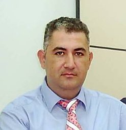 Yusuf Oner合作论文数Fac Engn, Pamukkale Univ3
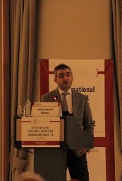 Abdulsamet Erden合作论文数Internal Medicine Department, Kayseri Training and Research Hospital3
 Galip Guz合作论文数3Erhan T. Ilgit合作论文数School of Medicine, Gazi University3Ayse Fadiloglu合作论文数2
 Esra Cataltepe合作论文数2Funda Yıldırım Borazan合作论文数2Basak Bolayir合作论文数2Rana Tuna Dogrul合作论文数2Turkane Fetullayeva合作论文数2Atiye Cenay Karabörk Kılıç合作论文数2İbrahim İleri合作论文数2Rıza Can Kardaş合作论文数2Eda Çeker合作论文数2Guldal Esendagli合作论文数2
 Akkan Koray合作论文数School of Medicine, Gazi University2




