Backscatter imaging is a reliable method of single-sided X-ray imaging where access may pose limitations on traditional transmission X-ray image capture. Because of physical challenges in its implementation, it has traditionally been regarded as unable to do material discrimination, a capability reserved for dual energy and transmission imaging by industry and academia. This research expanded on a novel method developed at the University of Florida, where a material calibration tool was used to calibrate a Viken Detection Nighthawk BTX in backscatter mode for postprocessing material discrimination. Namely, this novel approach identifies materials potentially found in explosive devices, i.e. lead, copper, steel, and plastic, as well as combinations of these materials through a fusion of filters. This research focused on expanding the previous work to determine if the novel approach worked at distances beyond the original use-based tests, as well as if source beam filtering and hardening would improve material resolution.
Advanced, real-time digital autoradiography (DAR) systems may provide rapid and selective identification of regions of interest on post-detonation samples. Data from these systems could aid in the creation of microsamples, enabling more timely sample analysis. One challenge is the high radioactivity of such samples, which can overwhelm many systems. Further, alpha emissions from actinides, of forensics interest, are overshadowed by colossal fission and activation products emitting beta and gamma-rays. DAR has the potential to quickly find targeted regions of interests where actinides and fission products are concentrated. This paper explores two advanced systems: the ionizing-radiation Quantum Imaging Detector (iQID), a real-time intensified scintillation camera, and large area Charge-Coupled Devices (CCDs) developed for dark-matter detection. These systems offer complementary features for identifying radioactive regions and particles of interest on a sample. This paper highlights measurement challenges with post-detonation samples, explores approaches such as use of gating and collimators to overcome quantitative measurement bottlenecks, and includes results of experiments to test those approaches. Concepts for measuring large samples with an iQID are also discussed.
Computed tomography (CT) is a central to three-dimensional medical imaging, yet CT-based artificial intelligence remains fragmented across task-specific models for segmentation, classification, registration, and report analysis. Here we present FlexiCT, a family of CT foundation models trained by agglomerative continual pretraining on 266,227 CT volumes from 56 publicly available datasets, forming a large-scale public resource for CT representation learning. FlexiCT uses agglomerative pretraining across three stages: two-dimensional axial pretraining, three-dimensional anatomical pretraining and report-guided semantic alignment. This training strategy supports slice-level, volume-level and vision-language analysis. Across five downstream task families (segmentation, classification, registration, vision-language understanding and clinical retrieval), FlexiCT matches or exceeds prior task-specific approaches on multiple benchmarks. Its embeddings further organize CT scans along gradients associated with various tumor stages, suggesting that CT foundation models can capture imaging features relevant to disease phenotype characterization. Code is available at https://github.com/ricklisz/FlexiCT
The characterization of materials is an essential aspect of nuclear forensics and safeguards. Verifying and examining materials is one way to help prevent proliferation. It is important to determine small changes that can indicate the diversion of special nuclear material. Conventionally, nuclear forensic examinations have been carried out through destructive imaging techniques. Because of this, the availability of a nondestructive image acquisition technique would help preserve samples for further examination and attribution. Traditional X-ray imaging has failed to differentiate between similar materials in a sample due to relying solely on transmission through the object. Gratings-based phase contrast X-ray imaging (GBX), on the other hand, allows for the extraction of three different signals: absorption, differential phase contrast that highlights edges in the material, and dark-field that measures scattering in the material. The application of GBX to characterize materials is explored. A sensitivity study was conducted on Monte Carlo N-Particle Transport Code (MCNP) in order to investigate the use of GBX on forensics applications.
Compton Backscatter Imaging (CBI) is a decades old approach to imaging surfaces where positioning of a detector and x-ray source for transmission imaging is either not possible or convenient. For that reason, it has been explored and utilized in areas of explosive detection and security applications, railway infrastructure integrity, and aerospace safety. Within these applications, much of the focus is on resolution for specific signatures within the target medium, particularly voids that indicate decreased structural integrity. The purpose of this research is to characterize the Modulation Transfer Function (MTF) signature of plastic, lead, steel, aluminum, and copper. MTF contrast analysis yields greater results in image clarity and post processing when compared to altering the image capture alone. Doing so would allow researchers to configure scanning equipment to detect key elements specific to CBI imaging of potential explosive devices. While past research has focused on an explosive penalty or contraband, the goal for this research is to target components of a device like the wiring or power source.
With advances in single sided handheld backscatter imaging and their incorporation into security applications, detector characterization is essential to operator feedback, image enhancement, and decision making. While a well-trained operator has the ability to discern contraband materials during inspections, understanding the limits of an imager’s resolution at varying distances inform additional inspections or advise threat scenarios. This research characterizes the detector response of a Nighthawk handheld backscatter imaging device developed by Viken Detection in laboratory conditions utilizing High-Density Polyethylene (HDPE) phantoms. The Nighthawk uses a collimated millimeter-sized pencil x-ray beam that scans 150 times per second laterally along a fan arc to generate a series of 1D linear images and then compile to a 2D image of the phantom. Sampling occurred using line pair bar slits of different interval frequencies and the straight edges of the HDPE phantoms at different separation distances from the NightHawk front surface ranging from 10 centimeters to 1 meters. The Nighthawk was placed on a moving table while phantom was kept stationary to maintain consistent scan speed during image capture. Image processing relied on analyzing the contrast visibilities of line pair phantoms and edge responses of a slanted edge phantom. The slanted edge image allowed over-sampling a sharp edge and then an Edge Spread Function and Modulation Transfer Function were derived to characterize system resolutions at different imaging distances.
Backscatter radiography, a form of x-ray inspection, has been used for better detection of flaws and degradation in the inspection of crossties. The method utilizes the portion of photons that scatter off the target object back towards the source. Current systems detect flaws and degradation in wood crossties but have been limited to the area of the tie between the rails. To advance the technology, this investigation seeks to evaluate the portion of the tie underneath the tie plate where x-ray attenuation severely impacts the image quality and penetration depth of the photons. Laboratory experiments were conducted to simulate scanning through tie plate materials. Scanning through the tie plate material is the best-case scenario for adapting current practices. Results show that backscatter radiography is an effective non-destructive testing method for crosstie evaluation and scanning through a tie plate is possible, but limitations exist with regards to fidelity and image resolution.
Explosive devices typically consist of several common materials, most of which have differing densities. When these materials are imaged through x-ray imaging, the resultant image displays differing contrast intensities within due to the scattering effects of these materials. These intensities provide shape and definition to the operator conducting the imaging, informing their decision making as to parts and components within the suspected device. A power source the size of a battery, or a piece of shrapnel, such a ball bearing, would have a noticeably different appearance than that of plastic explosives. The purpose of this research is to enhance single sided Compton Backscatter Imaging (CBI) of suspected explosive devices to support operators investigating a suspected device. Previous research by the team at UF in material discrepancy utilized Modulation Transfer Functions (MTF) derived from bar patterns of decreasing size to characterize a linear detector array and determine the maximum resolution in a CBI configuration. The previous research expanded into Edge Spread Functions (ESF) of these materials as a secondary method of characterizing material differences. This research explores the Slant Function, a derivative of the ESF, of differing materials to improve the ESF characterization. This occurs through a single edge analysis along every pixel of a linear detector array, comparing local intensities of the imaged edge to determine the cumulative effect on the overall ESF. This will allow for better MTF development, thereby enhancing filters developed for image post processing.
The health effects of galactic cosmic radiation are a serious impediment to crewed exploration of the solar system. OLTARIS, an interface for the 3DHZETRN deterministic radiation transport code, was used to assess the response of aerospace materials to this constant radiation exposure. Traditional aerospace structural materials like aluminum can, after a certain mass, increase the health effects of such radiation. However, materials with lower atomic mass may mitigate this build-up in secondary radiation with increasing areal density. As such, lower atomic mass structural alloys of magnesium and magnesium-lithium are promising candidates. These alloys may reduce the mass of structures when substituted for aluminum alloys. Reinforcement with boron carbide could further reduce atomic mass while also improving the mechanical properties of such lightweight alloys. This study found that the lower atomic mass of these materials increased nuclear fragmentation upon cosmic radiation interactions, leading to a softening of the secondary (neutron) radiation spectra. This softened spectra reduced the effective dose equivalent, a measure of health effects, for magnesium(-lithium) alloys and their boron carbide-reinforced composites when compared to aluminum.
Nuclear science and engineering is a field increasingly dominated by computational studies resulting from increasingly powerful computational tools. As a result, analytical studies, which previously pioneered nuclear engineering, are increasingly viewed as secondary or unnecessary. However, analytical solutions to reduced-fidelity models can provide important information concerning the underlying physics of a problem and aid in guiding computational studies. Similarly, there is increased interest in sensitivity analysis studies. These studies commonly use computational tools. However, providing a complementary sensitivity study of relevant analytical models can lead to a deeper analysis of a problem. This work provides the analytical sensitivity analysis of the one-dimensional (1D) cylindrical mono-energetic neutron diffusion equation using the forward sensitivity analysis procedure (FSAP) developed by Cacuci. Further, these results are applied to a reduced-fidelity model of a spent nuclear fuel cask, demonstrating how computational analysis might be improved with a complementary analytic sensitivity analysis.
Pulse shape discrimination (PSD) capable detectors, such as He-4, that respond to neutron and gamma-ray interactions have a threshold deposited energy value below which n/γ discrimination vanishes when using conventional PSD algorithms. Recent attempts in applying supervised learning based artificial neural networks for PSD use the pulses in the separated regions to train the networks so they can be used to classify another set of separated pulses. In doing so, pulses previously indistinguishable are not recovered for classification, which would have increased the number of neutron and gamma-ray pulses that could be used for further analysis. Assuming the reason why conventional PSD algorithms have unseparated regions is because the parameter space of the algorithms fail to capture the intrinsic (but subtle) distinguishing behavior of some of the neutron and gamma-ray pulses, a cycle-consistent generative adversarial network (CycleGAN) was trained to amplify those differences and extract well separated neutron and gamma-ray clusters. Results show that, once the network is trained with pulses from separated and unseparated regions, it was able to transform the pulses in the unseparated region to improve the PSD. Subsequent n/γ classification was performed using deep residual network (ResNet) that takes pulses with 512 data points as an input. Two different ResNets were explored – simple ResNet and modified ResNet which takes segmented pulse inputs in the first layer and the corresponding time axis values in the last hidden layer. The later approach enables the network to extract time correlated pulse features to enhance its ability to capture the pulse behaviors relevant for PSD. Although it achieves slightly lower accuracy, 99.41% versus 99.89%, based on simply counting the number of correct n/γ labels assigned, compared to the simple ResNet, the modified ResNets architecture was able to decreases the cross-entropy loss function by half, which implies that the correct n/γ labels assigned are less likely to be accidental. PSD parameter distributions based on n/γ classification by ResNet before and after transforming unseparated pulses using CycleGAN show that by enhancing the separation between neutrons and gamma-rays, the transformation helps improve the performance of classifier networks that are trained using labeled dataset. The enhancement of neutron and gamma-ray separation by the CycleGAN increased the PSD figure of merit (FOM) by up to 70% in some regions. The results show that, if a given detector achieves clear separation between neutron and gamma-ray pulses in any energy region, such neural network approaches can help lower the energy threshold for the separation and increasing the number of neutron and gamma-ray pulses that can be used for further analysis.
In the past when faced with solving a non-tractable problem, scientists would make tremendous efforts to simplify these problems while preserving fundamental physics. Solutions to the simplified models provided insight into the original problem. Today, however, the affordability of high-performance computing has inverted the process for analyzing complex problems. In this paradigm, results from detailed computational scenarios can be better assessed by “building down” the complex model through simple models rooted in the fundamental or essential phenomenology. This work demonstrates how the analysis of the neutron flux spatial distribution behavior within a simulated Holtec International HI-STORM 100 spent fuel cask is enhanced through reduced complexity analytic and computational modeling. This process involves identifying features in the neutron flux spatial distribution and determining the cause of each using reduced complexity computational and/or analytic model. Ultimately, confidence in the accuracy of the original simulation result is gained through this analysis process.
Compton backscatter imaging is a single-sided imaging technique and can be used as an excellent NDE tool for various applications. To develop an efficient and a high-resolution x-ray backscatter imaging system, a low-cost data acquisition electronic board to work with scintillation detectors in current mode is developed and tested. This in-house built current mode data acquisition board allows for higher count rate and to collect more information about the energy deposited in the detector. The board provides four independent data measuring channels running in current mode. Each channel can be configured with 500 integration selections and 2500 gain selections manually and digitally. The data from all four channels are sampled by a 32-bit micro-controller with onboard 16-bit high-speed ADCs with sample rates up to 1200 ksps/channel. The board also provides 22 general I/Os, which can be used to control mechanical components such as raster scanning tables, linear motion stages and rotating collimators. The total cost of this unique data acquisition board is less than $150. With an open system design, other modules can be added to this board to increase functionalities. This low-cost current mode data acquisition board has been tested on the backscatter imaging system at UF under different experiment setups. From the backscattered images of the concrete contrast tool, the minimum detectable depth of the defect is 1.5 mm with the RSD system and the data acquisition board. With its undeniable low price and from the results, this data acquisition board shows great potential in x-ray backscatter imaging and signal measurement applications such as non-destructive inspection of CFAT materials.
The energy spectrum and spatial distribution of the neutron beam from a reactor is an essential component of designing experimentations utilizing the reactor as a radiation source. Obtaining such information requires the use of detailed MCNP model of the reactor and experimental validation procedures. After updating the MCNP model of the University of Florida Training Reactor (UFTR) to align it with the current core arrangement that uses low enriched uranium (LEU), both the radial neutron flux toward the beam ports and the energy spectrum at the reactor core were generated using the model. This was followed by experimental validations based on neutron activation analysis (NAA) by irradiating multiple activation foils. A constant ratio between MCNP and measured neutron flux values was obtained at multiple locations along the reactor's beam ports, showing that the model captures the spatial distribution of neutron flux profiles without a significant systematic error. The energy spectrum at the core was extracted using the Unfolding with MAXED and GRAVEL (UMG) software. The spectrum unfolded by both MAXED and GRAVEL algorithms display features expected in thermal reactor neutron energy distribution, i.e., thermal, fast or fission, and intermediate or slowing down regions. The thermal and epithermal neutron fluxes at the reactor core calculated from the spectrum unfolded by the MAXED algorithm are 1.56 ± 0.12 × 10¹²n/cm2s and 1.98 ± 0.01 × 10¹²n/cm2s, whereas the corresponding values calculated from the spectrum unfolded by the GRAVEL algorithm are 1. 50 ± 0. 12 × 1012n/cm2s and 3.74 ± 0.21 × 10¹² n/cm2s. Following the n/cm2s. experimental validation, the full geometry MCNP model of the reactor has been utilized for a neutron tomography research project and it will continue to benefit future projects using the reactor facility.
Premise High‐resolution cameras are very helpful for plant phenotyping as their images enable tasks such as target vs. background discrimination and the measurement and analysis of fine above‐ground plant attributes. However, the acquisition of high‐resolution images of plant roots is more challenging than above‐ground data collection. An effective super‐resolution (SR) algorithm is therefore needed for overcoming the resolution limitations of sensors, reducing storage space requirements, and boosting the performance of subsequent analyses. Methods We propose an SR framework for enhancing images of plant roots using convolutional neural networks. We compare three alternatives for training the SR model: (i) training with non‐plant‐root images, (ii) training with plant‐root images, and (iii) pretraining the model with non‐plant‐root images and fine‐tuning with plant‐root images. The architectures of the SR models were based on two state‐of‐the‐art deep learning approaches: a fast SR convolutional neural network and an SR generative adversarial network. Results In our experiments, we observed that the SR models improved the quality of low‐resolution images of plant roots in an unseen data set in terms of the signal‐to‐noise ratio. We used a collection of publicly available data sets to demonstrate that the SR models outperform the basic bicubic interpolation, even when trained with non‐root data sets. Discussion The incorporation of a deep learning–based SR model in the imaging process enhances the quality of low‐resolution images of plant roots. We demonstrate that SR preprocessing boosts the performance of a machine learning system trained to separate plant roots from their background. Our segmentation experiments also show that high performance on this task can be achieved independently of the signal‐to‐noise ratio. We therefore conclude that the quality of the image enhancement depends on the desired application.
An update is provided on the dissertation underway and what has been learned thus far. Work thus far: Developed analytic models for each region in the spent fuel cask – Used to identify and explain physical processes which create features in detailed casks; Developed simplified computational models to identify details not seen in analytic models; SC were calculated in the fuel region; The difference in SC’s between the analytic model and the simplified computational model were identified. Outstanding issues: Analysis of analytic models in stainless steel and carbon steel – These materials are thin and have few features (just the slope); Discrepancies between absorption SC’s in fuel; Create a test problem to show the effects of the high energy resonances in the fuel region. Future Work: Sensitivity analysis needs to be continued through the cask – The detailed model will be added to the remaining materials; Sensitivity analysis paper; and, Addressing outstanding issues.
 Andreas Enqvist合作论文数Oak Ridge National Laboratory;Chalmers University of Technology;Department of Nuclear Engineering;Oak Ridge National Laboratory, Chalmers University of Technology14
 Kelly A. Jordan合作论文数Paul Scherrer Institut13
 Juan Claudio Nino合作论文数Department of Materials Science and Engineering, University of Florida12
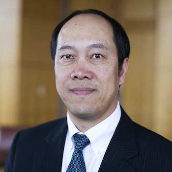 Zhong He合作论文数Department of Nuclear Engineering & Radiological Sciences, College of Engineering, University of Michigan11
 J. Nimmagadda合作论文数10
 Glenn Sjoden合作论文数University of Florida10
 Sasmit S. Gokhale合作论文数Brookhaven National Laboratory (BNL)7Gabriel Ghita合作论文数6Yuan Gao合作论文数Dept Radiat Oncol, Emory Univ6
 Shuang Cui合作论文数Changchun Institute of Optics Fine Mechanics and Physics, Chinese Academy of Sciences Chinese Academy of Sciences6Hannah Gardiner合作论文数5Rebecca S. Detwiler合作论文数5Christopher R. Greulich合作论文数Oak Ridge National Laboratory5Benjamin S Mcdonald合作论文数5Wei Qiu合作论文数5Travis R. Barker合作论文数4
 Paul Johns合作论文数4Lucas M. Rolison合作论文数Los Alamos Natl Lab4Christopher R. Hughes合作论文数Nuclear Engineering Program, University of Florida4HyukSu Han合作论文数4Y. Liang合作论文数4Detwiler R合作论文数4Robert C. Runkle合作论文数4
 Azaree Lintereur合作论文数Pacific Northwest National Laboratory4Tyler J. Remedes合作论文数3Randy R Kirkham合作论文数3S. Samant合作论文数3Scott D. Ramsey合作论文数XTD Appl Phys, Los Alamos Natl Lab3Keillor Martin E合作论文数3Brian D Milbrath合作论文数3
 Alina Zare合作论文数Department of Electrical & Computer Engineering, University of Florida;Artificial Intelligence and Informatics Research Institute, University of Florida3Carolyn E. Seifert合作论文数3Walter K. Hensley合作论文数2Timothy K. Mueller合作论文数2Jeremy P. Rishel合作论文数2Dudley F. Emer合作论文数2Gabriel Sandler合作论文数2Ting Zhu合作论文数2Surafel Woldegiorgis合作论文数2
 A. L. Richard合作论文数Facil Rare Isotope Beams, Michigan State Univ2Leah M. Arrigo合作论文数2H. Ray合作论文数2
 minki hong合作论文数Kyulux North Amer Inc2
 Cody E. Parker合作论文数2
 Soumitra Sulekar合作论文数Dept & Mat Res Lab, Univ Illinois2Seifert, A.合作论文数2Warnick J. Kernan合作论文数2Chandra, R.合作论文数2John E. Schweppe合作论文数2




