This study presents a reproducible methodological framework for constructing a longitudinal low-dose computed tomography (LDCT) screening image database and demonstrates its implementation using 11 years of real-world data from a single region in Japan. The framework integrates hierarchical identifier design, deterministic metadata linkage, structured anonymization, and a minimal metadata specification to enable consistent organization of longitudinal imaging data. The implemented database comprises 45,337 LDCT examinations from 23,065 examinees, with 47.0
The Japanese Society of Radiological Technology (JSRT) chest radiograph database (JSRT-DB), developed in 1998, has been widely used in medical imaging research for nearly three decades and has been cited more than 800 times. Although recent advances in artificial intelligence have shifted the focus toward large-scale datasets, the JSRT-DB has played an important role in establishing methodological standards for diagnostic performance evaluation, particularly in lung nodule detection. This review summarizes the historical background, design philosophy, and significant contributions of the JSRT-DB to observer performance studies, computer-aided diagnosis, and radiological education. It also discusses the lessons learned from the JSRT-DB that remain relevant to the development and utilization of large-scale medical imaging databases.
Somatostatin receptor scintigraphy (SRS) is an essential examination for the diagnosis of neuroendocrine tumors (NETs). This study developed a method to individually optimize the display of whole-body SRS images using a deep convolutional neural network (DCNN) reconstructed by transfer learning of a DCNN constructed using Gallium-67 ( 67 Ga) images. The initial DCNN was constructed using U-Net to optimize the display of 67 Ga images (493 cases/986 images), and a DCNN with transposed weight coefficients was reconstructed for the optimization of whole-body SRS images (133 cases/266 images). A DCNN was constructed for each observer using reference display conditions estimated in advance. Furthermore, to eliminate information loss in the original image, a grayscale linear process is performed based on the DCNN output image to obtain the final linearly corrected DCNN (LcDCNN) image. To verify the usefulness of the proposed method, an observer study using a paired-comparison method was conducted on the original, reference, and LcDCNN images of 15 cases with 30 images. The paired comparison method showed that in most cases (29/30), the LcDCNN images were significantly superior to the original images in terms of display conditions. When comparing the LcDCNN and reference images, the number of LcDCNN and reference images that were superior to each other in the display condition was 17 and 13, respectively, and in both cases, 6 of these images showed statistically significant differences. The optimized SRS images obtained using the proposed method, while reflecting the observer's preference, were superior to the conventional manually adjusted images.
Kiken-yochi Training (KYT) has been introduced in many hospitals as medical safety education in many departments, with the exception of radiology. KYT is also not used in the contents of medical safety lectures in the education of students for radiological technologists. One of the reasons for this is that the images for KYT (KYT images) cannot be created at each hospital or easily obtained on the website. The purpose of this research is to construct a database of KYT images (KYTDB) that can be used for medical safety education at hospitals and educational facilities. We also investigate the usefulness of KYTDB in FROC observer experiments. The KYT images were taken in various scenarios of routine medical examination (or treatment) in the radiology department of the two hospitals. A total of 367 KYT images were taken in seven sections of the radiology department, some containing dangerous (inappropriate) locations and languages, and some containing normal scenes. In addition, educational training materials in PowerPoint with audio were created for self-study of KYT. A FROC observer study was conducted using sample images extracted from the KYTDB that were not used in the educational materials, and figures of merit values were used to quantify the predictive risk reduction capacity of KYT. The results demonstrated that the KYTDB is useful for KYT education.
PURPOSE:The present study aimed to investigate the current situation of radiation protection education for designated radiation workers in hospitals.METHODS:A web-based questionnaire survey was conducted at 1,883 hospitals nationwide with 200 or more beds.RESULTS:Responses from 186 hospitals were included in the analysis. Seven hospitals (6.7%) regulated by the Act on the Regulation of Radioisotopes and six hospitals (7.4%) regulated by only the Ordinance on Prevention of Ionizing Radiation Hazards did not implement radiation protection education. In approximately 6% of the hospitals, designated radiation workers-including physicians, nurses, and radiological technologist-did not attend the education program. The education program attendance rate of physicians was lower than that of nurses. In more than 90% of the hospitals, the frequency of the periodical education program was once every year and lecture time spanned one or less than one hour. The topics of lecture in more than 90% of the hospitals were health effects of radiation and methods of radiation protection for occupational exposure. The radiological technologist was the instructor of the education program in approximately 70% of the hospitals.CONCLUSION:The implementation of radiation protection for designated radiation workers varied from hospital to hospital, and some hospitals did not comply with laws and regulations. Effective and efficient radiation protection education models should be implemented in hospitals.
Purpose: To develop a deep learning model to estimate lung age based on low-dose chest computed tomography (CT) images and investigate the feasibility of its application in health management. Materials and Methods: Deep learning models (InceptionV3, DenseNet, VGG16, and ResNet50) were trained on a set of low-dose chest CT images of 4,646 non-smokers. They were adjusted for an equal number of participants in each age group to estimate the chronological age and functional lung age. They were further tested on a dataset of 5,841 non-smokers and 16,709 smokers. The estimated ages were evaluated based on the mean absolute error, correlation coefficients with the actual ages, and Bland-Altman analysis. In addition, we tested whether the estimated age was higher in the group with a longer the smoking history. Results: There was a correlation between the estimated and actual ages in all models (chronological/functional lung ages): r=0.74/0.54, r=0.75/0.53, r=0.68/0.50, and r= 0.67/0.34 for InceptionV3, DenseNet, VGG16, and ResNet50, respectively. The InceptionV3 model showed the best performance with an estimation error of +/- 4.02 and +/- 11.18 years for chronological and functional lung ages, respectively. Furthermore, among smokers, the longer the smoking history, the higher the estimated chronological age, suggesting that the trained model can predict lung changes other than aging, such as the effects of smoking and signs of disease onset. Conclusion: We developed an age estimation model based on low-dose chest CT images that can quantify lung damage due to smoking as an increase in predicted age. The feasibility of CT image-based lung age estimation for health care was demonstrated.
In this study, we developed a method for generating quasi-material decomposition (quasi-MD) images from single-energy computed tomography (SECT) images using a deep convolutional neural network (DCNN). Our aim was to improve the detection of cholesterol gallstones and to determine the clinical utility of quasi-MD images. Four thousand pairs of virtual monochromatic images (70 keV) and MD images (fat/water) of the same section, obtained via dual-energy computed tomography (DECT), were used to train the DCNN. The trained DCNN can automatically generate quasi-MD images from the SECT images. Additional SECT images were obtained from 70 patients (40 with and 30 without cholesterol gallstones) to generate quasi-MD images for testing. The presence of gallstones in this dataset was confirmed by ultrasonography. We conducted a receiver operating characteristic (ROC) observer study with three radiologists to validate the clinical utility of the quasi-MD images for detecting cholesterol gallstones. The mean area under the ROC curve for the detection of cholesterol gallstones improved from 0.867 to 0.921 ( p = 0.001) when quasi-MD images were added to SECT images. The clinical utility of quasi-MD imaging for detecting cholesterol gallstones was showed. This study demonstrated that the lesion detection capability of images obtained from SECT can be improved using a DCNN trained with DECT images obtained using high-end computed tomography systems.
In this study, we propose a method for obtaining a new index to evaluate the resolution properties of computed tomography (CT) images in a task-based manner. This method applies a deep convolutional neural network (DCNN) machine learning system trained on CT images with known modulation transfer function (MTF) values to output an index representing the resolution properties of the input CT image [i.e., the resolution property index (RPI)]. Sample CT images were obtained for training and testing of the DCNN by scanning the American Radiological Society phantom. Subsequently, the images were reconstructed using a filtered back projection algorithm with different reconstruction kernels. The circular edge method was used to measure the MTF values, which were used as teacher information for the DCNN. The resolution properties of the sample CT images used to train the DCNN were created by intentionally varying the field of view (FOV). Four FOV settings were considered. The results of adapting this method to the filtered back projection (FBP) and hybrid iterative reconstruction (h-IR) images indicated highly correlated values with the MTF 10% in both cases. Furthermore, we demonstrated that the RPIs could be estimated in the same manner under the same imaging conditions and reconstruction kernels, even for other CT systems, where the DCNN was trained on CT systems produced by the same manufacturer. In conclusion, the RPI, which is a new index that represents the resolution property using the proposed method, can be used to evaluate the resolution of a CT system in a task-based manner.
Purpose: The necessity of image retakes is initially determined on a preview monitor equipped with an operating system; therefore, some image blurring is only noticed later, on a high-resolution monitor. The purpose of this study is to investigate blur detection performance on radiographs via a deep learning approach compared with human observers. Approach: A total of 99 radiographs (blurry 57, nonblurry 42) were independently observed and rated by six observers using preview and diagnostic liquid crystal displays (LCDs). The deep convolution neural network (DCNN) was trained and tested using ninefold cross-validation. The average areas under the ROC curves (AUCs) were calculated for each observer with LCDs and by stand-alone DCNN for each test session and then statistically tested using a 95% confidence interval. Results: The average AUCs were 0.955 for stand-alone DCNN and 0.827 and 0.947 for human observers using preview and diagnostic LCDs, respectively. The DCNN revealed a high performance for image motion blur on digital radiographs (sensitivity 94.8%, specificity 96.8%, and accuracy 95.6%), along with the capability to detect a slight motion blur that was overlooked by human observers with a preview LCD. There were no cases of motion blur overlooked by the stand-alone DCNN, of which some were incorrectly recognized as nonblurry by human observers. Conclusions: The deep learning-based approach was capable of distinguishing slight motion blur that was unnoticeable on a preview LCD, and thus, is expected to aid the human visual system for detecting blurred images in the initial review of digital radiographs.
In many digital X-ray imaging systems, although air kerma on a surface of each detector is used, a standardized dose index called an exposure index (EI) has been proposed by the IEC, which is expected to be utilized for dose management. In clinical practices, EI is effectively utilized using a deviation index (DI), which is a deviation between a target EI (EIT) set for each imaging region and an EIT of the acquired image. However, an important issue in clinical uses of EI is a suppression of excessive doses. It is difficult to achieve a reliable reduction in exposure doses by indicating DI. In this study, physical image characteristics of detectors, visual detectability by charts, and observer experiments using a chest phantom were examined to determine upper (DImax) and lower (DImin) limits of the EIT and DI to achieve a reliable dose reduction in chest examinations. As the result, the tolerance ranges indicated by DImax and DImin, which were set based on the results of physical and visual evaluations, proved to be almost consistent with the distribution of EI values in 735 clinical images taken with a photo-timer control in real clinical practices.
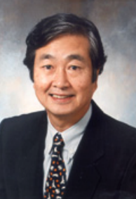 Kunio Doi合作论文数Department of Radiology, University of Chicago71
 Feng Li合作论文数Department of Radiology, The University of Chicago23
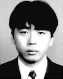 Yoshikazu Uchiyama合作论文数19
 Heber Macmahon合作论文数Department of Radiology, University of Chicago16
 Shigehiko Katsuragawa合作论文数16Chisako Muramatsu合作论文数Graduate School of Medicine, Gifu University16Hatagawa Masakatsu合作论文数15Roger Engelmann合作论文数15Robert A. Schmidt合作论文数14Kishimoto Kenji合作论文数13
 Rie Tanaka合作论文数13Qiang Li合作论文数13Akane Utsunomiya合作论文数11
 Hiroshi Fujita (藤田廣志)合作论文数Faculty of Engineering, Gifu University;Zhengzhou University;Shiga University of Medical Science10
 Fuminori Moriyasu合作论文数Center for Cancer Ablation Therapy at Sanno Hospital, International University of Health and Welfare;Tokyo Medical University9
 Takeshi Hara合作论文数Graduate School of Medicine, Gifu University9
 Katsutoshi Sugimoto合作论文数9Daniel Appelbaum合作论文数9Kenji Suzuki合作论文数8Shinichi Watanabe合作论文数8
 Yonglin Pu合作论文数Department of Radiology, The University of Chicago8Hiroyuki Abe合作论文数8S Sone合作论文数7Gillian M. Newstead合作论文数7Daisaku Tatsumi合作论文数6Goto Makoto合作论文数6M Aoyama合作论文数6Katsuhiro Ichikawa合作论文数6Kazuyoshi Tsuda合作论文数5
 Yoshie Kodera合作论文数5Ichida Takao合作论文数5Nose Hideo合作论文数5Tosiaki Miyati合作论文数5
 Nobuyuki Kanematsu (兼松伸幸)合作论文数Department of Accelerator and Medical Physics, National Institutes for Quantum Science and Technology;Institute for Quantum Medical Science, National Institutes for Quantum Science and Technology5Ryohei Fukui合作论文数4Yamaya Taiga合作论文数4Satoshi Kasai合作论文数4Fujio Araki合作论文数4KAZUYA YAMASHITA合作论文数4Higashida Mitsuji合作论文数4Tsushima Hiroyuki合作论文数4Shigeru Sanada合作论文数4Takeji Sakae合作论文数University of Tsukuba4Daisuke Fukuoka合作论文数4Nobuko Hanaoka合作论文数3Nobuhiko Shiizaki合作论文数3




