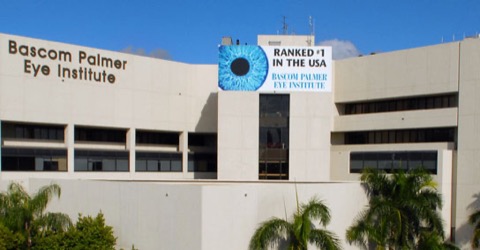
Bascom Palmer Eye Institute, of the University of Miami Leonard M. Miller School of Medicine, is a center for ophthalmic care, research, and education. Faculty and staff treat patients from around the world in facilities in Miami-Dade County, Broward County, Palm Beach County, and Collier County. The Institute's full-time faculty and researchers encompass many ophthalmic sub-specialties and has been consistently ranked as the #1 eye hospital and vision research center in the country by US News & World Report.The Institute's clinical faculty treats more than 250,000 patients each year, provides 24-hour emergency care, and is the only community-based ophthalmic care for indigent and low-income patients of Miami-Dade County.

Purpose:To quantify nonperfusion areas (NPA) and intraretinal microvascular abnormalities (IRMAs) and evaluate their diagnostic performance in diabetic retinopathy (DR) using ultrawide-field (UWF) swept-source optical coherence tomography angiography (SS-OCTA). Methods:In this cross-sectional study, nonperfusion index (NPI) and IRMA counts were analyzed on single-capture 29 × 24 mm optical coherence tomography angiography (OCTA) images. Regional analyses across the central zone (CZ), intermediate zone (IZ), and outer zone (OZ) were performed. Area under the receiver operating characteristic analysis evaluated the ability of regional and total metrics to discriminate DR severities. Results:A total of 165 eyes from 120 patients (73 male; mean age, 57.0 ± 11.1 years) across DR severities were included. Total NPI and IRMA counts increased with advancing DR severity (P < 0.001). NPI increased significantly from CZ to IZ across all DR stages (P < 0.001), with further extension into OZ in proliferative DR (PDR) (P < 0.001). IRMA counts were highest in the IZ in moderate nonproliferative DR (NPDR), severe NPDR, and PDR (P < 0.001). OZ NPI performed comparably with total NPI for distinguishing PDR from NPDR (area under the receiver operating characteristic, 0.88 vs. 0.86; P = 0.21), whereas IZ NPI and IRMA counts performed comparably with their respective total field measurements for distinguishing advanced DR from early DR (NPI, 0.95 vs. 0.95 [P = 0.83]; IRMA, 0.86 vs. 0.89 [P = 0.12]). Conclusions:Single-capture UWF SS-OCTA supports a regional biomarker framework for DR. More peripheral ischemia may add value for identifying neovascular transformation, whereas imaging extending into the intermediate retina may retain useful diagnostic value for distinguishing advanced from early DR when full UWF imaging is unavailable.
To describe the clinical indications for tube revision with a Tube Extender and to characterize the eyes in which they were used. Single-center retrospective cohort of eyes undergoing Tube Extender implantation (2015–2023). Clinical data were collected from medical records, and Fisher’s exact test assessed associations between glaucoma type and revision indication. Twenty-two eyes from 22 patients were included. The mean age at tube revision was 53 years, and 68
PURPOSE:To evaluate the effectiveness of ChatGPT-4o (OpenAI) in enhancing the readability and quality of Spanish patient educational materials (PEMs) on childhood glaucoma, and to determine whether they are written above the recommended 6th-grade reading level (per American Medical Association recommendation). METHODS:This cross-sectional comparative study analyzed 10 original Spanish-language PEMs on "glaucoma infantil." Each was inputted into ChatGPT-4o using a Spanish-language prompt requesting improved readability to a score of 70 or greater on the INFLESZ scale (adaptation of the Flesch-Szigriszt Index for Spanish-language texts), which is equivalent to a 4th- to 6th-grade reading level. Original and revised PEMs were compared using the INFLESZ scale. Additionally, three native Spanish-speaking ophthalmologists assessed each PEM's quality using a published 15-point Likert scale evaluating helpfulness, truthfulness, and harmlessness. RESULTS:Original PEMs had a mean INFLESZ score of 56.6 ± 4.9 (8th-grade level). ChatGPT-4o significantly improved readability to 69.6 ± 2.3 (4th- to 6th-grade level, P < .001). Revised PEMs had fewer syllables, words, and complex words without a significant difference in sentence count. Quality remained high across both groups (median score = 15, P = .74), and no hallucinations were observed in artificial intelligence-generated text. CONCLUSIONS:ChatGPT-4o can be used as a supplemental tool by health care professionals to improve the readability of existing Spanish PEMs on childhood glaucoma, without sacrificing the quality, accuracy, or reliability of their content.
Purpose: This case report describes a patient who developed a second episode of endophthalmitis in the same eye following intravitreal injections administered 2 years apart. Observations: An 83-year-old pseudophakic male with a history of neovascular age-related macular degeneration and diabetes mellitus complained of decreased visual acuity and pain in the left eye 1 day after receiving intravitreal aflibercept 2 mg. Best-corrected visual acuity (BCVA) was light perception. The clinical examination revealed signs of endophthalmitis including fibrin and hypopyon while B-scan ultrasonography demonstrated vitreous opacities and membranes. The patient underwent vitreous tap and injection with intravitreal vancomycin, ceftazidime, and dexamethasone. Pars plana vitrectomy with posterior hyaloid detachment was performed the following day due to a relative lack of clinical improvement. Vitreous cultures ultimately demonstrated no growth and BCVA improved to baseline. Anti-VEGF therapy was resumed 3 months after the initial diagnosis of endophthalmitis. Two years later the patient presented with evidence of a second episode of endophthalmitis 4 days after receiving aflibercept 8 mg. Vitreous tap and injection was again performed and cultures revealed growth of Staphyloccoccus epidermidis. The patient demonstrated marked improvement with no evidence of residual inflammation at 2 weeks. Best-corrected visual acuity was 20/50 at 7-month follow-up examination. Conclusion and importance: Endophthalmitis is a rare complication of intravitreal injection. The current report describes a patient who developed a second occurrence of endophthalmitis associated with an intravitreal injection 2 years later.





