Being the basic building blocks of chromatin, nucleosomes and their stability determine the genome accessibility for different DNA-dependent proteins. This characteristic is labile under all cell-life processes. One of the abundant DNA-binding proteins, which is important for genome compaction, is poly(ADP-ribose)polymerase1 (PARP1). Despite the extensive experimental data on the chromatin compaction regulation under ADP-ribosylation, the details of the interplay of nucleosome with PARP1 in the absence of protein activation remain unclear. In this study, we analyzed the changes in the nucleosome wrapping upon PARP1 interaction using a single-molecule approach — optical tweezers. We demonstrate that PARP1 binding leads to weakening of the contacts that support the nucleosome core.
DNA damage repair by the base excision repair (BER) mechanism is a complex, multistage process that requires precise coordination and regulation of activities of enzymes at each step of DNA repair. The central stage in this process is DNA synthesis in the gap formed by removal of the damaged nucleotide. DNA polymerases β (Pol β) and λ (Pol λ) of the X family possess all properties necessary for the DNA repair synthesis. Pol β is the major enzyme in DNA synthesis in BER, which may be due to the influence of other BER factors regulating its activity. The scaffold protein XRCC1 and poly(ADP-ribose) polymerases 1 and 2 (PARP1 and PARP2) are essential for the formation of functional BER complexes. We investigated the effect of XRCC1, PARP1, PARP2, and poly(ADP-ribosyl)ation on the activity of Pol β and Pol λ in the single-nucleotide gap filling reaction in BER. XRCC1 stimulated the activity of Pol λ to a significantly greater extent than the Pol β activity. PARP1 and PARP2 inhibited both DNA polymerases; the inhibitory effect of PARP1 was more pronounced on DNA with a tetrahydrofuran-phosphate group at the 5′ end of the gap, while PARP2 effect was more significant on DNA containing a 5′ phosphate group. XRCC1 partially restored the DNA polymerase activity, especially that of Pol β. It can be assumed that the XRCC1–Pol β complex competes with PARP1/2 for DNA binding more strongly than the XRCC1–Pol λ complex. Addition of NAD+ to PARP2 led to a more efficient restoration of Pol β (but not Pol λ) activity. These differences may be one of the causes why Pol β is the main DNA polymerase in BER.
Topotecan is widely used against various malignancies inhibiting topoisomerase 1 (TOP1). It traps TOP1 in the cleavage complex bound to the DNA, which potentially may be repaired by a tyrosyl‑DNA phosphodiesterase 1 (TDP1). The usnic acid derivative OL9‑116, that inhibits TDP1, increases the antitumor efficacy of topotecan and is a promising adjunct for combination therapy. However, its safety and the optimal administration schedules when used with topotecan remain uncharacterized. We demonstrated that OL9‑116 has no genotoxic and epigenetic effects using Ames and CBMN tests, as well as HeLa trichostatin A‑inducible cells with an epigenetically repressed GFP gene. Intraperitoneal administration was found to provide higher bioavailability than intragastric administration. We showed on the RShM‑5 cervical cancer model that repeated topotecan administration in a clinically relevant low single‑dose regimen was more effective than a single injection, and addition of topotecan at the Tmax of OL9-116 enhanced tumor growth inhibition. In the Lewis lung carcinoma model, the OL9‑116–topotecan combination reduced primary tumor mass and lung metastases, enhancing both antitumor and antimetastatic effects. Monitoring of hematological parameters, liver, and spleen indices revealed no clinically significant systemic toxicity. Moreover, peripheral blood parameters improved with the OL9‑116–topotecan combination compared with topotecan monotherapy. Collectively, these findings support OL9‑116 as a promising safe TDP1 inhibitor for combination with topotecan.
FUS participates in the formation of biomolecular condensates associated with PARP1-dependent synthesis of poly(ADP-ribose) (PAR). HPF1 regulates auto- and hetero-PARylation activities of PARP1 and PARP2 and may influence the formation of FUS compartments during PARP1 or PARP2 auto-PARylation. In this study, we used atomic force microscopy in combination with biochemical assay to investigate the formation of FUS compartments under activation of PARP1 and PARP2, when HPF1 modulates their activity. Similar to PARP1, FUS and PARylated PARP2 form DNA-rich compartments, indicating that PARP2 PARylation is sufficient for the formation of such compartments. The excess of HPF1 over PARP1 diminishes PARP1 activity and reduces the size of DNA-rich compartments. However, an excess of HPF1 over PARP2 does not significantly affect PARP2 activity and the size of compartments. Furthermore, HPF1 stimulates hetero-PARylation of FUS; this modification is stronger with PARP2 than with PARP1. HPF1-dependent intensive PARylation of FUS catalyzed by PARP1 or PARP2 impairs the assembly of DNA-rich compartment. These data provide a basis for investigating the effect of HPF1 on the formation of PAR-dependent condensates involving RNA-binding proteins like FUS, which interact effectively with PAR and show the ability to be targets of PARylation to regulate condensate formation at DNA damage sites.
DNA polymerase β (Polβ) is a central player of base excision repair (BER), performing gap-filling synthesis on damaged DNA. While nucleosome core particles (NCPs) are known to impede activity of BER enzymes, the regulation of this process in linker DNA adjacent to nucleosomes remains unclear. Here we demonstrate an unexpected stimulation of Polβ-catalyzed gap-filling and strand-displacement synthesis in linker DNA by the adjacent NCP. Notably, the nucleosomal context reinforces the regulatory modulation of Polβ activity by PARP1/PARP2 and FEN1. While linker histone H1 restricts strand-displacement synthesis at the nucleosome entry/exit site, PARP1 and PARP2 modulate Polβ function through competitive binding to DNA gaps or nicks and via poly(ADP-ribosyl)ation (PARylation). At the same time, PARPs binding differentially regulates BER sub-pathway choice, and PARylation alleviates H1-mediated inhibition. These findings reveal a multi-layered regulatory system wherein the nucleosome acts as a dynamic platform coordinating Polβ activity and its interplay with chromatin-associated factors, influencing the balance between short- and long-patch BER. The research advances understanding of chromatin-mediated control of BER DNA repair synthesis and the functional specialization of PARP1 and PARP2 in maintaining genome stability.
DNA-dependent nuclear enzymes poly(ADP-ribose) polymerases 1 and 2 (PARP1 and PARP2) are involved in the regulation of multiple DNA repair pathways, including base excision repair (BER). After activation by binding to damaged DNA, these enzymes synthesize negatively charged poly(ADP-ribose) (PAR) and covalently attach to amino acid residues of target proteins, including PARPs themselves. PARP2 activity is influenced by the nature of DNA lesion; for example, it is efficiently stimulated by DNA breaks flanked by phosphate group. However, it remains unclear which stages of the auto-PARylation reaction are most sensitive to the structure of damaged DNA. In this study, we investigated how PARP2 activity depends on the presence and position of a single-nucleotide gap in DNA (either free or in the context of nucleosome) at different stages of the automodification reaction conducted in the absence of the histone PARylation factor HPF1. The obtained results suggest that the presence of the gap affects the affinity of PARP2 for DNA/nucleosomes, thereby determining the number of catalytically active enzyme molecules and the efficiency of PARylation initiation. In contrast, PAR elongation was affected by the lesion location in the DNA/nucleosome structure, namely, its distance from the blunt DNA ends, and the environment of histone tails. Therefore, the damaged DNA structure can influence both the amount and the length of PAR synthesized by PARP2.
Being the basic building blocks of chromatin, nucleosomes and their stability determine the genome accessibility for different DNA-dependent proteins. This characteristic is labile under cell-life processes. One of the abundant DNA-binding proteins, which is important for genome compaction, is poly(ADP-ribose) polymerase1 (PARP1). Despite the extensive experimental data on the chromatin compaction regulation under ADP-ribosylation, the details of the interplay of nucleosome with PARP1 in the absence of protein activation remain unclear. In this study, we discovered unusual changes of the nucleosome wrapping strength upon PARP1 interaction using a single-molecule approach—optical tweezers. We demonstrated that PARP1 binding leads to weakening of the contacts of inner DNA turn in nucleosome.
Tyrosyl-DNA phosphodiesterase 1 (TDP1) is a key enzyme for the repair of stalled topoi-somerase 1 (TOP1)-DNA complexes. We have previously developed a TDP1 inhibitor, compound OL9-116, which is capable of enhancing the action of the anticancer drug topotecan (TPC), a TOP1 poison, in vitro and in vivo. In this study, the inhibition mode of OL9-116 (uncompetitive) was investigated. We have shown that N-terminal domain of TDP1, which is important for the cell function of TDP1 but is not involved in catalysis directly, reduced the inhibitory potency of OL9-116 probably by influencing the conformation of the enzyme. OL9-116 did not reduce cell viability and did not affect mitochondrial membrane potential. OL9-116 enhanced the cytotoxic/antiproliferative effect of TPC on the panel of tumor cells. This effect was not observed on nontumor cells or TDP1-deficient cells. OL9-116 and TPC had different effects on TDP1 and TOP1 gene expression detected by PCR depending on the cell type and the presence of functional TDP1. The direct relation between the effects of the compounds on the gene expression and cell survival was not found. The obtained data indicated a synergistic effect of OL9-116 and TPC, which appeared to be mediated by TDP1 inhibition rather than by an effect on TDP1 gene expression.
The maintenance of genome stability and the prevention of genotoxic damage to DNA require immediate DNA repair. In the cell, the repair process is usually preceded by a release of DNA from complexes with chromatin proteins accompanied by nucleosome sliding, relaxing or disassembly. Base excision DNA repair (BER) corrects the most common DNA lesions, which does not disturb the DNA helix dramatically. Notably, small DNA lesions can be repaired in chromatin without global chromatin decompaction. One of the regulatory mechanisms is poly(ADP-ribosyl)ation, leading to the relaxation of the nucleosome. In our work, we demonstrated that recently a discovered protein, HPF1, can modulate the efficiency of one of the key BER stages—DNA synthesis—via the regulation of total poly(ADP-ribosyl)ation. Accordingly, we investigated both short-patch and long-patch DNA synthesis catalyzed by DNA polymerase β (pol β; main polymerase in BER) and showed that HPF1’s influence on the poly(ADP-ribosyl)ation catalyzed by PARP1 and especially by PARP2 results in more efficient DNA synthesis in the case of the short-patch BER pathway in nucleosomes. Additionally, HPF1-dependent poly(ADP-ribosyl)ation was found to positively regulate long-patch BER.
Tyrosyl DNA phosphodiesterases 1 and 2 (TDP1 and TDP2), which are enzymes involved in the repair of DNA, are regarded as promising targets for the development of new anticancer drugs. In this study, a series of imidazolidine-2,4-diones, 2,4,5-triones, and 2-thioxoimidazolidine-4,5-diones based on dehydroabietylamine (DHAAm) were synthesized. The inhibitory activity of the new compounds against TDP1 and TDP2, as well as their cytotoxic characteristics, were evaluated. All types of heterocyclic DHAAm derivatives demonstrated effective inhibition of TDP1 in the micromolar range, with IC50 values in the range of 0.63-4.95 µM. It was observed that only the 2-thioxoimidazolidine-4,5-diones were TDP2 inhibitors, representing the first class of dual TDP1/TDP2 inhibitors among DHAAm derivatives. The findings of this study may contribute to an enhanced comprehension of the subsequent design of novel dual TDP1/TDP2 inhibitors for the further development of new antitumor agents.
Poly(ADP-ribose) polymerases are critical enzymes contributing to regulation of numerous cellular processes, including DNA repair and chromatin remodelling. Within the PARP family, PARP1 and PARP2 primarily facilitate PARylation in the nucleus, particularly responding to genotoxic stress. The activity of PARPs is influenced by the nature of DNA damage and multiple protein partners, with HPF1 being the important one. Forming a joint active site with PARP1/PARP2, HPF1 contributes to histone PARylation and subsequent chromatin relaxation during genotoxic stress events. This study elucidates interrelation between the presence and location of a one-nucleotide gap within the nucleosome core particle (NCP) and PARP activities in automodification and heteromodification of histones. Utilizing a combination of classical biochemical methods with fluorescence-based technique and a single-molecule mass photometry approach, we have shown that the NCP architecture impacts the efficiency and pattern of histone ADP-ribosylation and binding to the histones-associated damaged DNA more significantly for PARP2 than for PARP1. Analysis based on existing studies of HPF1-dependent ADP-ribosylome and NCP structural dynamics allows to suggest that the DNA damage location and the conformational flexibility of histone tails modulated by post-translational modifications are crucial for delineating the distinct roles of PARP1 and PARP2 during genotoxic stress responses.
Taking into account involvement of the RNA-binding proteins in regulation of activity of poly(ADP-ribose) polymerase 1 (PARP1), a key factor of DNA repair, the effect of the intrinsically disordered protein Sam68 (Src-associated substrate during mitosis of 68 kDa) on catalytic activity of this enzyme was studied. Plasmid containing coding sequence of the Sam68 protein was obtained. Using the obtained construct, conditions for the Sam68 expression in Escherichia coli cells were optimized and procedure for protein purification was developed. It was found that Sam68 is able to regulate catalytic activity of PARP1, stimulating auto-poly(ADP-ribosyl)ation of PARP1, interacting with the damaged DNA and purified poly(ADP-ribose) (PAR). Based on the experimental data, a hypothesis on the mechanism of PARP1 activity stimulation by the Sam68 protein was proposed, which involves formation of a complex of Sam68 with poly(ADP-ribosyl)ated PARP1. Sam68 interacts with PAR, shielding its negative charge, which increases the time of PARP1 in the complex with damaged DNA and the overall yield of PAR synthesized by this enzyme.
DNA damage results in distortion of the B-form structure of the DNA double helix. Recognition of such distortion by DNA repair proteins is an important stage in the process initiation. Nucleosome structure imposes restrictions on mobility and plasticity of DNA geometry. Under interaction of repair proteins with nucleosomal DNA, the main issue is the implementation of the DNA structure that characterizes the damage itself in a specific context. In addition, the DNA duplex in a nucleosome has a regular profile of contacts with histones corresponding to the pitch of DNA helix. Changes in the DNA structure could be assessed through the changes in this profile. This profile correlates with availability of the corresponding nucleotides for interaction with the DNA-binding proteins. In our work, it has been shown using footprinting assay that the presence of an AP site within the second or third turn from the 5'-end of the nucleosomal DNA does not significantly affect the profile of DNA contacts with histones.
In the present study, we developed and validated three liquid chromatography-tandem mass spectrometry (LC-MS/MS) methods for quantification of the agent OL9-116, a Tdp1 inhibitor based on usnic acid, in murine lungs, liver and kidney, respectively. Additionally, a semi-quantitative method was developed for quantification of the agent in the primary tumor node of Lewis lung carcinoma. Tissue samples were prepared using ultrasonic homogenization and QuEChERS methodology. The quantification of the compound was performed using SCIEX 6500 QTRAP mass spectrometer in MRM mode following a chromatographic separation on a C8 reversed-phase column. The methods were validated in terms of selectivity, calibration curve, accuracy, precision, recovery, matrix factor and stability of the prepared sample, and subsequently applied for quantification of the agent OL9-116 in the organs of healthy and tumor-bearing mice following a single intragastric administration of the substance at a dose of 150 mg/kg. A comparison of the distribution of OL9-116 in the organs of the animals demonstrated that the presence of the tumor significantly altered the pharmacokinetics of the substance, reducing its bioavailability, which should be taken into account when developing tumor treatment strategies.
Tyrosyl-DNA phosphodiesterase 1 (TDP1) is a key enzyme for the repair of stalled topoisomerase 1 (TOP1)-DNA complexes. Previously, we obtained HEK293A cells with homozygous knockout of the TDP1 gene by the CRISPR/Cas9 method and used them as a cell model to study the mechanisms of anticancer therapy and to investigate the effect of TDP1 gene knockout on gene expression changes in the human HEK293A cell line by transcriptome analysis. In this study, we investigated the effect of a TDP1 inhibitor ((R,E)-2-acetyl-6-(2-(2-(4-bromobenzyliden) hydrazinyl) thiazol-4-yl)-3,7,9-trihydroxy-8,9b-dimethyldibenzo[b,d] furan-1(9bH)-one, OL9-119, an usnic acid derivative), capable of potentiating the antitumor effect of topotecan, as well as its combination with topotecan, on the transcriptome of wild-type and TDP1 knockout HEK293A cells. OL9-119 was found to be able to reduce cell motility by decreasing the expression of a number of genes, which may explain the antimetastatic effect of this compound. Differentially expressed genes (DEGs) related to electron transport, mitochondrial function, and protein folding were also identified under TDP1 inhibitor treatment.
ADP-ribosyl-transferases (ADP-ribose polymerases) PARP1 and PARP2 are critical players in DNA damage response in the nucleus. Being activated by a genotoxic stress, these enzymes utilize NAD+ to attach ADP-ribose chains to wide variety of proteins; ribosomal proteins (RPs) have been identified among the major targets of the modification in different cell lines. However, little remained known concerning the peculiarities of the reaction of RPs ADP-ribosylation itself. Here, we study ADP-ribosylation of human RPs within the large (60S) and small (40S) ribosomal subunits and those isolated from the subunits, with PARP1 and PARP2 in vitro using radioactively labeled NAD+. We fail to detect the modification of ribosome-bound RPs but observed ADP-ribosylation of certain ribosome-free RPs when we use total protein isolated from the subunits. RPs from the 60S subunit were globally more modified than those from the 40S subunit, and ADP-ribosylation of several 60S RPs (but not 40S) was considerably enhanced in the presence of histone PARylation factor 1 (HPF1). With all kind RPs, HPF1 switches the modification preferentially to their serine/tyrosine residues. Major targets of the 60S RPs ADP-ribosylation were identified as RPL4 (uL4), RPL6 (eL6) and RPL13A/RPL15 (uL13/eL15). The modification levels of particular RPs differently depend on the concentration of total RP; the most selective HPF1-dependent ADP-ribosylation occurs in RPL6 (eL6). When present simultaneously with histones, RPs win linker histone H1 in the competition for both PARPs; in contrast, core histones strongly compete with RPs for ADP-ribosylation. Possible functional assignments of ADP-ribosylation of RPs are discussed. Bullet points ### Competing Interest Statement The authors have declared no competing interest. Russian Science Foundation, https://ror.org/03y2gwe85, 25-74-30006, 25-74-10045 Russian state-funded project for ICBFM SB RAS, 121031300041-4
Novel monoterpenoid derivatives of 7-hydroxycoumarine containing an isoxazole linker were synthesized, 2,3-dichloropropene having been the synthetic equivalent of propargyl chloride at the step of isoxazole moiety construction. The conjugates demonstrated promising inhibitory activity against TDP1, an important target for complex anticancer therapy, with IC50 values in the low micromolar or submicromolar concentration range. Me Me Me IC50([DP1) = 0.8 +/- 0.5 mu M O N O O O
Tyrosyl-DNA phosphodiesterase 1 (TDP1) is an important DNA repair enzyme and its functioning is considered as one of the possible reasons for tumor resistance to topoisomerase 1 (TOP1) poisons such as topotecan. Thus, TDP1 inhibitors in combination with topotecan may improve the effectiveness of anticancer therapy. TDP1 acts somehow in a phospholipase manner, depleting the phosphodiester bond between lipophilic tyrosine residue and 3′ end of DNA; therefore, lipophilic molecules bearing aromatic substituents can interact with TDP1 and even possess high inhibitory activity, which is evidenced by data from the literature. Previously, we identified lipophilic nucleoside derivative (compound 6d, IC50 = 0.82 µM) as an effective inhibitor of the purified enzyme TDP1 that enhances the cytotoxic, DNA-damaging, and antitumor effects of topotecan. However, the role of TDP1 inhibition in this synergistic effect remained not fully understood. In the present study, we have tested the hypothesis of a TDP1-dependent mechanism of action for compound 6d, showing that it sensitizes wild-type A549 lung cancer cells, but not TDP1 knockout cells, to the cytotoxic effects of topotecan. The sensitizing effect was absent in non-cancerous HEK293A cells regardless of TDP1 status. Additionally, we analyzed the effect of compound 6d and topotecan on the expression level of TOP1 and TDP1 to determine whether the observed synergy was due to direct TDP1 inhibition and/or changes in regulation of these enzymes. The data obtained shows that compound 6d did not affect TDP1 gene expression level in HEK293A and A549 WT cells. Thus, compound 6d most probably does not suppress the transcription or mRNA stability of TDP1, and the synergistic action of 6d with topotecan is related to TDP1 inhibtion.
Modified oligonucleotides (oligos) are widely used as convenient tools in many scientific fields, including biomedical applications and therapies. In particular, oligos with lipophilic groups attached to the backbone ensure penetration of the cell membrane without the need for transfection. This study examines the interaction between amphiphilic DNA duplexes, in which one of the chains contains a lipophilic substituent, and several DNA repair proteins, particularly DNA-damage-dependent PARPs, using various biochemical approaches. DNA with a lipophilic substituent (LS-DNA) demonstrates more efficient binding with DNA damage activated poly(AD-ribose) polymerases 1-3 (PARP1, PARP2, PARP3) and DNA polymerase β. Chemically reactive LS-DNA derivatives containing a photoactivatable nucleotide (photo-LS-DNAs) or a 5′ deoxyribose phosphate (dRP) group in the vicinity of double-strand breaks (DSBs) are used for the affinity labelling of PARPs and other proteins in several whole-cell extracts of human cells. In particular, photo-LS-DNAs are used to track the level of Ku antigen in the extracts of neuron-like differentiated SH-SY5Y, undifferentiated SH-SY5Y, and olfactory epithelial cells. In vitro, PARP1–PARP3 are shown to be able to slowly excise the 5′ dRP group at DSBs. LS-DNAs can activate PARP1 and PARP2 for autoPARylation, albeit less effectively than regular DNA duplexes.
Fused in sarcoma (FUS) is involved in the formation of nuclear biomolecular condensates associated with poly(ADP-ribose) [PAR] synthesis catalyzed by a DNA damage sensor such as PARP1. Here, we studied FUS microphase separation induced by poly(ADP-ribosyl)ated PARP1WT [PAR-PARP1WT] or its catalytic variants PARP1Y986S and PARP1Y986H, respectively, synthesizing (short PAR)-PARP1Y986S or (short hyperbranched PAR)-PARP1Y986H using dynamic light scattering, fluorescence microscopy, turbidity assays, and atomic force microscopy. We observed that biologically relevant cations such as Mg2+, Ca2+, or Mn2+ or polyamines (spermine4+ or spermidine3+) were essential for the assembly of FUS with PAR-PARP1WT and FUS with PAR-PARP1Y986S in vitro. We estimated the range of the FUS-to-PAR-PARP1 molar ratio and the cation concentration that are favorable for the stability of the protein’s microphase-separated state. We also found that FUS microphase separation induced by PAR-PARP1Y986H (i.e., a PARP1 variant attaching short hyperbranched PAR to itself) can occur in the absence of cations. The dependence of PAR-PARP1-induced FUS microphase separation on cations and on the branching of the PAR structure points to a potential role of the latter in the regulation of the formation of FUS-related biological condensates and requires further investigation.
 Svetlana N Khodyreva合作论文数147
 A. L. Zakharenko合作论文数97Nadezhda S Dyrkheeva合作论文数67Mikhail M Kutuzov合作论文数55Nariman F Salakhutdinov合作论文数50
 Konstantin Volcho合作论文数44Nadejda I Rechkunova合作论文数44
 Olga Luzina合作论文数40
 Irina O Petruseva合作论文数36Maria V Sukhanova合作论文数35
 Natalia Lebedeva合作论文数I. M. Gubkin State Academy for Petroleum and Gas35
 Nina Alexandrovna Moor合作论文数Institute of Chemical Biology and Fundamental Medicine, 630090 Novosibirsk, Russia35VN ANKILOVA合作论文数33
 Olga Zakharova合作论文数33Nadejda I. Rechkunova合作论文数33
 Jóhannes Reynisson合作论文数School of Allied Health Professions and Pharmacy, Keele University29Arina A Chepanova合作论文数26
 Pavel Pestryakov合作论文数26Ekaterina S Ilina合作论文数25
 Rashid O Anarbaev合作论文数25
 Maria Vladislavovna Sukhanova合作论文数23GA NEVINSKY合作论文数23VN PODUST合作论文数22Н.Ф. Салахутдинов合作论文数21Natalia A. Lebedeva合作论文数21Na Moor合作论文数20Irina O. Petruseva合作论文数20Alain Favre合作论文数20
 Dmitry M. Kolpashchikov合作论文数Department of Chemistry, University of Central Florida20E. A. Maltseva合作论文数19E. A. Belousova合作论文数19
 Ekaterina Belousova合作论文数19Ekaterina A. Maltseva合作论文数18Asya S. Levina合作论文数16
 Alexey Evdokimov合作论文数15GA NEVINSKII合作论文数15Dg Knorre合作论文数14Iv Safronov合作论文数14Dina V Korchagina合作论文数14G. A. Nevinskii合作论文数14David Pastré合作论文数Laboratoire d’étude des Milieux Nanométriques, Université d’Evry14Evgeniy V Suslov合作论文数Vorozhtsov Novosibirsk Institute of Organic Chemistry, Russian Academy of Sciences14Inna A Vasil'eva合作论文数13
 Vladimir N Silnikov合作论文数Institute of Chemical Biology and Fundamental Medicine, Russian Academy of Sciences13NI RECHKUNOVA合作论文数12Elizaveta E Alemasova合作论文数12Loic Hamon合作论文数Univ Evry, Université Paris-Saclay12
 Anastasia Malakhova合作论文数12
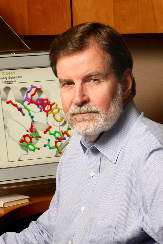 Samuel H. Wilson合作论文数Genome Integrity and Structural Biology Laboratory, National Institute of Environmental Health Sciences12




