Objective.Spinal muscular atrophy (SMA) is a genetic disorder that causes the progressive degeneration of spinal motor neurons, which can result in premature death. Restoring motor function in people with SMA is a serious issue. These individuals require lifelong motor rehabilitation to maintain their quality of life. A recent study showed that a two-week course of transcutaneous electrical spinal cord stimulation (tSCS) combined with physical therapy led to clinically significant improvements in Revised Upper Limb Module (RULM) and Hammersmith Functional Motor Scale Expanded (HFMSE), improved forced vital capacity (FVC), reduced joint contractures in people with SMA types 2 and 3 receiving gene therapy. It is unclear how long the benefits of a tSCS course last. This study aimed to determine the long-term effects of tSCS and to evaluate whether consecutive tSCS courses can improve or maintain motor activity in individuals with SMA types 2 and 3 receiving disease-modifying therapy.Approach.Case series involved nine individuals with SMA types 2 and 3 (aged 5-20 years). They underwent two consecutive courses of tSCS combined with motor task performance. Each course lasted two weeks, with a break of 3-15 months between courses. The RULM, HFMSE, FVC and a goniometric assessment of knee extension were recorded before and after each course.Main results.Significant improvements achieved during the first course did not deteriorate between the courses. The second course demonstrated significant improvements in HFMSE scores and FVC.Significance.This case series confirms the effectiveness of tSCS in motor rehabilitation for individuals with SMA types 2 and 3. It can be concluded that the effects of tSCS treatment last for 4-5 months. Consecutive courses with an interval of less than a year between them can likely improve motor activity in people with SMA types 2 and 3 who have received disease-modifying therapy.
(1) Background: We developed a novel technology that regulates human locomotion using transcutaneous electrical spinal cord stimulation to activate spinal locomotor networks and posterior root stimulation to activate leg flexor and extensor motor pools during swing and stance phases, respectively. This technology effectively restores walking in post-stroke individuals while forward propulsion in the stance phase and foot dorsiflexion in the swing phase are insufficient. In this study the effectiveness of regulating the stance and swing phases while healthy volunteers walked on a treadmill with transcutaneous electrical stimulation of the posterior roots, leg muscles, and their combined effects has been examined. (2) Methods: We analyzed the kinematic characteristics of stepping movements in healthy participants with spinal stimulation of the posterior roots and flexor/extensor leg muscles. (3) Results: Our findings clearly show that posterior root stimulation at T12 combined with tibialis anterior muscle stimulation during the swing phase effectively regulates foot dorsiflexion, whereas posterior root stimulation at L2 combined with hamstrings and medial gastrocnemius stimulation during the stance phase effectively regulates forward propulsion. (4) Conclusions: Combined stimulation in the stance and swing phases within the same gait cycle resulted in the most coordinated stepping, and effective control of forward propulsion and foot dorsiflexion.
Pupillometry can be used as a method for monitoring pain. There are experimental conditions under which standard pupillometry equipment cannot be used. Studying the effects of different pulse forms used for transcutaneous spinal cord stimulation (tSCS) is one such task. The aim was to create a system for recording pupil diameter based on a web camera because it can be synchronised with external equipment, which allows the diameter to be recorded simultaneously with other physiological signals. A markerless system for recording and analysing pupil diameter using deep neural networks was developed based on a commercially available web camera. The accuracy of this system was compared with the accuracy of measurements using manual analysis with ImageJ (version 1.54g). For validation, the system was tested in a study of the dependence of tolerance to tSCS on the shape of stimulating pulses, which involved volunteers (n = 12). The results of the developed pupillometry were compared with the pain rating scale traditionally used in such studies. The developed system is accurate in determining the pupil diameter, comparable to human accuracy. The pupillometry results reproduced those obtained using a subjective pain scale. This method was found to be a reliable method for recording nociceptive pupillary responses in electrophysiology.
The aim of the study was to investigate the spinal mechanisms involved in regulating postural balance in humans. Participants stood in a normal stance, with their spinal postural networks either non-invasively activated or not stimulated by electrical stimulation. Postural sway, muscle activity, joint kinematics and respiratory movements were monitored. Half of the participants depended on external sensory cues for controlling balance (field dependent), whereas the other half did not (field independent). Stimulation was performed at the T11-T12 or L1-L2 vertebral level using intensities below the motor threshold. When participants stood without stimulation, differences in body segment movement in the frontal plane were observed between subgroups, in addition to similarities in segment movement in the sagittal plane. Field-dependent participants demonstrated greater mediolateral sway and pelvic motion and relied more on hip involvement in frontal centre of pressure dynamics than field-independent participants. Stimulation at T11-T12 induced changes in muscle activity and body segment coordination in both groups. It led to a reduction in mediolateral sway and enhanced postural stability, but only in field-dependent individuals. Stimulation at L1-L2 altered muscle activity and joint kinematics in both subgroups without having an effect on postural stability. Stimulation did not affect respiratory movements or posture-respiratory coupling. In the human spinal cord, the interneuronal networks involved in postural regulation are located at the T11-T12 level and operate under supraspinal control. Interneuronal networks at the L1-L2 level also modulate muscle activity and body segment coordination; however, their specific role in regulating upright posture remains unclear.
It is known that neural networks of the human spinal cord can initiate the stepping pattern and control posture in the absence and with impaired supraspinal input. In the rehabilitation of children with spastic diplegia due to cerebral palsy, a new technology based on electrical transcutaneous spinal cord stimulation (tSCS) was used. Continuous and rhythmic tSCS was performed during walking. Continuous tSCS was performed at the level of C5–C6 and T11–T12 vertebrae. Rhythmic stimulation of the dorsal roots of the spinal cord was performed at the level of the T12 and L2 vertebrae to activate the motor pools of the flexor and extensor leg muscles in the swing and stance phases, respectively. Fourteen children with spastic diplegia, age 13 ± 2 years, participated in the study. Patients in the study were able to stand and walk independently with the help of a cane/walker or with the assistance of an adult. All patients received standard therapy and locomotor training (20 min per day, 10 days). During locomotor training, tSCS-based technology was used in patients in one group and no tSCS was used in patients in the other group. The effect of tSCS on the parameters of gait over flat surface (acute effect) was determined in all patients before the course. Before and after the course all patients were examined using clinical tests, kinematic characteristics of gait were analyzed. The acute effect of stimulation is manifested in a reduction in the duration of the stance phase, in an increase in the range of motion in the knee joint. After the course in the main group the scores on the motor function change assessment scale (GMFM-88) increased, spasticity decreased, and the distance passed in the 6-min walk test increased.
A study was conducted using the "Spinal neuroprosthesis neurostimulation device" specs 26.60.13-004-65248030-2021, which is designed to stimulate the dorsal roots of the cervical and lumbar spinal branches by rhythmic electrical stimulation during defined phases of the step cycle. The aim of the study was to evaluate the safety and efficacy of a neuroprosthesis to regulate motor functions in patients with the consequences of an acute cerebrovascular accident. Group 1 (main group): Patients who underwent transcutaneous electrical spinal cord stimulation with a neuroprosthesis and standard rehabilitation (therapeutic exercises, massage, physiotherapy). Group 2 (control): Patients who underwent electrical stimulation with a neuroprosthesis without current and standard rehabilitation. Primary efficacy point: improvement in walking performance in the 10-metre walk test and the 6-minute walk test. Secondary efficacy points: Improvement in performance tests on various scales: Fugl-Meyer, the Medical Research Council Scale for Muscle Strength , quantitative muscle strength assessment, Berg Balance Scale, Functional Independence Scale, modified Ashworth Scale and analysis of spatio-temporal and kinematic parameters assessed by laboratory methods, video recording of muscle activity. The rehabilitation course consisted of 12 daily procedures, each lasting 40-60 minutes and performed on a treadmill for 20 minutes. Tests were performed 1-2 days before the start of the rehabilitation course and the day after the rehabilitation course ended. The results of a clinical study showed that the use of a neuroprosthesis leads to a significant improvement in muscle strength, the ability to maintain balance, a decrease in spasticity and an increase in functional independence.
Objective.Transcutaneous spinal cord stimulation (tSCS) using kilohertz frequency carrier modulation has emerged as a non-invasive neuromodulation approach to improve motor recovery and reduce pain. Early application of 5-10 kHz modulated pulses for tSCS has shown promising results in spinal cord (SC) injury and post-stroke rehabilitation, but the mechanisms underlying these effects remain poorly understood.Approach.This narrative review synthesizes electrophysiological, computational and clinical evidence to assess how kilohertz modulation influences spinal and corticospinal excitability and analgesia. A total of 20 preclinical and clinical studies comparing the effects of kHz-modulated and conventional stimulation pulses were reviewed.Main results.The results indicate that kilohertz modulated tSCS increases tolerance to stimulation, but requires a higher charge to evoke motor responses in healthy participants and individuals with post-stroke motor disorder. Compared to conventional stimulation, modulated stimulation recruits afferents less efficiently at motor threshold intensity but appears to engage broader corticospinal circuits, especially near or below threshold. Frequency-specific effects include prolonged spinal inhibition, frequency-dependent modulation of supraspinal input, and selective activation of inhibitory interneurons in the dorsal horn. Computational study supports these observations, showing that kilohertz pulses produce delayed action potential initiation due to alternating depolarization cycles. A comparative functional study has shown that modulated tSCS improves motor function in individuals with SC injury more significantly than conventional stimulation.Significance.This narrative review highlights gaps in our understanding of the mechanisms of modulated tSCS, suggests directions for further research and will be useful in planning studies on the mechanisms behind tSCS with and without carrier frequency. It also holds engineering relevance for the optimal design of stimulation devices.
The combined effects of transcutaneous electrical stimulation (tES) of the spinal cord and affective sound stimulation on postural control were investigated to elucidate the involvement of spinal networks in the maintenance of vertical stability. Healthy volunteers (n = 27) underwent tES and sound stimulation separately and combined quasi-randomly. All participants were field-dependent; i.e., participants used the exteroceptive afferent stream for spatial orientation. Centre-of-pressure parameters were analysed to assess postural stability. Results showed that tES at the T11–T12 vertebrae stabilised posture, tES at the L1–L2 vertebrae had no postural effect, and sound stimulation from the left destabilised posture. To assess the role of spinal regulation of postural disturbances, we compared the effects of combined tES with sound stimulation to those of sound stimulation alone. Stimulation at the T11–T12 level reduced the lateral sway induced by affective sounds, whereas L1–L2 tES did not. These results suggest that, in healthy individuals, spinal networks located at the T11–T12 and L1–L2 vertebral levels have distinct roles in maintaining upright posture, both when a person is standing still and when they are actively stabilising their posture during destabilising perturbations. T11–T12 spinal networks stabilise upright posture when destabilising information is solely transmitted from the supraspinal level.
The study explored the effects of transcutaneous electrical spinal cord stimulation (tES) on postural control. Subjects were divided into field-dependent (FD) and field-independent (FI) groups according to their cognitive style. FD subjects use an exteroceptive afferent stream for spatial orientation, while FI subjects use an interoceptive stream. In darkness, vertical posture is maintained by head-trunk stabilization in FD subjects and by independent movements of body segments in FI subjects. Previously, we showed that tES at the L1-L2 vertebral level decreased postural stability in FD subjects. Now, stimulation was applied at the T11-T12 vertebral level (midline, above the left or right dorsal roots). Quiet standing was assessed using stabilometry in 18 FD and FI participants. Participants stood on a force platform in soundproof chamber with eyes closed during tES. Midline and left tES significantly improved postural stability by up to 28% in FD participants, while posture did not change significantly in FI participants. Pronounced differences between the effects of T11-T12 and L1-L2 stimulation are associated with selective topographical activation of proximal and distal leg muscles during tES of the lumbar enlargement. This study highlights the importance of considering cognitive style in postural control research.
Spinal muscular atrophy (SMA) is an orphan disease characterized by the progressive degeneration of spinal alpha motor neurons. In recent years, nusinersen and several other drugs have been approved for the treatment of this disease. Transcutaneous spinal cord stimulation (tSCS) modulates spinal neuronal networks, resulting in changes in locomotion and posture in patients with severe spinal cord injury and stroke. We hypothesize that tSCS can activate motor neurons that are intact and restored by medication, slow the decline in motor activity, and contribute to the development of motor skills in SMA patients. Thirty-seven children and adults with SMA types 2 and 3 participated in this study. The median duration of drug treatment was over 20 months. The application of tSCS was performed during physical therapy for 20–40 min per day for ~12 days. Outcome measures were specific SMA motor scales, goniometry of contractured joints, and forced vital capacity. Significant increases in motor function, improved respiratory function, and decreased contracture were observed in both type 2 and 3 SMA participants. The magnitude of functional changes was not associated with participant age. Further studies are needed to elucidate the reasons for the beneficial effects of spinal cord electrical stimulation on SMA.
(1) Background: Neurological deficits associated with coronavirus disease (COVID-19) exacerbate respiratory dysfunction, necessitating rehabilitation strategies that address both. Previous studies have demonstrated that spinal cord transcutaneous stimulation (scTS) can facilitate the excitation of respiratory spinal neural networks in patients with post-COVID-19 syndrome. This study evaluates the efficacy of combining scTS with respiratory training (RT) to improve respiratory function in individuals with post-COVID-19 pulmonary deficits; (2) Methods: In this before-after, case-controlled clinical trial, five individuals with post-acute COVID-19 respiratory deficits participated in two interventional programs: 10 daily sessions of respiratory training (RT), followed by 10 daily sessions of scTS combined with RT (scTS + RT). Forced vital capacity (FVC), peak inspiratory flow (PIF), peak expiratory flow (PEF), time-to-peak inspiratory flow (tPIF), and time-to-peak expiratory flow (tPEF) were assessed at baseline and after each program; (3) Results: Compared to RT alone, the scTS + RT intervention resulted in an average effect size that was twice as large, with significant increases in FVC and PEF, and a significant decrease in tPEF; (4) Conclusions: The scTS-induced activation of respiratory neuronal networks, when combined with respiratory training, offers a promising therapeutic approach for treating persistent respiratory deficits in patients with post-acute COVID-19 syndrome.
Neurorehabilitation of post-stroke patients with motor impairments is a significant and yet unresolved issue in restorative medicine. We propose a novel approach to rehabilitating such patients using transcutaneous electrical spinal cord stimulation (scTS), which targets the neural locomotor networks of the lumbar enlargement of the human spinal cord [1]. The Spinal Neuroprosthesis device was developed to control stimulation, providing noninvasive and phase-dependent activation of motoneuronal pools of flexors and extensors during a certain phase of the stepping cycle, combined with the activation of neuronal locomotor networks [2]. The aim of this study is to assess the efficacy of Spinal Neuroprosthesis in regulating locomotor functions among post-stroke patients with motor disorders. The study is designed to provide an objective evaluation of the medical device’s effectiveness. The study was conducted at the Russian Research Institute of Neurosurgery named after Prof. A.L. Polenov. The study enrolled 20 patients who had been experiencing severe motor disorders of the lower extremities in the form of hemiparesis. The duration of stroke among these patients ranged from 3 to 12 months. They were divided into two groups: control and experimental. The control group underwent sham (scTS-) stimulation during a motor rehabilitation session, while the experimental group received real scTS, establishing the difference between the groups. The rehabilitation program comprised 15 sessions of stimulation to the spinal cord. The treatment protocol comprised an initial evaluation of patients’ neurological and rehabilitation status and an investigation of spatial-temporal and kinematic parameters of walking. Subsequently, patients participated in rehabilitation sessions, which entailed walking on the treadmill and over-ground stepping with scTS. Finally, patients underwent a follow-up examination that included a re-evaluation of their neurological and rehabilitation status, as well as an investigation of spatial-temporal and kinematic parameters of walking. At the beginning of the program, the distance traveled by patients in the control and experimental groups during a six-minute walk test, according to the study results, did not differ significantly. However, following the treatment, patients in the experimental group demonstrated a substantially lengthier distance covered during the 6-minute test than the control group. Both groups of patients in the 10-meter walk test demonstrated an increase in distance walking speed, although the patients in the experimental group had a greater increase in speed compared to those in the control group. These improvements were more pronounced in patients from the experimental group. The results from neurological scales indicated an increase in muscular strength, improvement in balance functions, and an increase in functional independence in both groups. The findings provide evidence that the Spinal Neuroprosthesis effectively regulates stepping movements and restores locomotor function in patients post-stroke. Clinically significant improvements are observed within two weeks of neuroprosthesis use. Additionally, training increases patients’ exercise tolerance while walking, speed of movement, and functional independence.
Background: A growing number of studies have reported Coronavirus disease (COVID-19) related to both respiratory and central nervous system dysfunctions. This study evaluates the neuromodulatory effects of spinal cord transcutaneous stimulation (scTS) on the respiratory functional state in healthy controls and patients with post-COVID-19 respiratory deficits as a step toward the development of a rehabilitation strategy for these patients. Methods: In this before-after, interventional, case–controlled clinical study, ten individuals with post-acute COVID-19 respiratory deficits and eight healthy controls received a single twenty-minute-long session of modulated monophasic scTS delivered over the T5 and T10 spinal cord segments. Forced vital capacity (FVC), peak forced inspiratory flow (PIF), peak expiratory flow (PEF), time-to-peak of inspiratory flow (tPIF), and time-to-peak of expiratory flow (tPEF), as indirect measures of spinal motor network activity, were assessed before and after the intervention. Results: In the COVID-19 group, the scTS intervention led to significantly increased PIF (p = 0.040) and PEF (p = 0.049) in association with significantly decreased tPIF (p = 0.035) and tPEF (p = 0.013). In the control group, the exposure to scTS also resulted in significantly increased PIF (p = 0.010) and significantly decreased tPIF (p = 0.031). Unlike the results in the COVID-19 group, the control group had significantly decreased PEF (p = 0.028) associated with significantly increased tPEF (p = 0.036). There were no changes for FVC after scTS in both groups (p = 0.67 and p = 0.503). Conclusions: In post-COVID-19 patients, scTS facilitates excitation of both inspiratory and expiratory spinal neural networks leading to an immediate improvement of respiratory functional performance. This neuromodulation approach could be utilized in rehabilitation programs for patients with COVID-19 respiratory deficits.
We demonstrated previously that stress-induced glucocorticoids are gastroprotective hormones but not ulcerogenic ones (Filaretova et al., 2016). Recently electrical spinal cord stimulation began to be used for both experimental studies of motor functions regulation and rehabilitation of motor functions in patients with spinal cord injury (Gerasimenko et al., 2021; Moshonkina et al., 2016). The spinal cord stimulation directed to the activation of spinal locomotor related networks affected visceral systems as well. Recently in our teamwork we have shown that the electrical spinal cord stimulation can cause an increase in blood glucocorticoid level accompanied by a gastroprotective effect (Filaretova et al., 2022). The aim of the present work was to verify the hypothesis that the gastroprotective effect of the electrical spinal cord stimulation may be mediated by glucocorticoids produced in response to this stimulation. Gastric injury was induced in anesthetized rats by prolonged gastric ischemia/reperfusion (I/R, 30 min occlusion of celiac artery followed by 3 h of reperfusion) or indomethacin administration (IM, 35 mg/kg, sc). For spinal stimulation, stainless steel wire electrodes were fixed in fasciae between T11-12 and L1–L2 vertebrae of anesthetized animals. Motor evoked responses in hindlimbs muscles to spinal stimulation were recorded. Spinal stimulation at 30 Hz was carried out with a subthreshold current for inducing muscle contractions (80% of the threshold). Sham-stimulated (control) rats were subjected to the same manipulations except for the stimulation itself. To test the participation of glucocorticoids in the gastroprotective effect of the stimulation, two approaches were used: pretreatment by the inhibitor of glucocorticoid synthesis, metyrapone (30 mg/kg, i.p.) and the antagonist of glucocorticoid receptors RU-38486 (20 mg/kg, i.p.). The concentration of corticosterone in blood plasma was determined using commercial ELISA kits. In control rats the spinal cord stimulation resulted in an increase of blood corticosterone level 1 h later. The spinal stimulation, applied 1 h before the onset of ulcerogenic stimuli, also significantly attenuated gastric erosion formation induced by I/R and IM as well. Metyrapone injected shortly before the stimulation caused a fast inhibition of corticosterone response and reversed the gastroprotective effect stimulation. The gastroprotective effect of the stimulation was also prevented by the pretreatment rats with glucocorticoid receptor antagonist RU-38486. The obtained results indicate on contribution of glucocorticoids to gastroprotective effect of spinal cord stimulation. In conclusion, the findings support the hypothesis that the gastroprotective effect of the electrical spinal cord stimulation may be mediated by glucocorticoids produced in response to this stimulation. The study was supported by the Ministry of Education and Science of the Russian Federation (agreement No. 075-15-2020-921 for the creation and development of the world-class scientific center “Pavlov Center “Integrative Physiology - to medicine, high-tech healthcare and technologies of stress resistance”). This is the full abstract presented at the American Physiology Summit 2023 meeting and is only available in HTML format. There are no additional versions or additional content available for this abstract. Physiology was not involved in the peer review process.
Neural networks in the spinal cord can generate the walking pattern and control posture in the absence of supraspinal influences. A technology using transcutaneous electrical spinal cord stimulation (tSCS) was created. During walking, tSCS activated spinal locomotor networks, as well as leg flexor/extensor motor pools in the swing/stance phases, respectively. It was assumed that the use of this technology in subjects with locomotion disorders would improve walking. Patients with hemiparesis were studied 3–11 months after stroke, the duration of the course was 2 weeks. Patients of the main and control groups received standard therapy and rehabilitation using the technology; in the control group, sham tSCS was used. After the course, minimal clinically important differences in walking parameters were achieved in the main group, in contrast to the control group. The developed technology is an effective means of restoring walking in patients with hemiparesis.
Respiratory rate monitoring is fundamental in clinical settings, and the accuracy of measurement methods is critical. This study aimed to develop and validate methods for assessing respiratory rate and the duration leof respiratory cycle phases in different body positions using optoelectronic plethysmography (OEP) based on a motion capture video system. Two analysis methods, the summation method and the triangle method were developed. The study focused on determining the optimal number of markers while achieving accuracy in respiratory parameter measurements. The results showed that most analysis methods showed a difference of ≤0.5 breaths per minute, with R2 ≥ 0.94 (p < 0.001) compared to spirometry. The best OEP methods for respiratory rate were the abdominal triangles and the sum of abdominal markers in all body positions. The study explored inspiratory and expiratory durations. The research found that 5–9 markers were sufficient to accurately determine respiratory time components in all body positions, reducing the marker requirements compared to previous studies. This interchangeability of OEP methods with standard spirometry demonstrates the potential of non-invasive methods for the simultaneous assessment of body segment movements, center of pressure dynamics, and respiratory movements. Future research is required to improve the clinical applicability of these methods.
Transcutaneous electrical stimulation of the spinal cord is used to restore locomotion and body weight support in patients with severe motor disorders. We studied the effects of this non-invasive stimulation on postural control in healthy subjects. Stimulation at the L1–L2 vertebrae was performed to activate the extensor muscles of the lower limbs. Because postural regulation depends on the cognitive style, the effects of the stimulation were analyzed separately in field-dependent (FD) and field-independent (FI) participants. During the study, FD and FI participants (N = 16, 25 ± 5 years, all right dominant leg) stood on a force platform in a soundproof chamber with their eyes closed. Stimulation was applied in the midline between the L1 and L2 vertebrae or over the left or right dorsal roots of the spinal cord; under the control condition, there was no stimulation. Stimulation destabilized posture in healthy subjects, whereas patients with movement disorders usually showed an improvement in postural control. In the FD participants, left dorsal root and midline stimulation increased several postural parameters by up to 30%. Dorsal root stimulation on the side of the supporting leg reduced postural control, while stimulation on the side of the dominant leg did not. No significant changes were observed in the FI participants.
Maintaining a stable upright posture is a complex physiological process. Destabilizing this posture is one of the tools to study the mechanisms of upright postural regulation. Sound stimulation allows us to effect posture in both the sagittal and frontal planes of the body. The aim of this work was to evaluate postural modulation in response to listening affective audio signals from one of four directions in space. In healthy subjects ( n = 33), postural parameters were recorded in silence and with sound stimulation. The sound stimulation was of three types: long signals without pauses, long signals with pauses, and short signals with pauses. The sound sources were in front, behind, right, or left. The duration of stimulation and postural analysis was 30 s. There was an increase in postural parameters in response to affective sounds, which supports their destabilizing effect on posture. Comparison of the effects of three types of stimulation on posture showed that as the total duration of auditory stimuli during posture registration decreased the number of sound source positions at which significant changes in postural parameters were obtained decreased. Regardless of the type of stimulation, significant increases in postural parameters were found in all three cases when the sound source was located on the right side of the body. When the sound source was located in the sagittal plane (in front and behind), the effect was mainly observed for long duration sound signals, but it was less pronounced than for stimulation on the right side. No significant changes in postural parameters were found when stimulation was performed on the left side.
Multisegmental transcutaneous electrical stimulation of the spinal cord (sсTS), affecting spinal neural networks and motor pools of leg muscles, was used to correct treadmill walking in stroke patients. The study involved 15 patients in the recovery period after acute cerebrovascular accidents. A noninvasive spinal neuroprosthesis with a multichannel stimulator and a system for detecting phases of the walking cycle (“Cosima”, Russia) was used to activate motor pools of leg flexor muscles in the transfer phase, activation of motor pools of extensor muscles in the stance phase in combination with continuous activation of spinal locomotor networks. Using of sсTS during walking on a treadmill increased the amplitude of movements in the ankle joint and the stride length on the paresis side, as well as a decreased the asymmetry of both legs in the phases of the gait cycle. The clearance of the paretic leg lift increased in 80 of patients. With a combination of continuous and phase-dependent stimulation, the increase in the range of motion in the joints was maximal compared to phase-dependent or only constant stimulation. The obtained data demonstrate that the proposed algorithm of the sсTS modulates the parameters of walking movements in patients with the consequences of cerebral circulation accidents and can be considered as a promising method of motor rehabilitation.
An Erratum to this paper has been published: https://doi.org/10.1134/S0362119723970029
 Yu. P. Gerasimenko合作论文数32Gerasimenko Iu P合作论文数30
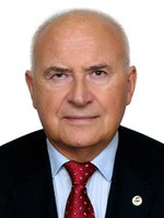 Yury Gerasimenko合作论文数Department of Physiology, School of Medicine, University of Louisville26R. M. Gorodnichev合作论文数19N. A. Shcherbakova合作论文数17A. A. Grishin合作论文数15
 Ирина Богачева合作论文数15Sergei V. Vissarionov合作论文数13Moiseev S A合作论文数12
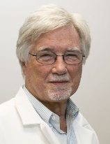 V. Reggie Edgerton合作论文数Department of Neurobiology, David Geffen School of Medicine, University of California, Los Angeles11A. A. Savokhin合作论文数9Kozlovskaia I B合作论文数9V. A. Selionov合作论文数8Ruslan Gorodnichev合作论文数8A. M. Pukhov合作论文数8
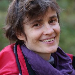 Natalia Pavlova合作论文数Centre for Neuroprosthetics and Brain Mind Institute, EPFL;St. Petersburg, Pavlov Institute of Physiology;Centre for Neuroprosthetics and Brain Mind Institute, Pavlov Institute of Physiology8
 Gennady I Lobov合作论文数8
 Inessa B Kozlovskaya合作论文数8
 Vsevolod Lyakhovetskii合作论文数Russian Acad Sci, Pavlov Inst Physiol7Baindurashvili A G合作论文数7Natalia Shamantseva合作论文数6Pivovarova E A合作论文数6
 Irina A Solopova合作论文数6I. A. Sukhotina合作论文数5N. D. Shandybina合作论文数5
 Tatiana Shigueva合作论文数5E. G. Gilerovich合作论文数5Vladimir A Otellin合作论文数5A. A. Savohin合作论文数4V. V. Umnov合作论文数4E. A. Fedorova合作论文数4Tomilovskaia E S合作论文数4
 Parag Gad合作论文数Univ. of California, Los Angeles;c;Univ. of California, Los Angeles4
 Dimitry Sayenko合作论文数Houston Methodist Department of Neurosurgery;Houston Methodist Research Institute;Center for Neuroregeneration, Houston Methodist4Selionov V A合作论文数4
 Elena Tomilovskaya合作论文数Russian Federation State Research Center, Institute for Biomedical Problems of the Russian Academy of Sciences4A. Z. Zakirova合作论文数3Stolbkov Yu. K.合作论文数3A V Miniaeva合作论文数3Юрий Петрович Герасименко合作论文数3
 s. N. Kaurkin合作论文数3I E Nikityuk合作论文数3
 Dmitry Skvortsov合作论文数Fed Res & Clin Ctr, Russias Fed Med Biol Agcy FNKC FMBA3Stolbkov Iu K合作论文数3
 E. V. Bobrova合作论文数3
 Elena Andreevna Vershinina合作论文数3
 Alexander V Ovechkin合作论文数3
 Irina a Andreeva合作论文数Institute of Geology of Ore Deposits, Petrography, Mineralogy, and Geochemistry Staromonetnyi per. 35, Russian Academy of Sciences3
 Roland R. Roy合作论文数Brain Research Institute, University of California, Los Angeles3




