Cancer immunotherapy has been revolutionized through the implementation of the state-of-the-art chimeric antigen receptor (CAR)-mediated therapies. CAR-based technologies, which encompass CAR T cells, CAR macrophages, and CAR NK cells, have shown great promise in the treatment of various cancers. Despite the success of CAR-based therapies in treating malignancies, they face numerous challenges, including dysfunction of effector innate and adaptive immune cells, immunosuppressive tumor microenvironment, antigen heterogeneity, and on-target/off-tumor bio-toxicity. The CD47/SIRPα axis is recognized as a critical innate immune checkpoint and is important in regulating myeloid-derived clearance of tumor cells and the cross-talk between innate and adaptive immune cells in cancer immunity. This signaling axis has risen as a promising target to enhance the CAR-based immunotherapies by overcoming phagocytic inhibition and modulating immune evasion. This narrative review explores the integration of CD47/SIRPα modulation as an adjunct to CAR therapies. CD47/SIRPα immune modulation revealed its potential to boost infiltration, persistence, and phagocytic activity of the immune cells. However, its blockade also poses challenges, including hematologic toxicities, CAR T-cell clearance, and compensatory escape pathways. Future work will depend on selective targeting, combinatorial checkpoint modulation, and engineered CAR designs that preserve safety while unlocking durable responses. Herein, we discuss preclinical and clinical advancements, safety considerations, and cutting-edge advancements.
Melanoma, an aggressive form of skin cancer, is responsible for more than 80
Autoimmune diseases arise from a complex interplay between genetic predisposition and environmental factors, including chemical exposures, infections, and psychological stress. These conditions occur when the immune system mistakenly targets the body's own healthy cells, resulting in tissue and organ damage. Both humoral and cellular immune mechanisms contribute to autoimmune disorders, leading to chronic inflammation and progressive tissue injury.Autoimmune diseases have diverse and often overlapping symptoms, significantly impacting patients' quality of life. Early diagnosis is crucial for effective disease management; however, the clinical similarity among autoimmune disorders and the lack of definitive diagnostic tests in many cases pose major challenges to timely identification.Notably, in many autoimmune conditions, oral manifestations are among the earliest or, in some cases, only clinical signs, as the oral epithelium is a frequent target of autoimmune responses. These manifestations often present as erosions and ulcers, causing pain and discomfort. Identifying oral lesions at an early stage can facilitate early diagnosis and improve patient outcomes. This chapter provides a comprehensive overview of autoimmune conditions and their oral manifestations.Since immune dysregulation is central to autoimmune disease pathogenesis, local and systemic corticosteroids are commonly prescribed to control symptoms, including oral lesions. In severe or refractory cases, immunosuppressive therapies may be essential to prevent disease progression and tissue destruction.
Common variable immunodeficiency disease (CVID) is the most prevalent symptomatic inborn errors of immunity, determined by defective B cell function, impaired antibody production, and susceptibility to frequent respiratory infections, enteropathy, autoimmunity, and malignancy. Due to the importance of autoimmunity in CVID and the probable role of regulatory B lymphocytes, we aimed to determine the frequency of B10 cells in CVID patients with and without autoimmunity. A total of 24 CVID patients and 12 healthy controls were enrolled in the study. Patients were divided into two equal groups, with and without autoimmunity. Peripheral blood cells were stained with monoclonal antibodies (mAbs) to identify CD24hiCD38hi B cells, CD27int CD38+ (plasmablasts), and CD24hiCD27+ B cells by flow cytometry. The percentages of B10, CD24hiCD27+ and CD27int CD38+ cells were significantly lower in total CVID patients, CVID patients with autoimmunity and CVID patients without autoimmunity compared to healthy controls (mean±standard deviation (SD) percentage of B10 cells: 6.36±9.21(total CVID), 2.81±5.00 (CVID with autoimmunity), and 3.25±3.5 (CVID without autoimmunity) vs. 13.02±12.45 (healthy controls); CD24hiCD27+ cells: 2.39±3.89, 3±5.20 and 1.78±1.94 vs. 20.38±14.27; CD27int CD38+ cells: 6.80±18.49, 7.40±20.24 and 6.20±17.45 vs. 11.84±5.71). CVID patients without autoimmunity had a higher percentage of CD24hiCD38hi cells than CVID patients with autoimmunity (4.73±4.14 vs. 2.62±5.02). The defect of regulatory B cells plays a significant role in the pathogenesis of autoimmunity in CVID. Further multicenter studies with higher sample sizes are suggested to determine the role of Breg cells in the clinical course of autoimmunity.
INTRODUCTION/OBJECTIVE:Autoimmune lymphoproliferative syndrome-like (ALPS-like) disorders are inherited conditions caused by non-FAS pathway mutations that clinically mimic ALPS, presenting with lymphoproliferation, autoimmunity, and cytopenias. This study describes a patient with STAT3-related ALPS-like disease that was initially misdiagnosed as ALPS and compares his clinical, immunological, and molecular features with those of previously reported cases to highlight diagnostic distinctions from classical ALPS. METHODS:Clinical data were obtained from direct examination and medical records. Whole-Exome Sequencing (WES) identified the causative mutation. A literature review using PubMed, Web of Science, and Scopus retrieved previously reported ALPS-like cases with STAT3 mutations for comparative analysis of clinical, immunological, and molecular findings. RESULTS:We report a 10-year-old boy with bicytopenia (thrombocytopenia and neutropenia), autoimmune thrombocytopenic purpura, refractory lymphadenopathy, splenomegaly, and recurrent infections, initially misdiagnosed as ALPS. Elevated double-negative T cells and vitamin B12 levels were detected. WES revealed a heterozygous de novo STAT3 mutation (p.L666V). A reduced frequency of Treg cells was observed in our case. The patient responded well to JAK inhibitor therapy. Review of reported STAT3-mutant ALPS-like cases (66 cases) showed that immune thrombocytopenia was the most common cytopenia, often accompanied by autoimmune hemolytic anemia, variable hypogammaglobulinemia, reduced Treg cells, and increased double-negative T cells. DISCUSSION:STAT3 gain-of-function-associated ALPS-like disease can closely mimic classical ALPS due to overlapping clinical and immunological features, including elevated double-negative T cells, which may lead to diagnostic challenges. CONCLUSION:This case highlights that STAT3 GOF ALPS-like disease can closely mimic classical ALPS, and that integrating genetic analysis with immunological assessment is essential for accurate diagnosis and guiding targeted therapy.
Experimental models play a fundamental role in bridging basic scientific discoveries with clinical applications in humans. Integrating in vivo studies into oncology research approaches has the potential to improve therapeutic innovation and support translational strategies. The remarkable similarities between animals and human immune systems have enabled researchers to better clarify immune pathways involved in cancer development and therapy. Moreover, ongoing preclinical investigations are still required to engineer immunomodulatory drugs and other emerging therapeutic approaches. Despite the considerable contributions of in vivo models, particularly murine systems, their future use in experimental theragnostics has become a subject of debate. Ethical concerns and questions regarding the extent to which these models accurately replicate human disease have prompted growing interest in alternative platforms, including organoids, organ-on-chip technologies, and advanced two- and three-dimensional cell culture systems. In the present review, we summarize current experimental evidence on immuno-oncology experimental models used across various cancer types and examine the evolving role of these models. We also discuss whether animal models are gradually becoming less satisfactory due to emerging technologies or biological limitations, or whether their continued application remains justified in certain circumstances.
Isocitrate dehydrogenase (IDH)-wildtype glioblastoma is an aggressive brain tumor characterized by limited therapeutic options and poor survival outcomes. Beyond well-established genomic alterations, epigenetic mechanisms such as DNA methylation have gained attention for their roles in glioblastoma pathogenesis. Tumor suppressor genes are frequently silenced through promoter hypermethylation, whereas global hypomethylation of repetitive elements contributes to genomic instability and oncogenic progression. Clinically, O6-methylguanine-DNA methyltransferase (MGMT) promoter methylation remains an important biomarker for predicting patient response to alkylating chemotherapies. Advances in single-cell epigenomic sequencing have characterized the remarkable heterogeneity of DNA methylation within glioblastoma. Techniques including single-cell bisulfite sequencing (scBS-seq) and single-cell reduced representation bisulfite sequencing (scRRBS) provide high-resolution insights into the methylation landscapes of individual tumor cells. These analyses reveal that epigenetic diversity underlies subclonal expansion, therapy resistance, and tumor recurrence. Moreover, integrating single-cell methylation data with other modalities such as transcriptomics and proteomics offers a multidimensional view of tumor evolution and microenvironmental dynamics. Despite ongoing challenges in cost, data interpretation, and large-scale integration, single-cell epigenomics has potential applications in refining glioblastoma classification and guiding personalized therapeutic strategies in the future. This review explores emerging roles of DNA methylation in glioblastoma alongside cutting-edge single-cell techniques and translational prospects.
Recent advances in mouse models of experimental asthma and improvements in respiratory physiology assessment have fundamentally improved preclinical research. These innovations now enable both higher translational value and more detailed mechanistic insight. Despite mouse models can reproduce discrete clinical phenotypes and endotypes, no single model can fully replicate the complexity of human asthma, thus careful selection of protocols is required to address specific research questions. Here, we review current murine protocols for modeling of asthma, compare commonly used allergens and exacerbation paradigms, and discuss how administration method, mouse strain, and scheduling influence immunophenotype and remodeling outcomes. conventional terminal readouts (bronchoalveolar lavage fluid cytology, histology, serology, plethysmography) provide only a snapshot of disease progression. Therefore, we evaluated in vivo imaging techniques—such as micro-computed tomography, MRI, PET/SPECT, and optical imaging—as a complementary approach for longitudinal, noninvasive assessment of structure, function and molecular signatures in asthma experimental models. In conclusion, refined murine models that better recapitulate distinct human endotypes, combined with validated and standardized imaging biomarkers, will bridge the translational gap and accelerate the identification of therapeutics for more effective, patient-centered asthma care.
Oral health is increasingly recognized as an important factor in the regulation of systemic immune responses and neurological health. Disruption of immune homeostasis in oral tissues, particularly in chronic conditions such as periodontitis, has been linked to the development and progression of neurodegenerative diseases, such as Alzheimer's disease (AD), Parkinson's disease (PD), and multiple sclerosis (MS). This chapter explores how chronic inflammatory conditions in the oral cavity, oral dysbiosis, and the presence of specific periodontopathogens contribute to systemic immune activation, microglial priming, and blood-brain barrier dysfunction. The discussion addresses multiple mechanistic pathways underlying these relationships, such as hematogenous spread of microbial components with subsequent systemic immune stimulation, neural signaling via trigeminal and vagal pathways, and alterations in gut-brain communication through the integrated oral-gut-brain axis. The chapter also reviews findings from clinical and experimental studies demonstrating correlations between oral inflammation and cognitive impairment, structural brain alterations, and elevated inflammatory and neurodegenerative biomarkers. Oral health disturbances are considered not only as secondary outcomes of neurological disease but also as a potential contributing factor in their pathogenesis. The emphasis is on the importance of incorporating oral health assessment and maintenance into preventive and therapeutic strategies for neurodegenerative conditions, while highlighting the necessity for well-designed mechanistic investigations to further explain these complex relationships.
This chapter explores the intricate interplay between immune regulation and craniofacial development, emphasizing the critical role of the immune system in both normal and pathological conditions. The process of craniofacial development, from the formation of pharyngeal arches and facial prominences to palate formation, is outlined, highlighting the susceptibility of these structures to maternal immune disruptions.Key immune interactions at the maternal-fetal interface are discussed, focusing on immune tolerance mechanisms that safeguard the fetus from maternal immune rejection. Dysregulation of this immune balance, whether through maternal infections, inflammation, or nutritional deficiencies, can lead to significant craniofacial defects such as cleft lip and palate. The chapter delves into the teratogenic effects of infections, such as Campylobacter rectus, rubella, and cytomegalovirus (CMV), which disrupt gene expression and interfere with crucial developmental pathways.The chapter further examines immune-mediated craniofacial disorders, including spondyloenchondrodysplasia (SPENCD), cherubism, autoimmune polyendocrinopathy-candidiasis-ectodermal dystrophy (APECED), Langerhans Cell Histiocytosis (LCH), and juvenile idiopathic arthritis (JIA), explaining how immune dysfunction leads to abnormal bone remodeling and developmental anomalies. Finally, the chapter addresses preventive strategies, emphasizing the importance of maternal health optimization, adequate nutritional support, and early management of infections to mitigate the impact of immune dysregulation on craniofacial development. This comprehensive exploration provides a foundation for understanding the critical role of the immune system in oral and craniofacial development within the broader context of oral immunity.
Background: Severe combined immunodeficiencies (SCIDs) are a group of disorders with variable clinical phenotypes, usually presenting with life-threatening infections. This type of immunodeficiency results from defective differentiation of hematopoietic stem cells into mature T lymphocytes leading to various identified affected genotypes of severe immunodeficiency. Omenn syndrome is an autosomal recessive immunodefi- ciency disorder characterized by generalized erythroderma, lymphoadenopathy, and eosinophilia. The aim of this study was to provide specific information about the clinical, immunological, and genetic character- istics in this context. Methods: A retrospective case review was conducted at Shahid Beheshti, Children Medical Center and Azad University Hospitals of Tehran so that the patients with a previously diagnosis of Omenn syndrome, admitted between years 2016 and 2023, were selected and included in this study. Results: Eleven patients with known Omenn syndrome were included in our study. The mean age of onset in the patients was 45 days old. Six (54.5%) were female and 5 (45.5%) were male. There was a history of parental consanguinity in 10 out of 11 studied children (91%). BCG dissemination, erythroderma, hepatos- plenomegaly, lymphadenopathies, failure to thrive, recurrent infections, and gastrointestinal manifestations were more prominent. Other presentations in order of frequency were failure to thrive (90.9%), recurrent infections (63.6%), erythroderma (63%), hepatosplenomegaly (45.5%), lymphadenopathy (36.4%), and BCG dissemination (27.3%). Conclusion: As Omenn syndrome is a type of SCID and a pediatric immunologic emergency, awareness about the various clinical manifestations of the disease among people of different ethnicities is highly essen- tial for timely and accurate diagnosis, treatment, and family counseling
Maternal high-fat diet (MHFD) alters the offspring's gut microbiome, impairs intestinal integrity, and increases susceptibility to ulcerative colitis (UC) later in life. As Bacillus coagulans ( B. coagulans ) is known to decrease intestinal inflammation, this study investigated whether its early-life administration could reverse MHFD effects and confer protection against dextran sulfate sodium (DSS)-induced UC. Female NMRI mice were fed a normal chow diet (NCD) or high-fat diet (HFD) during gestation and lactation. Male offspring from the MHFD group received a daily oral gavage of B. coagulans or phosphate-buffered saline (PBS) from postnatal day 21 until week 8. MHFD altered the gut microbiome, decreased expression of tight junction proteins (ZO-1 and Claudin-5), and exacerbated UC severity after DSS induction. Early-life administration of B. coagulans restored the gut microbiome, improved gut integrity, decreased pro-inflammatory cytokines (IL-1β and TNF-α), and protected the MHFD offspring against UC. We conclude that MHFD impairs offspring intestinal integrity and increases UC susceptibility, while early-life B. coagulans intervention restores the gut microbiome, improves intestinal health, and confers protection against UC later in life.
Inflammatory bowel disease (IBD) causes chronic suffering from gastrointestinal inflammation and dysfunction that can progress to colon cancer1,2. The prevalence of the disease is increasing, and there is an urgent need to better understand its pathogenic mechanisms to improve treatment. We show that GPR15-a G-protein-coupled receptor expressed in immune cells and described previously as an entry co-factor for human and simian immunodeficiency viruses3-is a marker and homing receptor for a subset of intramucosal GPR15-guided regulatory CD8+ T lymphocytes (CD8+ TIGR cells). Deleterious GPR15 gene variants in humans cause defective homing of CD8+ TIGR cells and are associated with severe early-onset IBD. Moreover, CD8+ TIGR cells are reduced in the intestinal mucosa of individuals with sporadic IBD. In mice, GPR15 deficiency impairs colonic homing of CD8+ TIGR cells, leading to accumulation of inflammatory macrophages and increased susceptibility to colitis. CD8+ TIGR cells potently kill macrophages activated by intestinal damage or disease using Fas ligand and TNF-related weak inducer of apoptosis (TWEAK). The identification of CD8+ TIGR cells yields new insights into organ-specific immune regulation and potential therapeutics for IBD.
Head and neck squamous cell carcinoma (HNSCC) is an aggressive cancer that evades immune surveillance by impairing the NKG2D receptor on natural killer (NK) cells and cytotoxic T lymphocytes. NKG2D recognizes stress-induced ligands (e.g., MICA, MICB, ULBPs) on tumor cells, but HNSCC tumors counteract this by shedding soluble ligands and secreting immunosuppressive cytokines like TGF-β1, reducing NKG2D expression and function. This review explores therapeutic strategies targeting the NKG2D axis to enhance HNSCC treatment. Cytokine therapies, such as IL-2 or IL-15, boost NKG2D-dependent NK cell activation. Monoclonal antibodies targeting MICA/B or ULBPs prevent ligand shedding and induce antibody-dependent cellular cytotoxicity (ADCC). Engineered cell therapies, including NKG2D-based chimeric antigen receptor (CAR)-T and CAR-NK cells, show potent preclinical efficacy against HNSCC. Combining these approaches with EGFR-targeted antibodies (e.g., cetuximab) or checkpoint inhibitors enhances NKG2D-mediated immunity. Despite promising results, challenges persist, including heterogeneous ligand expression, persistent shedding, and the immunosuppressive tumor microenvironment, which limit clinical efficacy. Future directions include multimodal strategies like bispecific NK-cell engagers and integration with chemoradiotherapy to overcome these barriers. By restoring NKG2D-driven immunity, these therapies hold potential to improve outcomes for HNSCC patients, though further research is needed to optimize their clinical translation.
ABSTRACT Background Pancreatic cancer (PC) is a lethal malignancy with a 5‐year survival rate of 13% as the 7th leading cause of cancer‐related death worldwide. Most patients are ineligible for resection at diagnosis. Monotherapy with immune checkpoint inhibitors (ICIs) has shown limited success in PC, with objective response rates below 5% and median overall survival rarely exceeding 6 months. This systematic review evaluates the efficacy and safety of combining gemcitabine and nab‐paclitaxel with ICIs in pancreatic cancer. Objective To assess the efficacy and safety of combining gemcitabine and nab‐paclitaxel chemotherapy with ICIs in patients with pancreatic cancer. Methods A comprehensive search of PubMed, Scopus, and Web of Science was performed using predefined keywords. Eligible studies included original research articles employing interventional, cohort, case–control, or cross‐sectional designs. Case reports, case series, and review articles were excluded. Data extraction and quality assessment using the Mixed Methods Appraisal Tool (MMAT, 2018) were conducted independently by two reviewers. Results Of 1904 search results, seven studies met inclusion criteria: four clinical trials and three retrospective cohorts. Sample sizes ranged from 17 to 180 participants with advanced or locally advanced PC. Median overall survival was ~15 months (range: 9.8–16.7) versus 8–9 months for chemotherapy alone. Progression‐free survival ranged from 5.5–9 versus 3.5–5.5 months. Objective response rates varied between 18%–50% for combination therapy and 23%–29% for chemotherapy. Grade 3–4 adverse events were mainly hematologic (anemia, neutropenia) and neurologic (fatigue, peripheral neuropathy). ICIs included PD‐1 inhibitors (pembrolizumab, nivolumab, toripalimab, camrelizumab, tislelizumab, sintilimab), PD‐L1 inhibitor (durvalumab), and CTLA‐4 inhibitor (tremelimumab). Conclusion Combining ICIs with gemcitabine and nab‐paclitaxel appears feasible and safe, with signals of improved efficacy compared with chemotherapy alone. However, evidence remains limited, and further large‐scale trials are warranted to confirm survival benefits and optimize therapeutic strategies in pancreatic cancer.
Background and Aims:Major Depressive Disorder (MDD) is a highly common neuropsychiatric disorder globally. A variety of factors contribute to the neuropathology of MDD. Microbiome research in neuropsychiatric disorders such as MDD has recently attracted attention. Indeed, the gut-brain axis could influence the course of MDD through metabolites such as Gamma-Aminobutyric Acid (GABA), Quinolinate, and other factors. Such metabolites may modulate the balance of excitatory and inhibitory signals. Moreover, MDD features abundant hyperinflammatory bacteria, whereas anti-inflammatory butyrate-synthesizing genera are decreased. Methods:Despite mounting evidence on the implications for the microbiome in MDD, it is unclear whether a bidirectional or causal relationship is in effect. To overcome this challenge, researchers have utilized AI tools to investigate the complex association between the microbiome and MDD. Results:Additionally, there is no solid biomarker recognized for diagnosis and prognosis of MDD, while further application of AI using ML protocols, such as random forest, NNs, SVM, and DL models, could offer a rather solid and reliable comprehension of the complicated nature of microbiome-MDD interplay. Conclusions:The present article reviews microbiome alterations as well as inflammatory and metabolic pathways in MDD with a focus on AI technology including support vector machines (SVM), random forests (RF), deep neural networks (DNNs), and autoencoders, which are used to identify microbial biomarkers, predict treatment results, and support personalized medicine.
Serine-threonine kinase 4 (STK4) deficiency is a rare primary immunodeficiency disorder characterized by progressive T-cell lymphopenia, recurrent infections, cutaneous warts, abscesses, autoimmunity, and cardiac manifestations. This condition is associated with impaired immune surveillance, predisposing patients to severe infections and immune dysregulation. Herein, we report a consanguineous Persian family with two affected siblings presenting with clinical features consistent with STK4 deficiency. At presentation, patient 1, a 30-year-old male, exhibited markedly reduced CD4 + lymphocyte counts. He presented with chronic diarrhea, fever, and oral thrush during infancy, followed by recurrent episodes of pneumonia that required hospitalization since the age of 4 months. At 18 years of age, he developed progressively worsening localized warts on both hands and feet. He also experienced a prolonged episode of bacterial meningitis lasting one month. Despite these infections, his serum immunoglobulin levels remained within the normal range. Patient 2, his affected 20-year-old sister, similarly presented with reduced CD4 + lymphocyte counts. She had multiple hospitalizations during her first year of life due to pneumonia and chronic diarrhea. By the age of 11, she developed localized warts on her hands and feet. In contrast, their parents and four other siblings remained asymptomatic, with no history of recurrent infections or cutaneous warts. Next-generation sequencing (NGS)-based targeted sequencing and Sanger segregation analysis revealed a homozygous frameshift mutation in the STK4 gene (p.Leu275AlafsTer21) in both affected siblings. The parents were heterozygous carriers of the mutation, while the other siblings were either heterozygous or wild type. Functional studies would be needed to further elucidate the pathogenic mechanism of this mutation. Long-term follow-up of these patients is essential to monitor for any additional manifestations associated with STK4 deficiency and to provide timely interventions.
Colorectal cancer (CRC) remains a major cause of cancer-related mortality, particularly in advanced or metastatic disease. The Janus kinase/signal transducer and activator of transcription (JAK/STAT) pathway mediates cytokine-driven signaling, and its persistent activation contributes to tumor growth, invasion, immune escape, and therapeutic resistance. Suppressor of cytokine signaling 3 (SOCS3) is a key negative regulator of cytokine and growth factor signaling, especially the IL-6/JAK/STAT3 axis. This review summarizes the structure and physiological functions of SOCS3 and discusses its dysregulation in CRC initiation, progression, metastasis, prognosis, and treatment response. Current evidence indicates that SOCS3 is frequently downregulated in CRC through promoter methylation and post-transcriptional regulation by oncogenic microRNAs, leading to sustained STAT3 activation, increased proliferation, reduced apoptosis, and enhanced invasiveness. SOCS3 also interacts with MEK/ERK and PI3K/AKT signaling and influences the tumor microenvironment by regulating T-cell balance, PD-L1 expression, and macrophage activity. Clinically, reduced SOCS3 expression has been associated with lymph node metastasis, advanced TNM stage, and poorer prognosis, whereas higher SOCS3 levels may correlate with improved outcomes and chemosensitivity. Emerging therapeutic strategies include epigenetic modulation, JAK/STAT pathway inhibition, regulation of IL-6 signaling in adoptive T-cell therapy, AhR/IL-22 modulation, and FXR activation. Further translational studies are needed to validate SOCS3 as a biomarker and therapeutic target in CRC.
Exhaustion of T cells, characterized by their compromised function and cytokine production, was first discovered in chronic infections and later redefined in the context of immunosuppressed tumor microenvironment (TME). Exhaustion markers include the immune checkpoints PD-1, TIM-3, LAG-3, and TIGIT. Colorectal cancer (CRC), which ranks among the highest in global prevalence, has been associated with exhaustion of T cells. While many trials have focused on anti-PD1 therapeutics in clinical trials, results indicate suboptimal efficacy. A robust approach involved dual blockade of other immune checkpoints together with PD-1. Interestingly, novel exhaustion markers could be used as prognostic markers for the development and progression of CRC. The present work discusses the basics of T cell exhaustion markers and their signaling in CRC. Also, novel combination therapy approaches and challenges in the field are discussed.
 Asghar Aghamohammadi合作论文数Research Center for Immunodeficiencies, Tehran University of Medical Sciences307
 Hassan Abolhassani合作论文数Department of Medical Biochemistry and Biophysics, Karolinska Institutet;Pan Hammarström Laboratory, Karolinska Institutet;Research Center for Immunodeficiencies, Tehran University of Medical Sciences144
 Amene Saghazadeh合作论文数Tehran University of Medical Sciences135
 Masoud Movahedi合作论文数Tehran University of Medical Sciences79
 Aliakbar Amirzargar合作论文数Molecular Immunology Research Center, Tehran University of Medical Sciences78
 Reza Yazdani合作论文数74
 Nima Parvaneh合作论文数Department of Pediatrics, Tehran University of Medical Sciences74
 Mahsa Keshavarz-Fathi合作论文数Childrens Med Ctr, Univ Tehran Med Sci68
 Mohammad Gharagozlou合作论文数Department of Clinical Immunology and Allergy;Allergy Research Institute;Tehran University of Medical Sciences64
 Sadr Maryam合作论文数63Zahra Chavoshzadeh合作论文数Mofid Children Hospital, Shaheed Beheshti University62
 Gholamreza Azizi合作论文数Imam Hassan Mojtaba Hospital, Alborz University of Medical Sciences59Zahra Pourpak合作论文数Allergy Research Institute;Tehran University of Medical Sciences;Allergy Research Institute, Tehran University of Medical Sciences59
 Zoghi Samaneh合作论文数School of Medicine, Tehran University of Medical Sciences;School of Medicine, Tehran University of Medical Sciences;Tehran University of Medical Sciences, Tehran University of Medical Sciences56
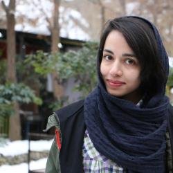 Niloufar Yazdanpanah合作论文数Universal Sci Educ & Res Network Usern, Network Immun Infect Malignancy & Autoimmun Niima, Tehran, Iran52
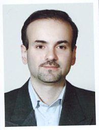 Mahdaviani Seyed Alireza合作论文数NRITLD, Shahid Beheshti Univ Med Sci52
 Mahdi Mahmoudi合作论文数Molecular Immunology Research Center, Tehran University of Medical Sciences52Maryam Mahmoudi合作论文数51
 Mohammad Nabavi合作论文数Semnan University of Medical Sciences51
 Sepideh Razi合作论文数Sch Med, Iran Univ Med Sci50
 Mostafa Moin合作论文数48
 Farhadi Elham合作论文数44
 Samaneh Delavari合作论文数Children's Medical Center, Tehran University of Medical Science43
 Farzaneh Rahmani合作论文数School of Medicine, Washington University43
 Saba Arshi合作论文数Rasoul Akram Hospital;Iran University of Medical Sciences;Rasoul Akram Hospital, Iran University of Medical Sciences43
 Vahid Ziaee合作论文数Department of Pediatrics;Sports Medicine Research Center;University of Tehran;Sports Medicine Research Center, University of Tehran42
 Hedayat Mona合作论文数School of Medicine, Tehran University of Medical Sciences41
 Hanaei Sara合作论文数40
 Parnian Jabbari合作论文数Universal Sci Educ & Res Network, USERN37
 Elham Rayzan合作论文数Res Ctr Immunodeficiencies RCID, Univ Tehran Med Sci35Rezaei Arezou合作论文数35
 Christoph Klein合作论文数Department of Pediatrics, Ludwig-Maximilians-University Munich;Gene Center Munich, Ludwig-Maximilians-University Munich34
 Shahkarami Sepideh合作论文数34
 Lennart Hammarström合作论文数Department of Medical Biochemistry and Biophysics, Karolinska Institutet34
 Kiarash Saleki合作论文数Student Res Comm, Babol Univ Med Sci33
 Taher Cheraghi合作论文数Children’s Medical Center, Tehran University of Medical Sciences33
 Sara Momtazmanesh合作论文数School of Medicine, Tehran University of Medical Sciences32
 Hirbod-Mobarakeh Armin合作论文数School of Medicine, Tehran University of Medical Sciences32
 Aryan Zahra合作论文数Cleveland Clinic, Respiratory Institute;Student's Scientific Research Center, Tehran University of Medical Sciences;Respiratory Institute, Tehran University of Medical Sciences32
 Parnian Shobeiri合作论文数Endocrinol & Metab Populat Sci Inst, Univ Tehran Med Sci30
 Behniafard Nasrin合作论文数Children's Medical Center, Tehran University of Medical Sciences30
 Kaan Boztug合作论文数CeMM Research Center for Molecular Medicine, Austrian Academy of Sciences;Medical University of Vienna30
 Tavakol Marzieh合作论文数Children's Medical Center, Tehran University of Medical Science29
 Sara Harsini合作论文数29
 Noosha Samieefar合作论文数USERN Office, Shahid Beheshti University of Medical Sciences28
 Nasser Ebrahimi Daryani合作论文数Department of Gastroenterology, Tehran University of Medical Sciences;Imam Khomeini Hospital, Tehran University of Medical Sciences28
 Fallahpour Morteza合作论文数Rasool e Akram Hospital, Iran University of Medical Sciences28
 Iraj Mohammadzadeh合作论文数School of Medicine, Babol University of Medical Sciences28Roya Sherkat合作论文数Isfahan University of Medical Sciences28




