2508 Background: Advanced hepatocellular carcinoma (HCC) remains a high unmet-medical-need malignancy with limited therapeutic options. Ori-C101 is a novel, armored, autologous GPC3-directed CAR-T cell therapy. Following promising results from early-phase trials (ChiCTR190028121; NCT05652920, BEACON study), we shall herein update outcomes from the BEACON study with focusing on long-term safety, durability of response, and survival after more than 2 years of follow-up. Methods: This is an open-label, multi-center, phase Ib dose-escalation and expansion study enrolled patients (pts) with GPC3 + advanced HCC who had progressed on ≥2 prior lines of systemic therapy (including ICIs and TKIs). A single dose of Ori-C101 was administered via hepatic arterial infusion to enhance regional cell delivery. Integrated analyses assessed safety, tolerability, PK, and efficacy (per RECIST v1.1),aiming to determine the RP2D. Results: As of Dec 24, 2025, 19 pts received Ori-C101 infusion across 4 dose levels (DLs). All pts had BCLC stage B/C disease and 31.6% (6/19) had extrahepatic metastases. Pts were previously treated with a median of 3 lines (range 2–8) therapies. Safety: Safety remained manageable; no late-onset nor cumulative toxicities were observed through the extended follow-up period. The most common ≥G3 TEAEs (≥10.0%) were transient hematologic toxicities and hepatic laboratory abnormalities. CRS occurred in 100.0% (19/19) of pts; while ≥G3 CRS observed in 42.1% (8/19). No ICANS occurred. One pt at DL4 experienced a DLT of G4 CRS complicated by secondary DIC. Efficacy: In 18 efficacy-evaluable pts, Ori-C101 demonstrated a robust dose-dependent response. Confirmed ORR was 50.0% (9/18); DCR was 77.8% (14/18). At RP2D (DL3), the confirmed ORR and DCR were 66.7% (6/9) and 88.9% (8/9), respectively. Critically, responses were not only rapid but also remarkably durable. 88.9% (8/9) of responders achieved objective response within 1.1 months; at M3, 83.3% (5/6) of responders at the RP2D remained PR. Notably, 1 pt at DL4 achieved CR with a duration exceeding 20 months. Preliminary overall survival data indicate a substantial long-term survival benefit with a median OS of 14.4 months (range 2.6–22.0). In addition, Dose-Exposure-Responses analysis showed dose-dependent CAR-T cells expansion, pharmacodynamic effects and improved tumor response. Conclusions: Ori-C101 demonstrates a manageable safety profile and compelling, durable anti-tumor activity in GPC3 + advanced HCC. The combination of high ORR and prolonged survival benefit distinguishes Ori-C101 as a potential paradigm-shifting therapy for patients who have failed standard-of-care treatments. A phase II/III study is currently underway to further confirm the efficacy and asses the safety of Ori-C101. Clinical trial information: NCT05652920 .
Hepatocellular carcinoma remains a leading cause of cancer mortality worldwide, with peritumoral microenvironment interactions playing a critical role in disease progression. This multi-omics study employed artificial intelligence-pathology, single-nucleus multi-omics, spatial transcriptomics, and metabolomics to characterize peritumoral ductular reactions. Ductular reaction scores strongly predicted poor clinical outcomes and correlated with cirrhosis severity. We identified three functionally distinct cholangiocyte subpopulations, with Small_duct_type_Cho exhibiting robust fibroblast interactions that promote stromal remodeling. Metabolomic profiling revealed tumor margin enrichment of cholic acid, which induced CD8+ T cell dysfunction via NR1H4-dependent PD1 upregulation. Importantly, NR1H4 inhibition synergized with anti-PD1 therapy in murine models, significantly suppressing tumor growth. These results position ductular reactions as both a prognostic biomarker and therapeutic target, with cholic acid/NR1H4 pathway inhibition representing a promising immunotherapeutic strategy for hepatocellular carcinoma patients.
BACKGROUND AND AIMS:Tertiary lymphoid structures (TLS) are associated with heterogeneous outcomes in hepatocellular carcinoma (HCC), but whether their anatomical context determines clinical impact remains unknown. We hypothesized that spatial compartmentalization of TLS influences their prognostic significance. METHODS:We developed SpatialDecoder, a deep learning pipeline that simultaneously segments TLS, classifies three maturation subtypes (Agg, Fol I, Fol II), and assigns spatial compartments (intratumoral, peritumoral, capsular), achieving accurate tissue segmentation (mean Dice similarity coefficient (DSC)=0.86), TLS segmentation (DSC=0.8609), and subtyping (macro Area Under the Receiver Operating Characteristic Curve (AUROC)=0.8810). Applied to 1188 resected HCC patients, it enabled large-scale spatial TLS mapping. RESULTS:Spatial mapping revealed a prognostic dichotomy: higher intratumoral TLS density independently predicted prolonged overall survival, whereas higher extratumoral (peritumoral and capsular) TLS density correlated with increased recurrence risk. Based on TLS distribution, we defined four spatial immune phenotypes: TLS-Enriched (high intra/low extra), TLS-Balanced (high/high), TLS-Excluded (low intra/high extra), and TLS-Deficient (low/low). These phenotypes stratified patients into a survival gradient (log-rank p<0.0001): TLS-Enriched conferred the best outcome (HR = 0.48), TLS-Excluded the worst (HR=1.80), with intermediate prognosis for the other two phenotypes. Multivariable analysis indicated independent prognostic value. Transcriptomic profiling uncovered distinct immune mechanisms across the four phenotypes. CONCLUSIONS:This study establishes TLS spatial context as a determinant of their dualistic role in HCC immunity, providing a large-scale demonstration of opposite prognostic roles based on TLS location. The SpatialDecoder pipeline and four-phenotype framework transform TLS assessment from a binary metric into a spatially informed approach for postoperative risk stratification.
To present the clinical course and outcome of a patient with advanced hepatocellular carcinoma (HCC) who received HBsAg-targeted T cell receptor-engineered T cells (TCR-T). A 61-year-old man with advanced HCC developed cytokine release syndrome (CRS) after HBsAg-targeted TCR-T infusion, which met grade 3 criteria within 24 h and then rapidly improved to grade 2, alleviated after two weeks. Although hematologic toxicities were prominent, recovery was achieved and there were no severe consequences. At 6 weeks post-infusion, the patient's cervical lymph nodes and pulmonary metastases showed slight reduction, suggesting a possible transient therapeutic effect. However, by day 87 post-infusion, all lesions had progressed again. Meanwhile, the bone metastases demonstrated continuous progression. HBsAg-targeted TCR-T cell therapy may represent a potential therapeutic strategy for advanced HCC, demonstrating a manageable safety profile when carefully monitored for ≥ Grade 3 cytokine release syndrome and hematological toxicities.
The aim of this study was to determine the expression of Glypican-3(GPC-3) and AXL in hepatocellular carcinomas (HCCs). A total of 140 patients diagnosed with HCC were included in this study. All patients had undergone radical surgery and had complete clinical information. Formalin-fixed paraffin-embedded tissue blocks of HCC from these patients were collected, and the expression levels of GPC-3 and AXL were detected by immunohistochemical (IHC) staining. Immunohistochemical analysis showed that GPC-3 and AXL were diffusely expressed in HCCs. The positive expression rate of GPC-3 was 77.1
Background & Aims: Intratumour heterogeneity (ITH) drives the clinical trajectory of HCC, yet routine pathology relies on global classifications that often mask local architectural diversity. We developed an unsupervised artificial intelligence framework to deconvolute HCC histology into a comprehensive lexicon of morphotypes, link them to in situ proteomic programs, and derive spatial biomarkers for precision prognostication. Approach & Results: We developed Morphological Landscape Mapping (MLM), a deep-learning framework integrating multi-scale clustering with optimal-transport similarity. Applied to 1,448 whole-slide images across four independent HCC cohorts, MLM distilled the tumour landscape into 16 reproducible morphological phenotypes associated with distinct prognostic outcomes. Deep in situ proteomics (>8,000 unique proteins) linked MLM-derived morphotypes to specific molecular programs. Notably, an aggressive loss-of-adhesion morphotype exhibited hypoxia signalling activation and focal-adhesion downregulation, consistent with its fragmented architecture, whereas favourable trabecular morphotypes retained xenobiotic metabolic machinery. Building on these phenotypes, we developed the Morphotypic Spatial Entropy Index (MSEI) to quantify architectural complexity within the tumour ecosystem. In multivariable Cox models, MSEI remained an independent predictor of survival in two large HCC cohorts (adjusted HR 2.57, 95% CI: 1.66-3.98; and adjusted HR 2.21, 95% CI 1.28-3.81) after accounting for tumor stage and vascular invasion, and it further stratified risk within early-stage (BCLC 0-A) disease. Conclusions: MLM transforms routine H&E slides into quantitative, molecularly characterized maps of HCC heterogeneity. By coupling morphology with underlying biology and spatial organization, this framework provides a scalable, cost-effective foundation for morphology-guided precision oncology. ### Competing Interest Statement The authors have declared no competing interest. ### Funding Statement This work was supported by Noncommunicable Chronic Diseases-National Science and Technology Major Project (No. 2023ZD0502000); Natural Science Foundation of China (No. 82103037, 82172860, 82090053, 82130077, 82473034, 82273187 and 81961128025); Science and Technology Commission of Shanghai Municipality (No. 19XD1420700 and 23JS1410200); National Key Research and Development Program of China (2022YFC2505100); Fundamental Research Funds for the Central Universities (No. 20199215296) and High-Performance Computing Platform of Xidian University. ### Author Declarations I confirm all relevant ethical guidelines have been followed, and any necessary IRB and/or ethics committee approvals have been obtained. Yes The details of the IRB/oversight body that provided approval or exemption for the research described are given below: This study was conducted in accordance with the Declaration of Helsinki and received ethical approval from the Ethics Committee of Zhongshan Hospital, Fudan University (B2024-262). Informed consent was obtained from all patients participating in the study. I confirm that all necessary patient/participant consent has been obtained and the appropriate institutional forms have been archived, and that any patient/participant/sample identifiers included were not known to anyone (e.g., hospital staff, patients or participants themselves) outside the research group so cannot be used to identify individuals. Yes I understand that all clinical trials and any other prospective interventional studies must be registered with an ICMJE-approved registry, such as ClinicalTrials.gov. I confirm that any such study reported in the manuscript has been registered and the trial registration ID is provided (note: if posting a prospective study registered retrospectively, please provide a statement in the trial ID field explaining why the study was not registered in advance). Yes I have followed all appropriate research reporting guidelines, such as any relevant EQUATOR Network research reporting checklist(s) and other pertinent material, if applicable. Yes The TCGA dataset used for validation is publicly available on the TCGA portal (https://portal.gdc.cancer.gov). The transcriptomic and proteomics data from Gao et al. and Yang et al. cohorts can be accessed through the National Omics Data Encyclopedia (NODE) at https://www.biosino.org/node under accession number OEP000321 and OEP002852. The processed spatial proteome data generated in this study can be accessed through the National Omics Data Encyclopedia (NODE) (https://www.biosino.org/node) with the accession number OEP00006347. Due to patient privacy and institutional review board requirements, the following data are not publicly available: the pathology images from both Gao et al., Shi et al. and Yang et al. cohorts, the raw spatial proteomics data from Gao et al. cohort, and the clinical follow-up data from Shi et al. cohort. Requests for access to these restricted raw data should be directed to the corresponding author.
The treatment of hepatocellular carcinoma (HCC) faces challenges of low response rates to targeted drugs and immune checkpoint inhibitors, which are influenced by complicated microenvironment of HCC. In this study, the complex tumor microenvironment was identified by using tissue microarray (TMA), spatial transcriptomes and single-cell sequencing. High expression of CC chemokine receptor 7 (CCR7) in tumor cells predicted lower Overall Survival (OS). Conversely, CRISPR-Cas9-mediated knockout of CCR7 enhanced the sensitivity of HCC to sorafenib in preclinical experiments, resulting from the inhibition of epithelial-mesenchymal transition through the AKT and ERK signaling pathways. Simultaneously, we revealed CCR7 expression in stromal cells, with increased infiltration of CCR7+ immune cells into the tumor mesenchyme associated with high CCL21 expression at tumor sites. Subsequently, VEGF-C was identified as an independent predictor of higher patient OS and showed a significant positive correlation with CCR7 signaling. Interestingly, exogenous VEGF-C was found to promote the formation of tertiary lymphoid structures (TLSs) by activating lymphatic angiogenesis and the CCL21/CCR7 axis. As a result, VEGF-C treatment enhanced the efficacy of anti-PD-1 immunotherapy. This study highlights the opposing effects of tumor cell-derived versus stromal cell-derived CCR7 expression and guides the precision treatment for HCC.
4084 Background: Previously, we reported the results of Ori-C101 from investigator initiated trial in China (ChiCTR1900028121). The data demonstrated that Ori-C101 owned a favorable safety profile and promising efficacy. Among 10 GPC3 + HCC patients (pts) treated with Ori-C101, 9 pts (90%) achieved disease control and 6 pts (60%) met partial response per RECIST 1.1. Two pts with PR attained progression-free survival of one and two years respectively, with an overall survival close to 3 years. These results implied that Ori-C101 potentially held significant clinical benefits. Subsequently, extensive optimizations and improvements in the manufacturing process were implemented to enhance its clinical efficacy and persistency. Hence, a multicenter registration study was launched in China (the BEACON study), and herein, we will present the preliminary results. Methods: This is an open-label, multi-center, dose-escalation (3+3 design) study. GPC3 + advanced HCC pts who failed at least 2 lines of systemic treatments received a single hepatic arterial infusion with a total dose of 0.9 to 6×10 8 CAR-T cells. Primary endpoints are rate of dose-limiting toxicities (DLTs) and safety with the aim to determine a recommended phase II dose (RP2D). Secondary endpoints are cellular kinetics, overall response rate by investigator assessment, duration of response, overall survival and overall safety. Results: As of Dec 17th, 2024, a total of 10 eligible pts received Ori-C101 infusion at 3 dose levels (DLs). All pts had BCLC stage B or C, with 20% (2/10) had extrahepatic metastasis. The median number of prior lines of therapy was 4.5 (range 2-9), 100% pts received immune checkpoint inhibitors and tyrosine kinase inhibitors. All pts were evaluable for safety. All adverse events were reported regardless of study drug relationship. Of 10 pts evaluable for safety, the most common ≥ grade (G) 3 AEs were lymphocyte count decreased (100%), neutrophil count decreased (60.0%), blood fibrinogen decreased (40.0%), transaminases increased (40.0%), platelet count decreased (20.0%), blood bilirubin increased (20.0%). CRS was observed in 10 (100%) pts with 3 (30.0%) ≥ G3 CRS. No ICANS was observed. One pt developed DLT event due to CRS and secondary disseminated intravascular coagulation. 9 pts were evaluable for efficacy per RECIST 1.1. While 6 pts (66%) achieved disease control at DL2 or higher, all pts at the DL3 achieved objective response. Particularly, one pt who achieved CR showed encouraging durability and no signs of relapse at 9 months follow up evaluation, and follow up is ongoing. Conclusions: These preliminary data showed Ori-C101 has manageable safety profile and exciting efficacy with encouraging sign of good durability. Currently, more pts have been enrolled at dose expansion to confirm the DLs of RP2D. More information will be presented at coming ASCO conference. Clinical trial information: NCT05652920 .
Dissecting the spatial heterogeneity of cancer-associated fibroblasts (CAFs) is vital for understanding tumor biology and therapeutic design. By combining pathological image analysis with spatial proteomics, we revealed two stromal archetypes in hepatocellular carcinoma (HCC) with different biological functions and extracellular matrix compositions. Using paired single-cell RNA and epigenomic sequencing with Stereo-seq, we revealed two fibroblast subsets CAF-FAP and CAF-C7, whose spatial enrichment strongly correlated with the two stromal archetypes and opposing patient prognosis. We discovered two functional units, one is the intratumor inflammatory hub featured by CAF-FAP plus CD8_PDCD1 proximity and the other is the marginal wound-healing hub with CAF-C7 plus Macrophage_SPP1 co-localization. Inhibiting CAF-FAP combined with anti-PD-1 in orthotopic HCC models led to improved tumor regression than either monotherapy. Collectively, our findings suggest stroma-targeted strategies for HCC based on defined stromal archetypes, raising the concept that CAFs change their transcriptional program and intercellular crosstalk according to the spatial context.
Chemokines play a key role in orchestrating the recruitment and positioning of myeloid cells within the tumor microenvironment. However, the tropism regulation and functions of these cells in hepatocellular carcinoma (HCC) are not completely understood. Herein, by scrutinizing the expression of all chemokines in HCC cell lines and tissues, we found that CCL15 was the most abundantly expressed chemokine in human HCC. Further analyses showed that CCL15 expression was regulated by genetic, epigenetic, and microenvironmental factors, and negatively correlated with patient clinical outcome. In addition to promoting tumor invasion in an autocrine manner, CCL15 specifically recruited CCR1 + cells toward HCC invasive margin, approximately 80% of which were CD14 + monocytes. Clinically, a high density of marginal CCR1 + CD14 + monocytes positively correlated with CCL15 expression and was an independent index for dismal survival. Functionally, these tumor‐educated monocytes directly accelerated tumor invasion and metastasis through bursting various pro‐tumor factors and activating signal transducer and activator of transcription 1/3, extracellular signal‐regulated kinase 1/2, and v‐akt murine thymoma viral oncogene homolog signaling in HCC cells. Meanwhile, tumor‐derived CCR1 + CD14 + monocytes expressed significantly higher levels of programmed cell death‐ligand 1, B7‐H3, and T‐cell immunoglobulin domain and mucin domain‐3 that may lead to immune suppression. Transcriptome sequencing confirmed that tumor‐infiltrating CCR1 + CD14 + monocytes were reprogrammed to upregulate immune checkpoints, immune tolerogenic metabolic enzymes (indoleamine and arginase), inflammatory/pro‐angiogenic cytokines, matrix remodeling proteases, and inflammatory chemokines. Orthotopic animal models confirmed that CCL15‐CCR1 axis forested an inflammatory microenvironment enriched with CCR1 + monocytes and led to increased metastatic potential of HCC cells. Conclusion: A complex tumor‐promoting inflammatory microenvironment was shaped by CCL15‐CCR1 axis in human HCC. Blockade of CCL15‐CCR1 axis in HCC could be an effective anticancer therapy.
ObjectiveBrown adipose tissue (BAT) development and function are essential for maintaining energy balance. However, the key factors that specifically regulate brown adipogenesis require further identification. Here, we demonstrated that the nuclear receptor subfamily 2 group F member 6 (NR2F6) played a pivotal role in brown adipogenesis and energy homeostasis.MethodsWe examined the differentiation of immortalized brown adipocytes and primary brown adipocytes when NR2F6 were deleted, and explored the mechanism through which NR2F6 regulated adipogenesis using ChIP-qPCR in vitro. Male wild type (WT) and Pdgfra-Cre-mediated deletion of Nr2f6 in preadipocytes (NR2F6-PKO) mice were fed with high fat diet (HFD) for 12 weeks, and adiposity, glucose intolerance, insulin resistance and inflammation were assessed.ResultsNR2F6 exhibited abundant expression in BAT, while its expression was minimal in white adipose tissue (WAT). Within BAT, NR2F6 was highly expressed in preadipocytes, experienced a transient increase in the early stage of brown adipocyte differentiation, and significantly decreased in the mature adipocytes. Depletion of NR2F6 in preadipocytes inhibited brown adipogenesis, caused hypertrophy of brown adipocytes, and impaired thermogenic function of BAT, but without affecting WAT development. NR2F6 transcriptionally regulated PPARγ expression to promote adipogenic process in brown adipocytes. Loss of NR2F6 in preadipocytes led to increased susceptibility to diet-induced metabolic disorders.ConclusionsOur findings unveiled NR2F6 as a novel key regulator of brown adipogenesis, potentially opening up new avenues for maintaining metabolic homeostasis by targeting NR2F6.
Polycystic ovary syndrome (PCOS), a prevalent reproductive disorder in women of reproductive age, features androgen excess, ovulatory dysfunction, and polycystic ovaries. Despite its high prevalence, specific pharmacologic intervention for PCOS is challenging. In this study, we identified artemisinins as anti-PCOS agents. Our finding demonstrated the efficacy of artemisinin derivatives in alleviating PCOS symptoms in both rodent models and human patients, curbing hyperandrogenemia through suppression of ovarian androgen synthesis. Artemisinins promoted cytochrome P450 family 11 subfamily A member 1 (CYP11A1) protein degradation to block androgen overproduction. Mechanistically, artemisinins directly targeted lon peptidase 1 (LONP1), enhanced LONP1-CYP11A1 interaction, and facilitated LONP1-catalyzed CYP11A1 degradation. Overexpression of LONP1 replicated the androgen-lowering effect of artemisinins. Our data suggest that artemisinin application is a promising approach for treating PCOS and highlight the crucial role of the LONP1-CYP11A1 interaction in controlling hyperandrogenism and PCOS occurrence.
Intrahepatic cholangiocarcinoma (iCCA) has a poor prognosis, and elucidation of the molecular mechanisms underlying iCCA malignancy is of great significance. Glycosylation, an important post-translational modification, is closely associated with tumor progression. Altered glycosylation, including aberrant sialylation resulting from abnormal expression of sialyltransferases (STs) and neuraminidases (NEUs), is a significant feature of cancer cells. However, there is limited information on the roles of STs and NEUs in iCCA malignancy. Here, utilizing our proteogenomic resources from a cohort of 262 patients with iCCA, we identified ST3GAL1 as a prognostically relevant molecule in iCCA. Moreover, overexpression of ST3GAL1 promoted proliferation, migration, and invasion and inhibited apoptosis of iCCA cells in vitro. Through proteomic analyses, we identified the downstream pathway potentially regulated by ST3GAL1, which was the NF-κB signaling pathway, and further demonstrated that this pathway was positively correlated with malignancy in iCCA cells. Notably, glycoproteomics showed that O-glycosylation was changed in iCCA cells with high ST3GAL1 expression. Importantly, the altered O-glycopeptides underscored the potential utility of O-glycosylation profiling as a discriminatory marker for iCCA cells with ST3GAL1 overexpression. Additionally, miR-320b was identified as a post-transcriptional regulator of ST3GAL1, capable of suppressing ST3GAL1 expression and then reducing the proliferation, migration, and invasion abilities of iCCA cell lines. Taken together, these results suggest ST3GAL1 could serve as a promising therapeutic target for iCCA.
Abstract Background The advances in deep learning-based pathological image analysis have invoked tremendous insights into cancer prognostication. Still, lack of interpretability remains a significant barrier to clinical application. Methods We established an integrative prognostic neural network for intrahepatic cholangiocarcinoma (iCCA), towards a comprehensive evaluation of both architectural and fine-grained information from whole-slide images. Then, leveraging on multi-modal data, we conducted extensive interrogative approaches to the models, to extract and visualize the morphological features that most correlated with clinical outcome and underlying molecular alterations. Results The models were developed and optimized on 373 iCCA patients from our center and demonstrated consistent accuracy and robustness on both internal (n = 213) and external (n = 168) cohorts. The occlusion sensitivity map revealed that the distribution of tertiary lymphoid structures, the geometric traits of the invasive margin, the relative composition of tumor parenchyma and stroma, the extent of necrosis, the presence of the disseminated foci, and the tumor-adjacent micro-vessels were the determining architectural features that impacted on prognosis. Quantifiable morphological vector extracted by CellProfiler demonstrated that tumor nuclei from high-risk patients exhibited significant larger size, more distorted shape, with less prominent nuclear envelope and textural contrast. The multi-omics data (n = 187) further revealed key molecular alterations left morphological imprints that could be attended by the network, including glycolysis, hypoxia, apical junction, mTORC1 signaling, and immune infiltration. Conclusions We proposed an interpretable deep-learning framework to gain insights into the biological behavior of iCCA. Most of the significant morphological prognosticators perceived by the network are comprehensible to human minds. Graphical Abstract
In recent years, significant advances have been achieved in liver cancer management with the development of artificial intelligence (AI). AI-based pathological analysis can extract crucial information from whole slide images to assist clinicians in all aspects from diagnosis to prognosis and molecular profiling. However, AI techniques have a “black box” nature, which means that interpretability is of utmost importance because it is key to ensuring the reliability of the methods and building trust among clinicians for actual clinical implementation. In this paper, we provide an overview of current technical advancements in the AI-based pathological analysis of liver cancer, and delve into the strategies used in recent studies to unravel the “black box” of AI's decision-making process.
PDF file - 656K, CXCL16 expression in HCC and its relationship with clinical outcome
PDF file - 856K, Effects of CXCL16-knockdown on HCC cell invasion and cytokine secretion in vitro, and tumor growth in vivo
PDF file - 481K, Kaplan-Meier analysis of OS and RFS for (A) MIL-Bs and (B)penetration of MIL-Bs through tumor capsule in the validation cohort.
PDF file - 1.6MB, The effects CXCR6-konckdown on expression of epithelial-to-mesenchymal transition markers and signaling molecules of hepatoma cells
 Qiang Gao (高强)合作论文数Institutes of Biomedical Sciences, Fudan University;Zhongshan Hospital, Fudan University58
 Jia Fan (樊嘉)合作论文数Zhongshan Hospital, Fudan University;Liver Cancer Institute, Fudan University;Shanghai Medical College, Fudan University47
 Xiaoying Wang (王晓颖)合作论文数44
 Zhichao Wang (王智超)合作论文数Shanghai Ninth People’s Hospital, Shanghai Jiao Tong University School of Medicine28Zhenbin Ding合作论文数Fudan University, Fudan University27
 Jian Zhou (周健)合作论文数Shanghai Sixth People’s Hospital Affiliated to Shanghai Jiao Tong University School of Medicine21Aiwu Ke合作论文数21
 Yinghong Shi (史颖弘)合作论文数Zhongshan Hospital, Fudan University;Liver Cancer Institute, Fudan University21
 Shuangjian Qiu (邱双健)合作论文数Zhongshan Hospital, Fudan University20
 Guo-Ming Shi (施国明)合作论文数Zhongshan Hospital, Fudan University19Duan Meng合作论文数18
 Xiaoyang Liu (杨柳晓)合作论文数18
 Ya Cao (曹亚)合作论文数Cancer Research Institute, XiangYa School of Medicine, Central South University14ZiLong Liu (刘子龙)合作论文数13Zhi Dai (代智)合作论文数Zhongshan Hospital, Fudan University12
 Shu Zhang合作论文数12
 Liangqing Dong合作论文数Key Lab Carcinogenesis & Canc Invas, Minist Educ, Fudan Univ10
 Xiaoming Zhang (张晓明)合作论文数Shanghai Instute of Materia Medica, Chinese Academy of Sciences;University of Chinese Academy of Sciences10Lijie Ma合作论文数10Guang-Yu Ding合作论文数8Yingjun Zhao合作论文数7
 Qiqun Tang (汤其群)合作论文数School of Basic Medical Sciences, Fudan University7
 Song Kang (康松)合作论文数7Fan Jia合作论文数6Yong YI (易勇)合作论文数6Min Zhihui合作论文数6
 Yuan Ji (纪元)合作论文数Zhongshan Hospital6
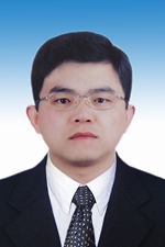 Yongsheng Xiao (肖永胜)合作论文数Zhongshan Hospital, Fudan University6
 Xianghuo He (何祥火)合作论文数Cancer Institute, Fudan University6Jiaqiang Ma合作论文数5
 Zhao Zhang (张朝)合作论文数School of Basic Medical Sciences, Fudan University5Bohao Zheng (郑博豪)合作论文数5
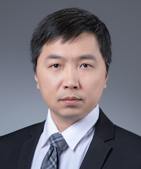 Ruibin Xi (席瑞斌)合作论文数School of Mathematical Sciences, Peking University;Center for Statistical Science, Peking University;Center for Data Science, Peking University5
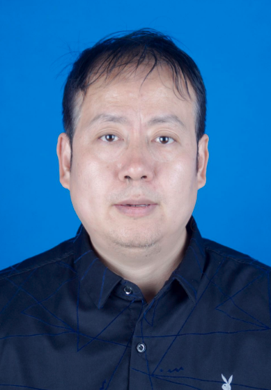 Xiyang Liu (刘西洋)合作论文数School of Computer Science and Technology, Xidian University5
 Guohe Song合作论文数4Xin-Yang Liu (刘歆阳)合作论文数4Yifei Cheng合作论文数4
 Kang Song (宋康)合作论文数4Zhao Yingjun合作论文数4
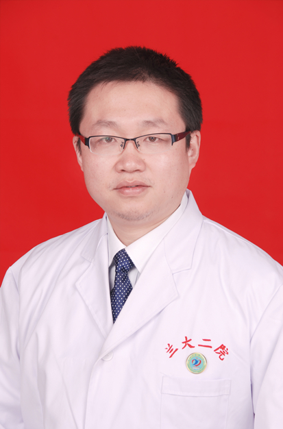 Jian Sun (孙健)合作论文数The Second Hospital, Lanzhou University4
 Xiaodong Wang (王晓东)合作论文数Beijing Cancer Hospital4
 Xiaowu Huang (黄晓武)合作论文数Zhongshan Hospital, Fudan University4Yi-fan Xia合作论文数3Zhihui Min (闵智慧)合作论文数3Lingyu Tian合作论文数3Youpei Lin合作论文数3




