Introduction Fractures are common injuries in abused children, second only to cutaneous bruising (1). Although fundamental to the documentation of abuse, the fractures are rarely life-threatening and few result in long-term deformity. Specific types of fractures are known to be associated with abuse, and their recognition is important for their accurate identification and in understanding their significance. Many reports of unexplained subdural hematomas (SDHs) in infants had appeared before Caffey’s historic 1946 article, but it was only after he associated these lesions with certain patterns of skeletal injury that the modern medical entity of child abuse was formulated (2). In a sense, recognition of the role of skeletal injuries in child abuse became the catalyst for the surge of interest in child maltreatment after Caffey’s original description. In the years that followed, most reports of child abuse focused mainly on the radiologic alterations associated with the skeletal trauma (3–14). The confident documentation of skeletal injury, facilitated by characteristic radiologic alterations, provided investigators the opportunity to study the multiple facets of child abuse. Eventually, the blend of the clinical and the radiologic findings led Kempe, Silverman, and others to bring these associations to the status of the “battered child syndrome” (15).
Juvenile xanthogranuloma (JXG) of the central nervous system (CNS) is a rare non-Langerhans cell histiocytosis. CSF1R mutations have been reported for peripheral JXG, but not in CNS JXG. A 3-month-old male presented with fever, lymphadenopathy, and macrocephaly with bulging fontanelles. Imaging demonstrated multiple dural-based masses causing obstructive hydrocephalus. The patient underwent right frontal endoscopic third ventriculostomy, choroid plexus cauterization, and craniotomy for resection and tissue diagnosis. Pathology was consistent with JXG, and NGS identified a somatic CSF1R mutation. Liquid imatinib monotherapy led to resolution of lesions and continued treatment response after 3 years without adverse events or drug interruptions.
Bone matrix-forming tumors are a group of neoplasms that exhibit differentiation toward any stage of osteoblast development. Their clinicopathologic features can resemble one another, yet their clinical management may vary significantly. Therefore, appropriate treatment requires accurate diagnosis, which can be challenging, especially with limited biopsy specimens. Recently, the driver genetic alterations underlying these neoplasms have been discovered, and their protein products can be targeted for diagnosis and therapy. Herein, we summarize the recent advances in our understanding of bone matrix-forming tumors and emphasize the integration of molecular genetics into their conventional clinicopathologic evaluation.
Conventional and dedifferentiated chondrosarcoma encompass a group of malignant neoplasms that produce cartilaginous matrix and arise within or on the surface of bone. Conventional chondrosarcomas are graded on a three-tiered scale, whereas dedifferentiated chondrosarcoma is typically not graded but is considered a high-grade sarcoma and represents the most aggressive subtype with a poor prognosis. IDH1 (isocitrate dehydrogenase-1) and IDH2 (isocitrate dehydrogenase-2) are the most commonly mutated genes in conventional and dedifferentiated chondrosarcoma, followed in frequency by COL2A1 and TP53. IDH1/2 driver mutations are also commonly found in enchondroma, considered a benign precursor lesion of chondrosarcoma, and other malignancies such as gliomas, cholangiocarcinoma, and acute myeloid leukemia. In acute myeloid leukemia, the presence of concurrent BCOR (BCL-6 corepressor) loss-of-function mutations has been linked to disease relapse and resistance to treatment with IDH inhibitors. After identifying an index case of conventional chondrosarcoma with unusually aggressive clinical evolution, we investigated the clinicopathological features of 12 cases of BCOR-mutated conventional and dedifferentiated chondrosarcomas against a control group of 15 BCOR-wildtype (WT) cases to determine whether BCOR-mutated tumors had patterns of biological progression different from tumors with intact BCOR. All identified BCOR alterations led to loss-of-function by either missense or nonsense mutations. The prevalence of BCOR mutations occurred in 5% of conventional and dedifferentiated chondrosarcoma, and these were associated with larger tumor size (p = 0.024), metastasis at the time of diagnosis (p ≤ 0.001) and higher T category (3-4 vs. 1-2) (p = 0.009). Although larger studies are necessary to clarify the full impact of BCOR mutations on patients with conventional and dedifferentiated chondrosarcoma, our data indicate that BCOR genetic aberrations are associated with adverse clinical features.
OBJECTIVES:Abdominal wall and intra-abdominal fibromatoses are locally aggressive, nonmetastasizing neoplasms. Surgery has been the mainstay of local control, but new forms of therapy have been developed that may influence the clinical course and morbidity. We studied the clinical features and outcomes of patients with abdominal and intra-abdominal fibromatoses over time. METHODS:Ninety-one patients-46 with abdominal wall and 45 with intra-abdominal fibromatosis-treated in our hospital systems between 2009 and 2023 were included. The patients were allocated to 1 of 2 groups based on the year of their initial treatment: before and including 2016 vs 2017-2023. Medical records and available histologic slides were reviewed. RESULTS:Forty-six patients were treated between 2009 and 2016, and 45 patients were treated between 2017 and 2023. Patient ages ranged from 1 to 85 years (median, 39 years), and most patients (70%) were women (2:2 men to women). Patients self-reported as Hispanic (49%), followed by White (28%), Black (20%), and Asian (3%). A subset (21%) had familial adenomatous polyposis (FAP)/Gardner syndrome. Individuals with intra-abdominal fibromatoses (37%) were more likely to have FAP than individuals with abdominal wall fibromatosis (4%) (P < .0001). The most common initial treatment before and during vs after 2016 was surgical excision (78% and 51% respectively; P = .02), followed by active surveillance with other medical intervention (9% and 18%, respectively; P = .28) and use of tyrosine kinase inhibitors (0% and 18%, respectively; P = .014). The rate of multivisceral transplant in patients with FAP/Gardner syndrome was 47% vs 4% in patients with sporadic disease (P < .001); most transplants (92%) were performed before and during 2016. The overall tumor recurrence/persistence rate in patients who had undergone surgery was 31%. The recurrence/persistence rate in patients treated before and during 2016 was 39% (median follow-up, 24 months), which fell to 13% (median follow-up, 18 months) in individuals treated after 2016 (P = .032). The overall recurrence/persistence rate in patients with FAP/Gardner syndrome was 64% vs 21% in patients with sporadic disease (P = .002). In patients with sporadic disease, there were recurrences in 29% of patients treated before and during 2016 and in 9% of patients treated thereafter (P = .086). Intra-abdominal vs abdominal wall lesions in patients with FAP and in patients with sporadic disease were more likely to recur (26% vs 10% and 16% vs 5%), but this occurrence did not reach statistical significance (P = .15). Most recurrent tumors were treated by surgical re-excision in both groups. CONCLUSIONS:Our data suggest that a combination of less morbid surgical approaches and the addition of nonsurgical approaches (active disease surveillance, use of tyrosine kinase inhibitors and other interventions) have resulted in substantially fewer surgical interventions over time for intra-abdominal and abdominal wall fibromatoses treated between 2009 and 2023. The overall probability of recurrences, however, in patients treated surgically remains similar.
The tissues removed in the surgical treatment of nonneoplastic musculoskeletal diseases represent the most frequent source of surgical pathology specimens in bone and soft tissue pathology. The pathologic changes in these specimens are often not appreciated because of the lack of familiarity with the pathophysiology of the disorders, the mechanisms in which they affect tissue, and their morphologic expressions. In this review, we highlight the important diagnostic features of selecting common nonneoplastic diseases of the musculoskeletal system that cause significant disability and morbidity.
Cartilage-forming tumors are a broad and diverse group of neoplasms frequently affecting the skeleton. Distinguishing between the members of this group is important because of significant differences in treatment and prognosis. Accurate diagnosis can be challenging because of similarities in their clinical, radiographic, and pathologic features. Immunohistochemistry and molecular tools are helpful in select instances. Therefore, careful evaluation and correlation of these features are essential in arriving at the correct diagnosis and appropriate patient management. This review provides an overview of the current literature, emphasizing helpful features in diagnosis.
e23544 Background: Leiomyosarcoma (LMS), a rare and aggressive malignancy, commonly arises from the uterus, retroperitoneum, and extremities, carrying a poor prognosis. This study investigates differences in clinical characteristics, treatment patterns, outcomes, and genomic data from next-generation sequencing (NGS) between Hispanic and non-Hispanic patients with non-metastatic and metastatic LMS. The goal is to identify disparities to inform personalized treatment strategies and improve outcomes for underrepresented populations. Methods: This single-center retrospective cohort study, conducted under an IRB-approved protocol, evaluated 190 patients with biopsy-proven LMS confirmed by sarcoma pathologists at the University of Miami Sylvester Cancer Center. NGS data were collected for patients tested between January 2009 and December 2024. Demographics, like sex, ethnicity, and age, along with clinical variables such as tumor location, size, grade, and recurrence, were gathered from electronic medical records and stored in REDCap. Kaplan-Meier and logistic regression models were used for survival analysis, adjusting for confounding variables. Patients with incomplete clinical histories were excluded. Results: The dataset included 190 patients (154 females, 33 males) with a median age of 55.8 years. Of these, 55 were metastatic, and 132 were nonmetastatic. Among nonmetastatic patients, 81 were high grade, including 55 non-Hispanic and 26 Hispanic patients. In the nonmetastatic, high-grade LMS cohort, disparities were observed: 5-year overall survival (OS) was 87.7% for non-Hispanics compared to 76.2% for Hispanics (p=0.045; hazard ratio: 1.96). Median disease-free survival (DFS) was shorter for Hispanics (14.3 months) than for non-Hispanics (50.7 months), indicating earlier recurrence among Hispanics (p=0.32). At 24 months, DFS was 39.4% for Hispanics versus 72.2% for non-Hispanics. Initial therapies, including radiation, surgery, and systemic treatments, showed no significant differences in recurrence rates, though non-Hispanic patients were more likely to receive radiation. Conclusions: These findings highlight survival disparities between Hispanics and non-Hispanics, potentially driven by earlier recurrence. NGS data from 38 patients' diagnostic tissue will be analyzed to identify potential drivers of these disparities and inform equitable treatment strategies. Clinical characteristics in patients with high grade, nonmetastatic leiomyosarcoma. Not Hispanic or Latino (N=55) Hispanic or Latino (N=26) Overall (N=81) Retroperitoneum 12.7% 11.5% 12.3% Trunk/Extremity 23.6% 34.6% 27.2% Uterine 63.6% 53.8% 60.5% First Therapy - Radiation 3.6% 0% 2.5% First Therapy - Surgery 76.4% 73.1% 75.3% First Therapy - Systemic 18.2% 19.2% 18.5% Had Recurrence 61.8% 65.4% 63.0% Had NGS 76.4% 65.4% 72.8%
This investigation describes the clinicoradiologic, pathologic, and molecular features of a unique soft tissue tumor characterized by a peripheral shell of bone and composed of bland myoid spindle and epithelioid cells that are keratin-positive. Our study cohort consists of 6 men and 6 women, with a mean age of 32 years. The tumors arose in the extremities (n = 9) and proximal limb girdle (n = 3) and were equally distributed between deep and superficial soft tissues. Patients reported dull painless masses of several months to >10 years duration (mean: 2.9 years). Imaging demonstrated a complete or partial peripheral shell of bone that could extend centrally, and the tumor’s mean size was 5.7 cm. Histologically, the tumors were composed of uniform, eosinophilic myoid spindled cells growing in sheets and intersecting fascicles, surrounded by mature lamellar and/or woven bone. Also present was an admixed component of intermediate-sized epithelioid cells with eosinophilic cytoplasm. Mitotic activity was consistently low. Immunohistochemistry showed strong multifocal staining for keratins, and 50% (5/10) showed focal staining for S100; however, all were negative for SMA, desmin, SOX10, ERG, and CD34. Genetic analysis by multiple targeted RNA sequencing panels was negative (n = 10); however, whole transcriptome sequencing (n = 8) revealed a recurrent and novel in-frame SRSF7::NFATC3 fusion in 4 tumors. Dual fluorescence in situ hybridization probes for SRSF7::NFATC3 successfully confirmed this fusion and identified a fifth case, which had not undergone whole transcriptome sequencing but was negative by a targeted RNA fusion panel. Methylation profiling (n = 8) demonstrated a shared epigenetic profile distinct from other entities. Clinical follow-up (n = 11) showed no evidence of recurrence after primary excision with a mean of 41.6 months. In summary, we describe a novel soft tissue tumor designated “ossifying spindled and epithelioid tumor” as a descriptive histologic term that also emphasizes its close radiologic mimic, ossifying fibromyxoid tumor. All cases have behaved in a benign fashion without recurrence following simple excision. Awareness of this entity is important, so that it can be distinguished from other neoplasms that have more aggressive biological potential.
INTRODUCTION:Conventional classification systems for giant cell tumors (GCTs) lack robust correlation with management and clinical outcomes. We propose a new radiologic classification system based on surgically relevant features to address this shortcoming. METHODS:This IRB-approved single-institution retrospective study involved 35 extremity GCTs from 2013 to 2023 with preoperative radiographs and cross-sectional imaging (MRI and/or CT). An experienced musculoskeletal (MSK) radiologist and orthopaedic oncologist independently assessed tumors according to the Campanacci or new grading system, defined on 1 to 3 scale: (1) intraosseous contained tumor, (2) intraosseous noncontained tumor with extraosseous implant accessible through single incision, and (3) intraosseous noncontained tumor with an extraosseous soft tissue implant nonaccessible from single incision alone. Interrater agreement was determined through the intraclass correlation coefficient. The two-way Friedman test with rater and grading system as factors was used to compare system grading similarity. RESULTS:Thirty patients underwent curettage, five underwent resection; 10 experienced local recurrence. Intraclass correlation coefficients between raters for the Campanacci and novel grading systems were 0.83 and 0.79, respectively. However, compared with the novel system, Campanacci grades were significantly higher by an average of 0.34 ± 0.68 and 0.46 ± 0.70 for the first and second raters, respectively (P = 0.003). None of the patients who underwent resection experienced local recurrence, but in patients who underwent curettage, recurrence rates were higher in Campanacci versus novel grade 1 tumors (29% vs. 17%). DISCUSSION:The novel GCT grading system demonstrates excellent interrater agreement, and classified more nonrecurrent curetted tumors as low grade, suggesting improved predictive performance compared with the Campanacci classification.
The accurate diagnosis of giant cell-rich tumors of bone is challenging, especially in limited tissue samples. This diverse group of neoplasms have similar and often ambiguous clinical presentations, radiologic features, and morphologic characteristics. During the last decade, the discovery of pathogenic recurrent genetic alterations has allowed the development of immunohistochemical surrogate markers and FISH assays that can help differentiate the entities of this broad group from one another. The correct diagnosis of these neoplasms is essential in the management of the affected patients.
Chordomas are rare notochordal malignancies typically occurring in the axial skeleton. Less than 100 extra-axial chordomas have been reported. We describe a unique chordoma of the anteromedial thigh. The clinical presentation of extra-axial chordomas is highly variable; thus, recognizing this entity in the differential diagnoses of extra-axial bone/soft tissue tumors is important.
Juvenile xanthogranuloma (JXG) of the central nervous system(CNS) is a rare non-Langerhans cell histiocytosis typically affecting children and young adults. CSF1R mutations have been reported for peripheral JXG but have not previously been reported in CNS JXG. Treatment of CNS JXG relies on maximal safe surgical resection followed by cytotoxic chemotherapy, radiotherapy and/or molecular targeted therapy involving MAPK signaling pathway. Imatinib has been effective in treating tenosynovial giant cell tumor with CSF1R mutations. Case Report A 3 month old male presented with fever, lymphadenopathy, and macrocephaly (45.5cm, 99.97%) with bulging fontanelles. Head ultrasound revealed dilated ventricles and brain MRI demonstrated multiple dural-based masses, the largest measuring 6.6 x 5.3 x 5.7 cm was located along the right tentorium and caused obstructive hydrocephalus. Patient underwent right frontal endoscopic third ventriculostomy and choroid plexus cauterization to treat hydrocephalus and right parietal craniotomy to resect the superficial dural-based mass for tissue diagnosis. Pathology was most consistent with a JXG that harbored a somatic CSF1R (NM_001288705.3) mutation. DNA Methylation profiling was unmatched. PET scan was negative for non-CNS lesions. Due to the multiple masses on presentation, the surgical risks of resection of the largest lesion, as well as having a targetable mutation, we opted for medical therapy. As Pexidartinib, a selective CSF1R inhibitor, was not available for infants, patient was started on liquid imatinib 340 mg/m2/day monotherapy. Three months after initiation of imatinib, the smaller parietal lesions resolved and after six months, the largest mass continued to shrink (6.3 x 2.9 x 2.9 cm). Treatment has been well tolerated without adverse events or drug interruptions. This is the first report of CNS JXG with a CSF1R mutation. Importantly, it demonstrates that imatinib is a potentially safe and effective form of monotherapy for this rare disease.
This review summarizes the clinicopathologic features of various lipomatous tumors of soft tissue and addresses some recent conceptual issues relating to adipocytic neoplasms, such as atypical spindle cell/pleomorphic lipomatous tumor and myxoid pleomorphic liposarcoma, and provides an update on the molecular aspects of these tumors. Recent advances in cytogenetic characterization and classification of lipomatous tumors are reviewed, and the genetic importance of distinct chromosomal aberrations are briefly discussed.
Neoplasms of the tongue are relatively common, and the vast majority are epithelial in phenotype. Although uncommon, a diverse and distinctive array of mesenchymal neoplasms arises in this anatomic site. To increase our understanding of these lesions, we reviewed our experience of MNs of the tongue and described their clinicopathologic features. The pathology archives from 2005 to 2021 and the consultation files of one of the authors were queried for all MNs of the tongue. We reviewed the histologic slides and ancillary studies and obtained clinical data from the available medical records. Ninety-three cases were identified, and they form the study cohort - to our knowledge, this is the largest series of mesenchymal neoplasms of the tongue. Forty-eight patients were female, and forty-five were male, with a mean age of 51 years (range: 1-94 years). The tumors included 43 (46.2%) hemangiomas, 14 (15%) granular cell tumors, 8 (9%) lipomas, 4 (4.3%) schwannomas, 4 (4.3%) solitary fibrous tumors - all with low risk of progression based on risk stratification criteria, 2 (2.2%) lymphangiomas, 3 (3.2%) Kaposi sarcomas, 2 (2.2%) chondromas, 2 (2.2%) myofibromas, 1 (1.1%) solitary circumscribed neuroma, 1 (1.1%) perineurioma, 1 (1.1%) neurofibroma, 1 (1.1%) ectomesenchymal chondromyxoid tumor, 1 (1.1%) atypical glomus tumor with a NOTCH2 rearrangement and TLL2 mutation, 1 (1.1%) spindle cell rhabdomyosarcoma, 1 (1.1%) pleomorphic fibroblastic sarcoma, 1 (1.1%) malignant rhabdoid tumor, 1 (1.1%) leiomyosarcoma, 1 (1.1%) angiosarcoma, and 1 (1.1%) alveolar soft part sarcoma. Most of the patients underwent surgical excision, and 1 patient (with hemangioma) underwent embolization. On follow-up, the patient with spindle cell rhabdomyosarcoma developed postoperative numbness at the surgical site and was disease-free through 17 months of follow-up. The patient with leiomyosarcoma declined adjuvant radiation and developed metastasis to the lung at 22 months. The patient with alveolar soft part sarcoma had metastases to the lung at the time of diagnosis and received adjuvant chemotherapy. The remaining patients had no local or distant recurrence. MNs of the tongue are usually benign and characterized by either endothelial, adipocytic, or schwannian differentiation. The mainstay of treatment is surgical excision with the extent of excision determined by tumor type. Adjuvant therapy is reserved for high-grade sarcomas.
Multidisciplinary tumor boards (MTBs) facilitate decision-making among subspecialists in the care of oncology patients, but the mechanisms by which they enhance outcomes remain incompletely understood. Our aim was to measure the agreement between sarcoma MTBs and radiology reports’ disease assessment and management recommendations. This single-center IRB-approved retrospective study evaluated cases presented at a weekly sarcoma MTB from 1 August 2020 to 31 July 2021. Cases without clinical notes, imaging studies, or radiology reports were excluded. The data collected included the patient’s clinical status at the time of the MTB, the treatment response assessment by the MTB and radiologists (stable disease; partial response; complete response; progressive disease/recurrence), and the recommendations of the radiology reports and of the MTB. The agreement between the initial radiologist review and MTB on disease assessment and recommendations was analyzed using kappa statistics. In total, 283 cases met the inclusion criteria. Radiology reports provided recommendations in 34.3% of cases, which were adhered to by the ordering providers in 73.2% of cases. The agreement between MTBs and radiology reports was moderate in disease assessment (86.2% agreement; κ = 0.78; p < 0.0001) and negligible in recommendations (36% agreement; κ = 0.18; p < 0.0001). Radiologists were more likely to assign progressive disease/recurrence than MTBs (54.4% vs. 44.4%; p < 0.001) and to recommend short-term imaging follow-up more commonly than MTBs (46.4% vs. 21.7%; p < 0.001). At a tertiary care center, radiologists’ isolated interpretations of imaging findings and management recommendations frequently differ from the MTB’s consensus, reflecting the value of multidisciplinary discussions incorporating the patient’s clinical status and the available treatment options into the final radiographic assessment.
Opinion statementHead and neck osteosarcoma (HNOS) is a rare subtype of sarcoma that most commonly arises in the mandible or maxilla. Treatment for HNOS typically involves a multidisciplinary and multimodal approach depending on the size, grade, and histological subtype. Surgery by sarcoma-experienced head and neck surgeons and orthopedic oncologists remains a crucial component of treatment in all subtypes of HNOS, particularly for those with low-grade histology, which can be treated definitively with surgical resection if negative margins are obtained. Negative surgical margins are of utmost prognostic importance, and neoadjuvant or adjuvant radiation should be considered in patients with positive (or anticipated positive) margins/residual postoperative disease. Current data favors the use of (neo)adjuvant chemotherapy in patients with high-grade HNOS to improve overall survival but must be individualized to weigh benefits and risks of the short- and long-term effects of treatment. Our center uses a multidisciplinary treatment plan and notes anecdotal improvement in treatment outcomes with a combined surgical and ifosfamide-containing chemotherapeutic approach with radiotherapy for local control if positive margins. Large volume cohorts and adequate randomized control trials assessing the efficacy of chemotherapy in HNOS are scant and additional research and multi-institutional collaboration are needed to study polychemotherapeutic and radiation treatment regimens and outcomes more adequately.
 G. Petur Nielsen合作论文数Division of Bone and Soft Tissue Pathology, Massachusetts General Hospital;Harvard Medical School185
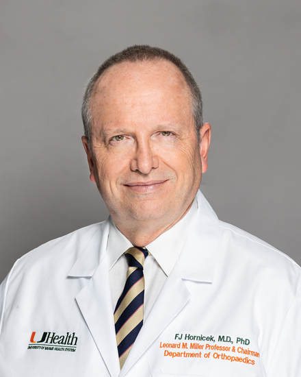 Francis John Hornicek合作论文数Sarcoma Research Laboratory, University of Miami88
 Susan Kattapuram合作论文数88
 Daniel Rosenthal合作论文数Department of Radiology, Massachusetts General Hospital83
 Henry J. Mankin合作论文数Harvard Medical School;Department of Orthopaedic Surgery, Massachusetts General Hospital68
 Vikram Deshpande合作论文数Department of Pathology, Beth Israel Deaconess Medical Center, Harvard Medical School65
 Jonathan C Trent合作论文数Sylvester Comprehensive Cancer Center, University of Miami;Division of Hematology, Department of Medicine, Miller School of Medicine, University of Miami45
 Robert Edward Scully合作论文数Harvard Medical School;Massachusetts General Hospital35
 Subhawong Ty K合作论文数30William F. Mcneely合作论文数From the Departments of Medicine, Boston University School of Medicine and Tufts University School of Medicine, Boston, Mass., USA30
 Diaz-Perez Julio A合作论文数Moores Cancer Center, University of California29Betty U. Mcneely合作论文数28
 Herman D. Suit合作论文数Department of Radiation Oncology, Massachusetts General Hospital;Harvard Medical School, Harvard University27Richard C. Cabot合作论文数26
 Eugene Jerome Mark合作论文数Department of Pathology, Massachusetts General Hospital;Harvard University24
 Jaylou M. Velez Torres合作论文数23John X. O'Connell合作论文数Department of Pathology, University of British Columbia23Jx Oconnell合作论文数22Kerr Darcy A合作论文数22Re Scully合作论文数19
 Thomas F. Delaney合作论文数Francis H. Burr Proton Therapy Center, Massachusetts General Hospital;Harvard Medical School, Harvard University19
 Breelyn Wilky合作论文数Department of Medicine-Medical Oncology, School of Medicine, University of Colorado Anschutz Medical Campus;University of Colorado Hospital17
 Paola S. Dal Cin合作论文数Department of Pathology, Harvard Medical School;Cytogenetics Laboratory, Brigham And Women's Hospital16
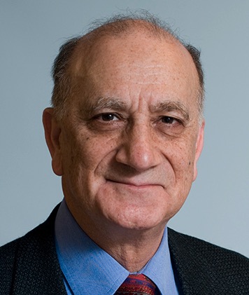 Atul Bhan合作论文数Department of Pathology, Massachusetts General Hospital;Harvard Medical School15Mc Gebhardt合作论文数14
 D'Amato Gina Z合作论文数14
 Jimmy Efird合作论文数Department of Radiation Oncology, School of Medicine, Case Western Reserve University14S B Keel合作论文数13Ehara Shigeru合作论文数13Christopher D. M. Fletcher合作论文数13Mark C. Gebhardt合作论文数13
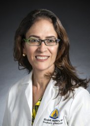 Elizabeth A. Montgomery合作论文数Department of Pathology, Miller School of Medicine, Medical Campus, University of Miami13IJ Spiro合作论文数13
 Cristina R. Antonescu合作论文数Memorial Sloan-Kettering Cancer Center13Emily Jonczak合作论文数12
 Aditi Dhir合作论文数12G. Richard Dickersin合作论文数12
 Edwin Choy合作论文数Departments of Medicine and Cell Biology, New York University School of Medicine12
 Kevin A. Raskin合作论文数Department of Orthopaedic Oncology, Massachusetts General Hospital12Ds Springfield合作论文数11Gr Dickersin合作论文数11
 Juan Abelardo Augusto Pretell合作论文数11
 Sheila Conway合作论文数11Germaine Cuff合作论文数11
 Carmen Gomez-Fernandez合作论文数Department of Pathology and Laboratory Medicine, Miller School of Medicine, University of Miami11David C. Harmon合作论文数10
 Zhang Yaxia合作论文数Department of Pathology, Cleveland Clinic10
 Lena M. Napolitano合作论文数Weil Institute, University of Michigan10Carrie Y. Inwards合作论文数10




