AIM:The underlying mechanisms contributing to cholestatic liver injury remain unclear. The pro-inflammatory leukocyte-restricted cytokine interleukin-17A (IL-17A) has been implicated in human cholestatic liver injury. However, mechanistic insights are lacking and require further exploration in preclinical models. Herein, we examined the effect of IL-17A genetic ablation in a mouse model of cholestatic liver injury. METHOD:Age and gender-matched littermate wild type (WT) and Il-17a-/- C57BL/6 mice were fed an intermittent 0.1% 3,5-diethoxycarbonyl-1,4-dihydrocollidine (DDC) diet for 21 days to induce cholestatic liver injury or a control diet. RESULTS:As compared to WT littermates, Il-17a-/- mice displayed more abundant desmin-positive myofibroblasts and increased fibrosis. NanoString analysis of intrahepatic leukocyte populations using a fibrosis-related gene panel identified upregulation of Tnfsf14 (encoding the protein LIGHT) in the DDC-fed Il-17a-/- mice. Although mass cytometry identified an increase in myeloid cells in both genotypes of the DDC-fed mice, we could not identify LIGHT expression in this cell lineage. Instead, the upregulation of LIGHT expression was largely restricted to a CD4+ T cell population as assessed by flow cytometry. Enhanced LIGHT expression was observed in a Th1+ CD4+ T cell population. LIGHT activated primary human hepatic stellate cells in vitro, suggesting that LIGHT stimulation of hepatic fibrogenesis may be direct. CONCLUSION:Taken together, these data suggest that IL-17A restrains expression of the profibrogenic cytokine, LIGHT, by Th1-polarized CD4+ T cells, and implicate a role for LIGHT in cholestatic fibrogenesis in DDC-fed mice; a finding which requires validation in additional models.
During cholestasis, cholangiocytes become activated, promoting macrophage-associated periductal infiltration and fibrosis. The cholangiocyte-specific mechanisms responsible for these processes are unclear. To gain insight into the cholangiocyte signaling mechanisms contributing to these pathophysiologic processes, mice were fed a 3,5-diethoxycarbonyl-1,4-dihydro-collidine (DDC) diet for 10 days to induce liver injury and then switched to a chow diet to permit recovery, designated as R days. Profiling of isolated intrahepatic leukocytes by mass spectrometry revealed an abundant CX3CR1+ macrophage population on the DDC diet that declined during the recovery period. This observation was confirmed using Cx3cr1GFP mice. Next, cholangiocytes were isolated from control, DDC, and R15 mice, and RNA sequencing (RNAseq) was performed. Cholangiocyte CX3CL1 expression, the cognate ligand for CX3CR1, increased in DDC-fed mice and returned to basal values by R15, implicating cholangiocytes in CX3CR1+ macrophage recruitment. Ingenuity pathway analysis (IPA) of the RNAseq data revealed upregulation of the pathogen-induced cytokine storm pathway in cholangiocytes activated from DDC fed mice, and resolution of this pathway in R15 isolated cholangiocytes. SCENIC regulon analysis identified that NF-Y, a transcription factor complex, was activated only on the DDC diet, but not in control or R15 mice. Finally, siRNA targeted suppression of NF-YA in normal human cholangiocytes (NHC) reduced cholangiocyte expression of the profibrogenic ligand TGFβ1. Consistent with this observation, Tgfβ1 was increased in cholangiocytes from DDC-fed animals that returned to control values at day R15. Collectively, these observations provide mechanistic insights into cholangiocyte pathobiology during cholestasis.NEW & NOTEWORTHY Cholangiocyte pathophysiological activation was examined in a model of murine cholestasis. CX3CR1+ macrophages are recruited to the periportal region, likely mediated by cholangiocyte expression of CX3CL1. Cholangiocyte transcriptomics from cholestatic mice display activation of a "pathogen-induced cytokine storm" pathway, and exhibit activation of the transcription factor NF-Y. In human cholangiocytes, NF-Y promotes expression of the profibrogenic ligand TGFβ1. These observations provide insights into the cholestatic cholangiocyte pathobiology contributing to periductal inflammation and fibrosis.
Ductular reactive (DR) cells exacerbate cholestatic liver injury and fibrosis. Herein, we posit that tumor necrosis factor-related apoptosis-inducing ligand (TRAIL) emanates from recruited macrophages and restrains DR cell expansion, thereby limiting cholestatic liver injury. Wild type (WT), Trailfl/fl and myeloid-specific Trail deleted (TrailΔmye) C57BL/6 mice were exposed to DDC diet-induced cholestatic liver injury, which induced hepatomegaly and liver injury as compared to control diet-fed mice. However, parameters of liver injury, fibrosis, and inflammation were all increased in the TrailΔmye mice as compared to the WT and Trailfl/fl mice. High dimensional mass cytometry indicated that cholestasis resulted in increased hepatic recruitment of subsets of macrophages and neutrophils in the TrailΔmye mice. Spatial transcriptomics analysis revealed that the PanCK+ cholangiocytes from TrailΔmye mice had increased expression of the known myeloid attractants S100a8, Cxcl5, Cx3cl1, and Cxcl1. Additionally, in situ hybridization of Cxcl1, a potent neutrophil chemoattractant, demonstrated an increased expression in CK19+ cholangiocytes of TrailΔmye mice. Collectively, these data suggest that TRAIL from myeloid cells, particularly macrophages, restrains a subset of DR cells (i.e., Cxcl1 positive cells), limiting liver inflammation and fibrosis. Reprogramming macrophages to express TRAIL may be salutary in cholestasis.
Intrahepatic macrophages in nonalcoholic steatohepatitis (NASH) are heterogenous and include proinflammatory recruited monocyte-derived macrophages. The receptor for advanced glycation endproducts (RAGE) is expressed on macrophages and can be activated by damage associated molecular patterns (DAMPs) upregulated in NASH, yet the role of macrophage-specific RAGE signaling in NASH is unclear. Therefore, we hypothesized that RAGE-expressing macrophages are proinflammatory and mediate liver inflammation in NASH. Compared with healthy controls, RAGE expression was increased in liver biopsies from patients with NASH. In a high-fat, -fructose, and -cholesterol-induced (FFC)-induced murine model of NASH, RAGE expression was increased, specifically on recruited macrophages. FFC mice that received a pharmacological inhibitor of RAGE (TTP488), and myeloid-specific RAGE KO mice (RAGE-MKO) had attenuated liver injury associated with a reduced accumulation of RAGE+ recruited macrophages. Transcriptomics analysis suggested that pathways of macrophage and T cell activation were upregulated by FFC diet, inhibited by TTP488 treatment, and reduced in RAGE-MKO mice. Correspondingly, the secretome of ligand-stimulated BM-derived macrophages from RAGE-MKO mice had an attenuated capacity to activate CD8+ T cells. Our data implicate RAGE as what we propose to be a novel and potentially targetable mediator of the proinflammatory signaling of recruited macrophages in NASH.
BACKGROUND:NASH is the progressive form of NAFLD characterized by lipotoxicity, hepatocyte injury, tissue inflammation, and fibrosis. Previously, Rho-associated protein kinase (ROCK) 1 has been implicated in lipotoxic signaling in hepatocytes in vitro and high-fat diet-induced lipogenesis in vivo. However, whether ROCK1 plays a role in liver inflammation and fibrosis during NASH is unclear. Here, we hypothesized that pathogenic activation of ROCK1 promotes murine NASH pathogenesis. METHODS AND RESULTS:Patients with NASH had increased hepatic ROCK1 expression compared with patients with fatty liver. Similarly, hepatic ROCK1 levels and activity were increased in mice with NASH induced by a western-like diet that is high in fat, fructose, and cholesterol (FFC). Hepatocyte-specific ROCK1 knockout mice on the FFC diet displayed a decrease in liver steatosis, hepatic cell death, liver inflammation, and fibrosis compared with littermate FFC-fed controls. Mechanistically, these effects were associated with a significant attenuation of myeloid cell recruitment. Interestingly, myeloid cell-specific ROCK1 deletion did not affect NASH development in FFC-fed mice. To explore the therapeutic opportunities, mice with established NASH received ROCKi, a novel small molecule kinase inhibitor of ROCK1/2, which preferentially accumulates in liver tissue. ROCK inhibitor treatment ameliorated insulin resistance and decreased liver injury, inflammation, and fibrosis. CONCLUSIONS:Genetic or pharmacologic inhibition of ROCK1 activity attenuates murine NASH, suggesting that ROCK1 may be a therapeutic target for treating human NASH.
Two Supplemental figures for manuscript: "Pathogenesis of Murine Cholestatic Liver Disease Associated with Sex-Related Differences in Hepatic Natural Killer Cell Subsets and Function"
We report for the first time that in wild-type (WT) mice, fast-food diet (FFD) induced a threefold increase in hepatic Sulf2 mRNA and a 2.2-fold increase in sulfatase 2 (SULF2) protein expression compared with WT mice on standard chow diet (SC). We showed that knockout of SULF2 ameliorates FFD-induced obesity, hyperlipidemia, steatohepatitis, and fibrosis. These data, along with work from other laboratories, suggest that SULF2 may be critical to the ability of the liver to progress to nonalcoholic steatohepatitis and fibrosis in conditions of overnutrition.
BACKGROUND AND AIMS:In cholestatic liver diseases, ductular reactive (DR) cells extend into the hepatic parenchyma and promote inflammation and fibrosis. We have previously observed that multidrug-resistant 2 (Mdr2-/- ) double knockout (DKO) mice lacking tumor necrosis factor-related apoptosis-inducing ligand receptor (Tr-/- ) display a more extensive ductular reaction and hepatic fibrosis compared to Mdr2-/- mice. This observation suggests that the magnitude of the DR-cell population may be regulated by apoptosis.APPROACH AND RESULTS:To examine this concept, we cultured epithelial cell adhesion molecule-positive reactive cholangioids (ERCs) obtained from wild-type (WT), Tr-/- , Mdr2-/- and DKO mice. Single-cell transcriptomics and immunostaining of both WT and DKO ERCs confirmed their DR-cell phenotype. Moreover, DKO ERCs displayed a unique translational cluster with expression of chemokines, indicating a reactive state. Incubation with the myeloid cell leukemia 1 (MCL1) inhibitor S63845, a proapoptotic BH3-mimetic therapy, significantly decreased DKO and Mdr2-/- ERC viability compared to WT. Intravenous administration of S63845 significantly reduced the DR-cell population and markers of inflammation and liver fibrosis in Mdr2-/- and DKO mice. Furthermore, DKO mice treated with S63845 displayed a significant decrease in hepatic B lymphocytes compared to untreated mice as assessed by high-definition mass cytometry by time-of-flight. Coculture of bone marrow-derived macrophages with ERCs from DKO mouse livers up-regulated expression of the B cell-directed chemokine (C-C motif) ligand 5. Finally, DR cells were noted to be primed for apoptosis with Bcl-2 homologous antagonist/killer activation in vitro and in vivo in primary sclerosing cholangitis liver specimens.CONCLUSIONS:DR cells appear to play a key role in recruiting immune cells to the liver to actively create an inflammatory and profibrogenic microenvironment. Pharmacologic targeting of MCL1 in a mouse model of chronic cholestasis reduces DR-cell and B-cell populations and hepatic fibrosis.
BACKGROUND & AIMS: Endoplasmic reticulum to nucleus signaling 1 (ERN1, also called IRE1A) is a sensor of the unfolded protein response that is activated in the livers of patients with nonalcoholic steatohepatitis (NASH). Hepatocytes release ceramide-enriched inflammatory extracellular vesicles (EVs) after activation of IRE1A. We studied the effects of inhibiting IRE1A on release of inflammatory EVs in mice with diet-induced steatohepatitis. METHODS: C57BL/6J mice and mice with hepatocyte-specific disruption of Ire1a (IRE1 alpha(Delta hep)) were fed a diet high in fat, fructose, and cholesterol to induce development of steatohepatitis or a standard chow diet (controls). Some mice were given intraperitoneal injections of the IRE1A inhibitor 4 mu 8C. Mouse liver and primary hepatocytes were transduced with adenovirus or adeno-associated virus that expressed IRE1A. Livers were collected from mice and analyzed by quantitative polymerase chain reaction and chromatin immunoprecipitation assays; plasma samples were analyzed by enzyme-linked immunosorbent assay. EVs were derived from hepatocytes and injected intravenously into mice. Plasma EVs were characterized by nanoparticle-tracking analysis, electron microscopy, immunoblots, and nanoscale flow cytometry; we used a membrane-tagged reporter mouse to detect hepatocyte-derived EVs. Plasma and liver tissues from patients with NASH and without NASH (controls) were analyzed for EV concentration and by RNAscope and gene expression analyses. RESULTS: Disruption of Ire1a in hepatocytes or inhibition of IRE1A reduced the release of EVs and liver injury, inflammation, and accumulation of macrophages in mice on the diet high in fat, fructose, and cholesterol. Activation of IRE1A, in the livers of mice, stimulated release of hepatocyte-derived EVs, and also from cultured primary hepatocytes. Mice given intravenous injections of IRE1A-stimulated, hepatocyte-derived EVs accumulated monocyte-derived macrophages in the liver. IRE1A-stimulated EVs were enriched in ceramides. Chromatin immunoprecipitation showed that IRE1A activated X-box binding protein 1 (XBP1) to increase transcription of serine palmitoyltransferase genes, which encode the rate-limiting enzyme for ceramide biosynthesis. Administration of a pharmacologic inhibitor of serine palmitoyltransferase to mice reduced the release of EVs. Levels of XBP1 and serine palmitoyltransferase were increased in liver tissues, and numbers of EVs were increased in plasma, from patients with NASH compared with control samples and correlated with the histologic features of inflammation. CONCLUSIONS: In mouse hepatocytes, activated IRE1A promotes transcription of serine palmitoyltransferase genes via XBP1, resulting in ceramide biosynthesis and release of EVs. The EVs recruit monocyte-derived macrophages to the liver, resulting in inflammation and injury in mice with diet-induced steatohepatitis. Levels of XBP1, serine palmitoyltransferase, and EVs are all increased in liver tissues from patients with NASH. Strategies to block this pathway might be developed to reduce liver inflammation in patients with NASH.
The tumor necrosis factor-related apoptosis-inducing ligand (TRAIL; TNFSF10) receptor (TR) is a proapoptotic receptor whose contribution to chronic cholestatic liver disease is unclear. Herein, we examined TRAIL receptor signaling in a mouse model of cholestatic liver injury. TRAIL receptor-deficient (Tnsf10 or Tr-/-) mice were crossbred with ATP binding cassette subfamily B member 4-deficient (Abcb4(-/-), alias Mdr2(-/-)) mice. Male and female wild-type, Tr-/-, Mdr2(-/-), and Tr-/- Mdr2(-/-) mice were assessed for liver injury, fibrosis, and ductular reactive (DR) cells. Macrophage subsets were examined by high-dimensional mass cytometry (time-of-flight mass cytometry). Mdr2(-/-) and Tr (-/-) Mdr2(-/-) mice had elevated liver weights and serum alanine transferase values. However, fibrosis was primarily periductular in Mdr2(-/-) mice, compared with extensive bridging fibrosis in Tr(-/-)Mdr2(-/-) mice. DR cell population was greatly expanded in the Tr(-/-)Mdr2(-/-) versus Mdr2(-/-) mice. The expanded DR cell population in Tr(-/-)Mdr2(-/-) mice was due to decreased cell loss by apoptosis and not enhanced proliferation. As assessed by time-of-flight mass cytometry, total macrophages were more abundant in Tr(-/-)Mdr2(-/-) versus Mdr2(-/-) mice, suggesting the DR cell population promotes macrophage-associated hepatic inflammation. Inhibition of monocyte-derived recruited macrophages using the CCR2/CCR5 antagonist cenicriviroc in the Mdr2(-/-) mice resulted in further expansion of the DR cell population. In conclusion, genetic deletion of TRAIL receptor increased the DR cell population, macrophage accumulation, and hepatic fibrosis in the Mdr2(-/-) model of cholestasis.
Introduction: Metabolic syndrome is a disorder characterized by a constellation of findings including truncal obesity, elevated blood pressure, abnormal cholesterol levels, and high blood glucose. Recent evidence suggests that metabolic syndrome may be associated with increased risk of age-related macular degeneration (AMD) and other eye diseases. Recently, C57BL/6J wild-type mice fed with a "fast food" diet consisting of high fat, cholesterol, and fructose-supplemented water showed unique systemic pathology consistent with metabolic syndrome and nonalcoholic steatohepatitis. Additionally, these mice showed higher levels of fibrosis, inflammation, endoplasmic reticulum stress, and mitochondrial dysfunction compared to mice fed with only a high-fat diet alone. Since similar pathways are activated in AMD, we sought to determine whether mice fed a "fast food" diet exhibited retinal changes.Methods: 3-month-old wild-type mice were randomized to a standard chow (n = 11) or a "fast food" (n = 18) diet and fed for 9 months. At 1 year of age, tissues were collected and retinas were analyzed using transmission electron microscopy. Quantitative measures of Bruch's membrane thickness and retinal pigment epithelium (RPE) cell counts were performed.Results: "Fast food" fed mice showed ocular pathology relevant to various stages of AMD including basal laminar deposits, focal thickening of Bruch's membrane, and a significant loss of RPE cells.Discussion/conclusion: A wild-type mouse model of metabolic syndrome fed a "fast food" diet developed changes to the retina similar to some of the pathologic features seen in AMD. Further investigations into this and similar animal models as well as further epidemiological studies are needed to more clearly define the association between metabolic syndrome and AMD.
In mouse models of biliary tract diseases, macrophages are recruited to the periductal milieu and promote injury and cholestasis. Although cell necrosis with release of biomolecules termed damage-associated molecular patterns (DAMPs) promotes recruitment and activation of macrophages, necrosis was not observed in these studies. Because extracellular vesicles (EVs) are important in cell-to-cell communication, we postulated that activated cholangiocytes may release EVs containing DAMPs as cargo. Both the human (NHC) and mouse cholangiocyte (603B) cell lines display constitutive activation with mRNA expression of chemokines. Proteomic analysis revealed that EVs from both cell lines contained the DAMP S100A11, a ligand for the receptor for advanced glycation end products (RAGE). Bone marrow-derived macrophages (BMDM) incubated with EVs derived from the mouse 603B cell line increased mRNA expression of proinflammatory cytokines. Genetic or pharmacologic inhibition of RAGE reduced BMDM expression of proinflammatory cytokines treated with EVs. RAGE signaling resulted in activation of the canonical NF-κB pathway, and consistently, proinflammatory cytokine expression was blunted by the IKKα/β inhibitor TPCA-1 in BMDM incubated with EVs. We also demonstrated that primary mouse cholangiocyte-derived organoids express chemokines indicating cholangiocyte activation, release EVs containing S100A11, and stimulate proinflammatory cytokine expression in BMDM by a RAGE-dependent pathway. In conclusion, these observations identify a non-cell death mechanism for cellular release of DAMPs by activated cholangiocytes, namely by releasing DAMPs as EV cargo. These data also suggest RAGE inhibitors may be salutary in macrophage-associated inflammatory diseases of the bile ducts.
BACKGROUND & AIMS:Hepatic recruitment of monocyte-derived macrophages (MoMFs) contributes to the inflammatory response in non-alcoholic steatohepatitis (NASH). However, how hepatocyte lipotoxicity promotes MoMF inflammation is unclear. Here we demonstrate that lipotoxic hepatocyte-derived extracellular vesicles (LPC-EVs) are enriched with active integrin β1 (ITGβ1), which promotes monocyte adhesion and liver inflammation in murine NASH.METHODS:Hepatocytes were treated with either vehicle or the toxic lipid mediator lysophosphatidylcholine (LPC); EVs were isolated from the conditioned media and subjected to proteomic analysis. C57BL/6J mice were fed a diet rich in fat, fructose, and cholesterol (FFC) to induce NASH. Mice were treated with anti-ITGβ1 neutralizing antibody (ITGβ1Ab) or control IgG isotype.RESULTS:Ingenuity® Pathway Analysis of the LPC-EV proteome indicated that ITG signaling is an overrepresented canonical pathway. Immunogold electron microscopy and nanoscale flow cytometry confirmed that LPC-EVs were enriched with activated ITGβ1. Furthermore, we showed that LPC treatment in hepatocytes activates ITGβ1 and mediates its endocytic trafficking and sorting into EVs. LPC-EVs enhanced monocyte adhesion to liver sinusoidal cells, as observed by shear stress adhesion assay. This adhesion was attenuated in the presence of ITGβ1Ab. FFC-fed, ITGβ1Ab-treated mice displayed reduced inflammation, defined by decreased hepatic infiltration and activation of proinflammatory MoMFs, as assessed by immunohistochemistry, mRNA expression, and flow cytometry. Likewise, mass cytometry by time-of-flight on intrahepatic leukocytes showed that ITGβ1Ab reduced levels of infiltrating proinflammatory monocytes. Furthermore, ITGβ1Ab treatment significantly ameliorated liver injury and fibrosis.CONCLUSIONS:Lipotoxic EVs mediate monocyte adhesion to LSECs mainly through an ITGβ1-dependent mechanism. ITGβ1Ab ameliorates diet-induced NASH in mice by reducing MoMF-driven inflammation, suggesting that blocking ITGβ1 is a potential anti-inflammatory therapeutic strategy in human NASH.LAY SUMMARY:Herein, we report that a cell adhesion molecule termed integrin β1 (ITGβ1) plays a key role in the progression of non-alcoholic steatohepatitis (NASH). ITGβ1 is released from hepatocytes under lipotoxic stress as a cargo of extracellular vesicles, and mediates monocyte adhesion to liver sinusoidal endothelial cells, which is an essential step in hepatic inflammation. In a mouse model of NASH, blocking ITGβ1 reduces liver inflammation, injury and fibrosis. Hence, ITGβ1 inhibition may serve as a new therapeutic strategy for NASH.
 Gregory J. Gores合作论文数Division of Gastroenterology and Hepatology, Department of Internal Medicine, Mayo Clinic39
 Michael R. Charlton合作论文数Center for Liver Diseases, The University of Chicago Medicine25
 Kimberly Viker合作论文数23
 Maria Eugenia Guicciardi合作论文数17
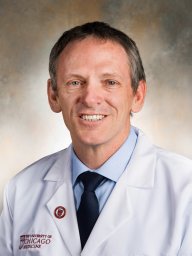 Michael Charlton合作论文数Madrigal Pharmaceuticals;University of Chicago;Mayo Clinic16Steven F. Bronk合作论文数14Schuyler O. Sanderson合作论文数12
 Petra Hirsova合作论文数Faculty of Medicine in Hradec Kralove, Charles University in Prague11
 Kevin D. Pavelko合作论文数Department of Immunology, Mayo Clinic8Azad Adiba合作论文数8
 Ype P. de Jong合作论文数Joan and Sanford I. Weill Department of Medicine, Weill Cornell Medicine, Cornell University6Marcus Dorner合作论文数Center for the Study of Hepatitis C, The Rockefeller University;Center for the Study of Hepatitis C, The Rockefeller University6Katsumi Tomohiro合作论文数6
 James M Swain合作论文数6Michiel C Mommersteeg合作论文数5Nazli Begum Ozturk合作论文数Istanbul Faculty of Medicine, Istanbul University5
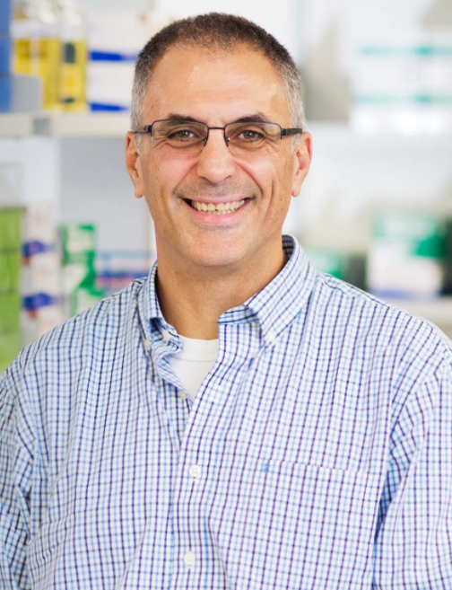 Luis A. Chiriboga合作论文数Department of Pathology, Grossman School of Medicine, New York University5
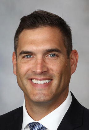 Nathan K. LeBrasseur合作论文数Department of Physical Medicine & Rehabilitation, Mayo Clinic;Department of Physiology & Biomedical Engineering, Mayo Clinic;Robert and Arlene Kogod Center on Aging, Mayo Clinic5
 Christy E Trussoni合作论文数5Xiao Jing W合作论文数Center for the Study of Hepatitis C, The Rockefeller University5
 Harmeet Malhi合作论文数Division of Gastroenterology and Hepatology, Department of Internal Medicine, College of Medicine, Mayo Clinic5
 Charles Rice合作论文数School of Kinesiology, Western University;Neuromuscular Lab, Western University5
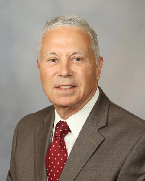 Nicholas F. Larusso合作论文数Departments of Gastroenterology and Hepatology, Mayo Clinic;Mayo Clinic Comprehensive Cancer Center5Thomas A White合作论文数4Bridget M Donovan合作论文数4
 Samar saber Ibrahim合作论文数4Takashi Kitagataya合作论文数4Hui Peng合作论文数4
 Wei Fan合作论文数Karsh Div Gastroenterol & Hepatol, Cedars Sinai Med Ctr4
 Alexander Ploss合作论文数Department of Molecular Biology, Princeton University;Rutgers Cancer Institute of New Jersey, Rutgers, The State University of New Jersey4
 Jalan-Sakrikar Nidhi合作论文数Department of Molecular Physiology and Biophysics, Vanderbilt University School of Medicine4Kendrick Michael L合作论文数4
 Watt Kymberly D合作论文数4Koehler Edith合作论文数4Labitt Rachael N合作论文数4Liu Ting合作论文数4
 Dennis Burton合作论文数Department of Immunology and Microbiology, The Scripps Research Institute;The Ragon Institute of MGH, MIT and Harvard;IAVI Neutralizing Antibody Center, The Scripps Research Institute4
 Michael G. Sarr合作论文数Department of Surgery, Mayo Clinic4Florencia G. Que合作论文数4Heping Yang合作论文数University of Southern California, University of California Los Angeles Research Center for Alcoholic Liver and Pancreatic Diseases4Geoffrey B. Thompson合作论文数4Andrea L. Mcconico合作论文数4Nathan W. Werneburg合作论文数4Tony W. H. Li合作论文数4
 Patrick Splinter合作论文数4
 Steven P O'Hara合作论文数Miles and Shirley Fiterman Center for Basic Research in Digestive Diseases, Mayo Clinic College of Medicine4
 Alejandro Benjamin Balazs合作论文数Harvard Medical School;Massachusetts General Hospital4
 David Baltimore合作论文数Division of Biology and Biological Engineering, California Institute of Technology4Jiaohong Wang合作论文数4




