One of the major conundrums of cancer research and treatment is that the metastases that lead to death in most patients do not appear to involve additional driver mutations. Previously, we reported widespread loss of heterochromatin with activation of pro-metastatic genes in the subset of cells of primary pancreatic tumors that gave rise to liver and lung metastases. Here we hypothesized that this change in chromatin could create unique vulnerabilities in distant metastases. Using a CRISPR screen of human patient-derived xenografts from metastases and primary tumors, we identified KLF5 as essential for metastatic cell proliferation but not primary tumor growth. Further, we found that KLF5 induced epigenetic modifier genes, including NCAPD2 and MTHFD1, which themselves facilitated expression of specific genes driving migration and epithelial-mesenchymal transition, including TGFBR2, VIM, EMP1, and ITGB1. Inhibition of expression of these modifier genes restored heterochromatin in the specific regions that distinguish the primary and metastatic tumors. We backed up this causal chain of evidence with rigorous additional knockdown experiments with the modifier genes, and single cell RNA and chromatin experiments, and we also replicated the main findings in a second set of paired primary and distant metastasis xenograft lines. Finally, KLF5 expression was strongly associated with patient survival and human PDAC cell plasticity in a dataset of 70 PDAC patients and KLF5 expression was increased in the majority of lung, liver and peritoneal metastases compared to the matched primary tumor, confirming its importance in PDAC metastasis and mortality. In summary, we have identified a cascade of epigenetic modulators, modifiers and mediators that maintains the widespread heterochromatin loss supporting metastatic cell proliferation in human pancreatic cancer (see Graphical Abstract).
Cancer is still largely interpreted through the lens of genetic mutations, which continues to shape most therapeutic strategies. Yet single cell analyses reveal limits to this view: phenotypic heterogeneity is pervasive even among genetically identical cancer cells, and many canonical driver mutations are also present in non-malignant tissues. These paradoxes can be reconciled by viewing cancer as a new tissue state characterized by aberrant cellular information processing, where mutations act as context-dependent modifiers of the signaling codes. We advance a framework in which input-specific signaling dynamics determine phenotypic outcomes, while oncogenic mutations bias and blur these dynamics rather than acting as simple "on-off" switches. In this view, therapeutic success depends on restoring the fidelity of dynamic signal encoding and decoding rather than merely inhibiting isolated pathway components.
Cells make hard calls under noise. When signaling is abnormal, those calls can go wrong and drive pathological conditions and diseases. In this research, we develop a Neyman-Pearson (NP) detection-theory framework that maximizes probability of detection (PD) for a chosen false alarm probability (PFA), without requiring prior probabilities, using experimental single-cell measurements of NF-κB responses to tumor necrosis factor (TNF), a critical pathway involved in cell survival, apoptosis, immune signaling, and stress response, in wild-type and A20-deficient fibroblasts. We model log-responses as (multi)variate Gaussian and compute optimal thresholds, PD-PFA trade-offs, and ROC curves at 30 minutes and 4 hours. The NP framework captures expected biology: PD increases with TNF dose; wild-type cells outperform A20-/- at matched conditions; and combining two time points (bivariate analysis) improves detection (e.g., for 0.0052 vs. 0.2 ng/mL, PD rises from 0.71 (30 minutes) and 0.42 (4 hours) to 0.80 at PFA = 0.1). The analysis recovers expected biology (higher TNF causes higher detectability; negative feedback lowers late responses) and flags cases where decision quality degrades (e.g., perturbations that blunt separation between conditions). The same recipe extends to multivariate readouts without changing the logic. A non-parametric (kernel-density) detector applied to the raw single-cell data reproduces these detection probabilities (|ΔPD| ≤ 0.092), confirming the conclusions do not rely on the Gaussian approximation. Overall, the NP detection framework provides a compact, quantitative score of pathway performance and failure. It turns noisy single-cell readouts into actionable decision metrics that compare doses, time points, and perturbations, and ultimately, can help explain when and how cellular decisions drift toward pathology.
Control of cell identity and number is central to tissue function, yet principles governing the organization of malignant cells remain poorly understood. Using genetically engineered mouse models and orthotopic allografts with dual WNT reporter systems, we discover that pancreatic ductal adenocarcinoma (PDAC) organizes in a stereotypical pattern, whereby PDAC cells responding to WNT signals (WNT-R) neighbor WNT-secreting cancer cells (WNT-S). Lineage tracing reveals that the WNT-R state is transient and gives rise to a stable WNT-S state. A subset of WNT-S cells expressing DLL1 forms a functional niche for WNT-R cells. The genetic inactivation of WNT secretion or Notch pathway components, or the cytoablation of WNT-S cells, disrupts PDAC tissue organization, suppressing tumor growth and metastasis. Analysis of human PDAC tissues confirms conservation of these populations. PDAC growth depends on an intricately controlled equilibrium of functionally distinct cancer cell states, revealing the fundamental principles governing solid tumor organization and therapeutic opportunities.
Pancreatic ductal adenocarcinoma (PDAC) is a devastating cancer with increasing incidence and a dismal prognosis. Here, we uncover serine protease inhibitor Kazal type 1 (SPINK1) as a putative determinant of PDAC progression with a previously unrecognized role in epigenomic regulation. We show that SPINK1 expression, which is highly dynamic across PDAC progression, is associated with key aggressive cancer phenotypic states and regulates cancer cell stemness and plasticity in both in vitro and patient samples. Mechanistically, our results suggest a new signaling axis where SPINK1 interacts with COL18A1 to promote its cleavage into endostatin, which then induces histone H3 modifications. These results reveal a new function of SPINK1 in PDAC and highlight the SPINK1-COL18A1-endostatin signaling axis as a potential therapeutic target to combat PDAC aggressiveness.
Are there general, systems-level principles guiding the evolution and design of natural or artificial sensory and signaling networks? Here, we argue that the signal transduction networks in living cells display important similarities in their organization and dynamical responses to both synaptic networks of brain cells and recent architectures of artificial neural networks. We propose that the key property of all of these networks-organization into multiple layers with hierarchically distributed timescales-is not accidental but rather reflects optimal processing of complex signaling and sensory inputs. We term this the hierarchical timescale hypothesis. We propose that the convergent evolution toward multi-step processing with "decreasing bandwidth" can also explain multiple properties of signaling networks, such as how a single input can control diverse outputs on different timescales and how noise and delay accumulation can be gracefully handled by the network.
Extracellular-signal-regulated kinase (ERK) integrates multiple growth factor and hormone stimuli to control essential cellular processes such as proliferation, survival, and migration. In electrically excitable cells, the ERK pathway also interfaces with intracellular Ca2+ dynamics to achieve non-canonical, cell-type specific functions, having been implicated in neuronal synaptic plasticity, cardiac hypertrophy, and pancreatic insulin secretion. Yet how the classical Ras/MEK/ERK cascade responds to and decodes dynamic Ca2+ signals at its multiple levels to regulate cellular function is poorly understood. Here, we investigated the dynamics of Ca2+-induced ERK pathway activation in a pancreatic β-cell line using genetically encoded fluorescent biosensors. By carefully manipulating Ca2+ input signals and directly monitoring the activity dynamics of individual ERK pathway components, we reveal that β-cell Ca2+ oscillations undergo sequential signal processing along the ERK cascade, mediated by the characteristic response kinetics at each pathway step. We further demonstrate that the ERK cascade and possibly other Ca2+-responsive pathways operate within a hybrid network architecture to achieve both hierarchical and parallel processing of β-cell Ca2+ oscillations, providing important insights into dynamic signal decoding by this crucial signaling network.
Delineating the mechanisms that control the movement of cells is central to understanding diverse physiological and pathophysiological processes. The transcriptional coactivator YAP is important during development and associated with cancer metastasis. Here, we found that YAP promoted cell migration by modulating a Rho family guanosine triphosphatase (GTPase) switch involving Rac1 and RhoA, which are key regulators of cytoskeletal dynamics. YAP transcriptionally transactivated the gene encoding the Rac1 guanine nucleotide exchange factor TRIO by directly binding to its intronic enhancer. This led to the activation of Rac1 and inhibition of RhoA, which increased cell migration and invasion in vitro and in vivo. This YAP-dependent program was observed across many cell types, including human breast epithelial cells and astrocytes, but it was particularly enhanced in a patient-specific manner in glioblastoma (GBM), the most common malignant brain tumor. Additionally, YAP-TRIO signaling activated STAT3, a transcription factor implicated in invasive growth in cancer, suggesting potential for cross-talk with this pathway to exacerbate invasive behavior. Clinically, hyperactivation of YAP, TRIO, and STAT3 gene signatures in GBM were associated with poor survival outcomes in patients. Our findings suggest that the YAP-TRIO-Rho-GTPase signaling network regulates invasive cell spread in both physiological and pathological contexts.
Pancreatic ductal adenocarcinoma (PDAC) is a devastating cancer with an increasing incidence and extremely dismal prognosis. Discovery and mechanistic understanding of the genetic and epigenetic drivers of PDAC is therefore of critical importance. Here, we uncover serine protease inhibitor Kazal type 1 (SPINK1) as a putative determinant of clinical progression and demonstrate that it can have a non-canonical function as a regulator of epigenomic states of PDAC cells and associated extensive changes in gene expression. We show that SPINK1 expression, which varies in PDAC, is associated with key aggressive phenotypic cancer states and is correlated with the expression of stemness markers both in vitro and in patient samples. Mechanistically, our results strongly suggest that SPINK1 acts through a new signaling axis, by interacting with COL18A1 in the Golgi apparatus, promoting endostatin release and eventually inducing extensive histone H3 modifications. These results reveal a new function of SPINK1 in PDAC and suggest a potential therapeutic route to combat PDAC aggressiveness by targeting the SPINK1-COL18A1-endostatin signaling.
The repertoire of neurons and their progenitors depends on their location along the antero-posterior and dorso-ventral axes of the neural tube. To model these axes, we designed the Dual Orthogonal-Morphogen Assisted Patterning System (Duo-MAPS) diffusion device to expose spheres of induced pluripotent stem cells (iPSCs) to concomitant orthogonal gradients of a posteriorizing and a ventralizing morphogen, activating WNT and SHH signaling, respectively. Comparison with single-cell transcriptomes from the fetal human brain revealed that Duo-MAPS-patterned organoids generated an extensive diversity of neuronal lineages from the forebrain, midbrain, and hindbrain. WNT and SHH crosstalk translated into early patterns of gene expression programs associated with the generation of specific brain lineages with distinct functional networks. Human iPSC lines showed substantial interindividual and line-to-line variations in their response to morphogens, highlighting that genetic and epigenetic variations may influence regional specification. Morphogen gradients promise to be a key approach to model the brain in its entirety.
The pleiotropic effects of human disease and the complex nature of gene-interaction networks require knock-in mice allowing for multiplexed gene perturbations. Here we describe a series of knock-in mice with a C57BL/6 background and with the conditional or constitutive expression of LbCas12a or of high-fidelity enhanced AsCas12a, which were inserted at the Rosa26 locus. The constitutive expression of Cas12a in the mice did not lead to discernible pathology and enabled efficient multiplexed genome engineering. We used the mice for the retrovirus-based immune-cell engineering of CD4+ and CD8+ T cells, B cells and bone-marrow-derived dendritic cells, for autochthonous cancer modelling through the delivery of multiple CRISPR RNAs as a single array using adeno-associated viruses, and for the targeted genome editing of liver tissue using lipid nanoparticles. We also describe a system for simultaneous dual-gene activation and knockout (DAKO). The Cas12a-knock-in mice and the viral and non-viral delivery vehicles provide a versatile toolkit for ex vivo and in vivo applications in genome editing, disease modelling and immune-cell engineering, and for the deconvolution of complex gene interactions. Knock-in mice conditionally or constitutively expressing Cas12 variants enable a wide range of ex vivo and in vivo applications requiring multiplexed genome engineering.
Responses of endothelial cells to elevated levels of vascular endothelial growth factor (VEGF), frequently accompanying local decrease in oxygen supply, include loosening of cell contacts, rearrangement of cells in vessel remodeling, and ultimately, angiogenic growth. How these complex processes, occurring on diverse time scales, are coordinated and how they are guided by a single key signaling input is still incompletely understood. Here, we show that the various phenotypic responses associated with VEGF signaling are controlled at different steps of a pathway involving sequential activation of Src, tumor endothelial marker 4 (TEM4), YAP, and components of pro-angiogenic Notch signaling. Notably, due to feedback regulation at different pathway levels, the functional outcomes are controlled by oscillations of the pathway components occurring on distinct time scales. Deeper pathway layers integrate faster upstream responses and control progressively slower phenotypic outcomes. This signal-decoding pathway organization can ensure a high degree of complexity in a vital physiological process. A record of this paper's transparent peer review process is included in the supplemental information.
Cells make hard calls under noise. When signaling is abnormal, those calls can go wrong and drive pathological conditions and diseases. In this research, we develop a Neyman-Pearson (NP) detection-theory framework that maximizes probability of detection (P D) for a chosen false alarm probability (P FA), without requiring prior probabilities, using experimental single-cell measurements of NF-κB responses to tumor necrosis factor (TNF), a critical pathway involved in cell survival, apoptosis, immune signaling, and stress response, in wild-type and A20-deficient fibroblasts. We model log-responses as (multi)variate Gaussian and compute optimal thresholds, P D-P FA trade-offs, and ROC curves at 30 minutes and 4 hours. The NP framework captures expected biology: P D increases with TNF dose; wild-type cells outperform A20-/- at matched conditions; and combining two time points (bivariate analysis) improves detection (e.g., for 0.0052 vs. 0.2 ng/mL, P D rises from 0.71 (30 minutes) and 0.42 (4 hours) to 0.80 at P FA = 0.1). The analysis recovers expected biology (higher TNF causes higher detectability; negative feedback lowers late responses) and flags cases where decision quality degrades (e.g., perturbations that blunt separation between conditions). The same recipe extends to multivariate readouts without changing the logic. Overall, the NP detection framework provides a compact, quantitative score of pathway performance and failure. It turns noisy single-cell readouts into actionable decision metrics that compare doses, time points, and perturbations, and ultimately, can help explain when and how cellular decisions drift toward pathology.
Cancer cells, including the most aggressive brain cancer, glioblastoma, can exhibit a high degree of phenotypic plasticity. However, it is not well understood whether and how distinct phenotypic states might be associated with or driven by specific signaling processes. Here, we use a novel approach to the identification of phenotype-specific signaling networks in cells undergoing the Go-or-Grow switch in populations of GBM cell lines. We find that the transition to invasive spread may be associated with the onset of DNA damage response, cell cycle arrest, and activation of the AKT-mTOR signaling. We reconstructed the large-scale signaling networks, revealing integration of diverse pathways and possible feedback interactions mediating phenotypic stability. We further show that phosphorylation outcomes may be preferentially mediated by 14-3-3 proteins, previously implicated in stress response and control of the cell cycle. Finally, we demonstrate that the phenotype-specific phospho-site signatures have predictive power for disease-free patient survival. This study paves the way for a more comprehensive understanding of phenotypic plasticity in GBM and other aggressive cancers. ### Competing Interest Statement The authors have declared no competing interest.
Tumor vascularization is critical to survival of cancer cells, but is frequently perturbed leading to disorganized angiogenesis and emergence of alternative means of delivery of oxygen and nutrients, such as vasculogenic mimicry (VM). Understanding of VM and its relationship to endothelial vascularization has been hampered by the lack of comprehensive combination of in vivo clinical data and relevant in vitro models. We address this challenge by analyzing glioblastoma (GBM) tumors and clinically isolated cancer cells. This analysis strongly suggests a key role of macrophage-induced controlled cell death in emergence of VM. The results further point to entosis of cancer cells as a critical intermediate state in this process, enabled by mechano-chemical cell heterogeneity. We find evidence that macrophages can regulate endothelial angiogenesis and VM as two alternative vascularization mechanisms. These results reveal mechanistic underpinnings of VM and pave the way to predictive analysis of tumor progression. ### Competing Interest Statement The authors have declared no competing interest.
The repertory of neurons generated by progenitor cells depends on their location along antero-posterior and dorso-ventral axes of the neural tube. To understand if recreating those axes was sufficient to specify human brain neuronal diversity, we designed a mesofluidic device termed Duo-MAPS to expose induced pluripotent stem cells (iPSC) to concomitant orthogonal gradients of a posteriorizing and a ventralizing morphogen, activating WNT and SHH signaling, respectively. Comparison of single cell transcriptomes with fetal human brain revealed that Duo-MAPS-patterned organoids generated the major neuronal lineages of the forebrain, midbrain, and hindbrain. Morphogens crosstalk translated into early patterns of gene expression programs predicting the generation of specific brain lineages. Human iPSC lines from six different genetic backgrounds showed substantial differences in response to morphogens, suggesting that interindividual genomic and epigenomic variations could impact brain lineages formation. Morphogen gradients promise to be a key approach to model the brain in its entirety.
In addition to frequently occurring stable all-or-none responses, live cells can display more complex response dynamics, e.g., oscillations in the activity/concentration of biomolecules. While the emergence and function of oscillatory dynamics have been heavily investigated, fewer efforts have been spent on whether cells can fine-tune different aspects of an oscillatory signal (e.g., peakwidth, duty cycle, and frequency) and whether this fine-tuning can allow oscillatory signals to convey different information. In this study, we investigate glucose-induced calcium (Ca2+) oscillation in MIN6 cells, finding that the spontaneous or induced changes in the phosphatidylinositol 4,5-bisphosphate (PIP2) level during Ca2+ oscillation can modulate the peakwidth of individual Ca2+ spikes. Additionally, using a combination of optogenetics and Ca2+ imaging, we demonstrate that variation of the widths and frequencies of Ca2+ spikes can exert strong influence on coupling between neighboring MIN6 cells. ### Competing Interest Statement The authors have declared no competing interest.
Angiogenesis is a morphogenic process resulting in the formation of new blood vessels from pre-existing ones, usually in hypoxic micro-environments. The initial steps of angiogenesis depend on robust differentiation of oligopotent endothelial cells into the Tip and Stalk phenotypic cell fates, controlled by NOTCH-dependent cell–cell communication. The dynamics of spatial patterning of this cell fate specification are only partially understood. Here, by combining a controlled experimental angiogenesis model with mathematical and computational analyses, we find that the regular spatial Tip–Stalk cell patterning can undergo an order–disorder transition at a relatively high input level of a pro-angiogenic factor VEGF. The resulting differentiation is robust but temporally unstable for most cells, with only a subset of presumptive Tip cells leading sprout extensions. We further find that sprouts form in a manner maximizing their mutual distance, consistent with a Turing-like model that may depend on local enrichment and depletion of fibronectin. Together, our data suggest that NOTCH signaling mediates a robust way of cell differentiation enabling but not instructing subsequent steps in angiogenic morphogenesis, which may require additional cues and self-organization mechanisms. This analysis can assist in further understanding of cell plasticity underlying angiogenesis and other complex morphogenic processes.
Tissue homeostasis is controlled by cellular circuits governing cell growth, organization, and differentation. In this study we identify previously undescribed cell-to-cell communication that mediates information flow from mechanosensitive pleural mesothelial cells to alveolar-resident stem-like tuft cells in the lung. We find mesothelial cells to express a combination of mechanotransduction genes and lineage-restricted ligands which makes them uniquely capable of responding to tissue tension and producing paracrine cues acting on parenchymal populations. In parallel, we describe a large population of stem-like alveolar tuft cells that express the endodermal stem cell markers Sox9 and Lgr5 and a receptor profile making them uniquely sensitive to cues produced by pleural Mesothelium. We hypothesized that crosstalk from mesothelial cells to alveolar tuft cells might be central to the regulation of post-penumonectomy lung regeneration. Following pneumonectomy, we find that mesothelial cells display radically altered phenotype and ligand expression, in a pattern that closely tracks with parenchymal epithelial proliferation and alveolar tissue growth. During an initial pro-inflammatory stage of tissue regeneration, Mesothelium promotes epithelial proliferation via WNT ligand secretion, orchestrates an increase in microvascular permeability, and encourages immune extravasation via chemokine secretion. This stage is followed first by a tissue remodeling period, characterized by angiogenesis and BMP pathway sensitization, and then a stable return to homeostasis. Coupled with key changes in parenchymal structure and matrix production, the cumulative effect is a now larger organ including newly-grown, fully-functional tissue parenchyma. This study paints Mesothelial cells as a key orchestrating cell type that defines the boundary of the lung and exerts critical influence over the tissue-level signaling state regulating resident stem cell populations. The cellular circuits unearthed here suggest that human lung regeneration might be inducible through well-engineered approaches targeting the induction of tissue regeneration and safe return to homeostasis.
Interactions between endothelial cells (EC) and mural pericytes (PC) are critical to maintaining the stability and function of the microvascular wall. Abnormal interactions between these two cell types are a hallmark of progressive fibrotic diseases, such as systemic sclerosis (SSc, scleroderma). However, the role that PCs play in signaling microvascular dysfunction remains underexplored. It is hypothesized that integrin-matrix interactions contribute to PC migration from the vascular wall and conversion into interstitial myofibroblasts. Using pro-inflammatory TNF alpha (TNFα) or fibrotic growth factor TGF beta-1 (TGFβ1), human PC inflammatory and fibrotic phenotypes were evaluated by assessing their migration, matrix deposition, integrin expression, and subsequent effects on endothelial dysfunction. Both TNFα and TGFβ1 treatment altered integrin expression and matrix protein deposition, but only fibrotic TGFβ1 drove PC migration in an integrin-dependent manner. In addition, integrin-dependent PC migration was correlated to changes in EC angiopoietin-2 levels, a marker of vascular instability. Finally, there was evidence of changes in vascular stability corresponding to disease state in human SSc skin. The work presented demonstrates that TNFα and TGFβ1 induce changes in PC integrin expression and matrix deposition that facilitate migration and reduce vascular stability, providing evidence that microvascular destabilization can be an early indicator of tissue fibrosis.
 Deok-Ho Kim合作论文数Department of Biomedical Engineering, Whiting School of Engineering, Johns Hopkins University;School of Medicine, Johns Hopkins University33
 Raymond Cheong合作论文数29
 Alfredo Quinones-Hinojosa合作论文数Department of Neurosurgery, Mayo Clinic22
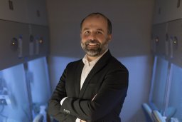 Hugo Guerrero-Cazares合作论文数Dept Neurosurg & Oncol, Johns Hopkins Univ21
 Park JinSeok合作论文数Department of Biomedical Engineering and Institutefor Cell Engineering, Johns Hopkins University18
 Kahp-Yang Suh合作论文数School of Mechanical and Aerospace Engineering, Seoul National University14
 Chris L. Smith合作论文数13C Joanne Wang合作论文数12Lin Benjamin合作论文数12Junaid Afzal合作论文数Department of Medicine,12
 Wijesekera Olindi合作论文数Neuro-Oncology Outcomes Laboratory, Johns Hopkins University School of Medicine12
 Yasir Suhail合作论文数Department of Biomedical Engineering, Johns Hopkins University12
 Sagar R. Shah合作论文数Cardiovascular Implant Research Laboratory, Clemson University12
 Kaisorn Chaichana合作论文数Johns Hopkins University School of Medicine11
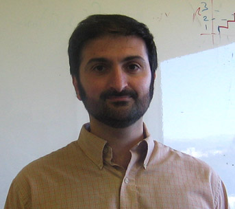 Alex Groisman合作论文数Department of Physics, Division of Physical Sciences, University of California10Ganesan Ambhighainath合作论文数10
 Ilya Nemenman合作论文数Joint Centers for Systems Biology;Columbia University;Joint Centers for Systems Biology, Columbia University10
 Saurabh Paliwal合作论文数Dept Biomed Engn, Johns Hopkins Univ10
 David A. Chesler合作论文数Departments of Neurological Surgery and Pediatrics, Stony Brook University School of Medicine, Stony Brook, NY, 11794-8122, USA10Joanna Shaofen Wang合作论文数10Sussan J. Salas合作论文数9
 Mingxin Zhu合作论文数9Vanaja Kiran Gireesan合作论文数Department of Biomedical Engineering, Johns Hopkins University9Smith Chris L合作论文数9
 Kang Tae-Yun合作论文数Department of Mechanical Engineering, Pohang University of Science and Technology (POSTECH),9
 Jin Zhang合作论文数University of California, San Diego9Colette Aprhys合作论文数8Li Qian合作论文数8
 Tomasz Lipniacki合作论文数8Kshitiz Gupta合作论文数8
 Ali Abdi合作论文数Advanced Communication and Signal Processing Research Lab, Department of Electrical & Computer Engineering, New Jersy Institute of Technology8
 Nathaniel D. Tippens合作论文数Dept Biol Stat & Computat Biol, Cornell Univ7
 Eun Hyun Ahn合作论文数Biomedical Engineering, School of Medicine, Johns Hopkins University7
 Kshitiz合作论文数7
 Kim Hong Nam合作论文数School of Mechanical and Aerospace Engineering, Seoul National University7
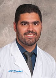 Hubbi Maimon E合作论文数Vasc Program, Johns Hopkins Univ7Wang Joanne合作论文数7
 Andrew (Andy) Feinberg合作论文数Department of Biomedical Engineering, Whiting School of Engineering, Johns Hopkins University7
 Gregg L. Semenza合作论文数Institute for Cell Engineering, Johns Hopkins University;Department of Pathology, School of Medicine, Johns Hopkins University7Steven S. An合作论文数7
 Sung Hoon Lee合作论文数School of Mechanical and Aerospace Engineering, Seoul National University7
 Ahmed Mohyeldin合作论文数Department of Neurological Surgery, University of California6
 Pablo A. Iglesias合作论文数Department of Electrical and Computer Engineering, Johns Hopkins University6
 Paula Schiapparelli合作论文数6
 Steven Larson合作论文数Sloan Kettering Institute, Memorial Sloan Kettering Cancer Center;Department of Medicine, Memorial Sloan Kettering Cancer Center;Weill Cornell Medical College, Cornell University6
 Gunter Wagner合作论文数Department of Ecology & Evoultionary Biology, School of Medicine, Yale University6
 Onur Kilic合作论文数Edward L. Ginzton Laboratory;Stanford University;Edward L. Ginzton Laboratory, Stanford University6
 Andrew Mugler合作论文数Dept Phys & Astron, Purdue Univ6Rhoda M. Alani合作论文数5




