Esophagogastroduodenoscopy is essential to evaluate symptoms of suspected eosinophilic esophagitis (EoE), assess endoscopic findings, obtain biopsy specimens for histopathologic evaluation, perform esophageal dilation, confirm the diagnosis, and monitor the condition. The American Society for Gastrointestinal Endoscopy (ASGE) previously provided consensus recommendations on the approach to endoscopy in EoE across topics of endoscopic diagnosis, endoscopic grading, and esophageal dilation. Because additional areas of endoscopy still required guidance, we performed an independent modified Delphi process focusing on pediatric considerations, disease assessment, and disease monitoring. A core group of EoE experts reviewed published guidelines and developed a set of patient-centered recommendation statements informed by literature review. A multidisciplinary group of adult and pediatric international EoE experts then voted on the statements over 2 Delphi rounds. All statements with 80% agreement were accepted for inclusion. This process yielded 28 consensus statements. Pediatric-specific statements covered when to suspect EoE and perform endoscopy, how to grade endoscopic severity, and when and how to perform esophageal dilation in children. Statements across all age ranges addressed the role of less-invasive monitoring, performing diagnostic endoscopy off treatment, the need to consider symptoms, endoscopic features, and histologic findings when assessing disease activity, treatment-based monitoring intervals, and the approach to esophageal biopsies during monitoring. Coupled with the original consensus work, we provide a comprehensive endoscopic approach to EoE as well as practical guidance for procedure-related aspects in the field to facilitate high-quality endoscopic care to patients with EoE.
BACKGROUND:Non-specific esophagitis has been recently described in patients with dysphagia not fulfilling criteria for eosinophilic esophagitis (EoE). Whether it is a distinct entity and how dysphagia can develop in the absence of eosinophilic infiltration remains unknown. OBJECTIVE:In this study, we comprehensively characterized non-specific esophagitis aiming to elucidate eosinophil-independent fibrosis mechanisms. METHODS:We cross-sectionally and longitudinally analyzed treatment-naïve patients presenting with non-specific esophagitis defined by esophageal dysfunction and low-grade lymphocytic infiltration (< 30 lymphocytes/hpf) in the absence of tissue eosinophilia and reflux disease (GERD). We compared clinical, endoscopic, (immuno)-histological disease activity to EoE, lymphocytic esophagitis, and GERD. RNA sequencing was performed to investigate disease mechanisms. RESULTS:We identified 19 patients (6 males, median age 48 years), with a follow-up of 20 months (IQR 10-41). Clinical disease burden was considerable, contrasting mild endoscopic activity (EREFS 0, IQR 0-2). Immunostaining of esophageal biopsies did not reveal increased numbers of eosinophils or mast cells as contributors to the disease. Lymphocytic infiltration (15/hpf, IQR 9-24) was not increased compared to healthy controls (9/hpf, IQR 9-10) or GERD patients (13/hpf, IQR 9-23); 14 (73.6%) patients were treated with topical steroids (symptomatic response in 85.7%) and 6 patients (31.6%) underwent endoscopic dilation. Progression to EoE was observed in 1 patient, while esophageal eosinophilia (< 15 eos/hpf) developed in 3 patients. RNA sequencing (n = 10) revealed a distinct transcriptomic profile, with highly enriched fibrosis-related pathways. Immunostaining for E-Cadherin/Vimentin as a marker for epithelial-mesenchymal transition (EMT, n = 10) confirmed increased EMT compared to controls. CONCLUSION:Non-specific esophagitis appears to be a distinct non-eosinophilic phenotype with fibrotic traits. Our data suggest eosinophil-independent development of fibrosis.
BACKGROUND:Eosinophilic esophagitis (EoE) is a chronic type 2 inflammatory disease of the esophagus that progresses to a fibrotic phenotype when left untreated. Current treatment options aim to control clinical, endoscopic, and histological disease activity. However, as of yet, it remains elusive if achieving disease control, particularly the long-term control of histological disease activity, can prevent the development of disease complications. OBJECTIVE:We aimed to assess the impact of ongoing histological disease activity on the development of disease complications in adult patients with EoE. METHODS:We evaluated prospectively included patients in the Swiss EoE cohort. Data on all patients with ongoing maintenance treatment, and at least 2 follow-up visits, but without concomitant gastroesophageal reflux or strictures at baseline, were analyzed. We compared patients with ongoing histological disease activity versus patients with disease control, with regard to development of disease complications over time (strictures, bolus impactions, and need for treatment escalation). Histological disease activity was defined by a peak eosinophil count of ≥15 eosinophils during all follow-up visits. RESULTS:We included a total of 151 patients with a median follow-up of 56.0 months (70.9% male, median age 39.0 years). A total of 93 patients (61.6%) were classified as having disease control during follow-up, whereas 58 patients (38.4%) showed ongoing histological disease activity. Development of complications occurred in a total of 108 patients (71.5%), significantly more often in patients with ongoing histological activity compared with patients with disease control (89.7% vs 60.2%, P < .001). This difference was mainly due to higher rates of stricture formation and the need for treatment escalation. Multivariate Cox regression models revealed ongoing histologic disease activity as a significant predictor of the development of complications in the follow-up (hazard ratio [HR]: 2.45, P < .001), particularly for the need for treatment escalation (HR: 2.63, P < .001) and development of strictures (HR: 3.16, P = .025). CONCLUSIONS:Ongoing histological disease activity predicts development of complicating disease course in patients with EoE. Current treatment strategies should aim to control both clinical and histological disease activity to prevent disease complications.
Esophagogastroduodenoscopy is essential to evaluate symptoms of suspected eosinophilic esophagitis (EoE), assess endoscopic findings, obtain biopsy specimens for histopathologic evaluation, perform esophageal dilation, confirm the diagnosis, and monitor the condition. The American Society for Gastrointestinal Endoscopy (ASGE) previously provided consensus recommendations on the approach to endoscopy in EoE across topics of endoscopic diagnosis, endoscopic grading, and esophageal dilation. Because additional areas of endoscopy still required guidance, we performed an independent modified Delphi process focusing on pediatric considerations, disease assessment, and disease monitoring. A core group of EoE experts reviewed published guidelines and developed a set of patient-centered recommendation statements informed by literature review. A multidisciplinary group of adult and pediatric international EoE experts then voted on the statements over 2 Delphi rounds. All statements with 80% agreement were accepted for inclusion. This process yielded 28 consensus statements. Pediatric-specific statements covered when to suspect EoE and perform endoscopy, how to grade endoscopic severity, and when and how to perform esophageal dilation in children. Statements across all age ranges addressed the role of less-invasive monitoring, performing diagnostic endoscopy off treatment, the need to consider symptoms, endoscopic features, and histologic findings when assessing disease activity, treatment-based monitoring intervals, and the approach to esophageal biopsies during monitoring. Coupled with the original consensus work, we provide a comprehensive endoscopic approach to EoE as well as practical guidance for procedure-related aspects in the field to facilitate high-quality endoscopic care to patients with EoE. (Gastrointest Endosc 2026;103:396-417.)
BACKGROUND:Both eosinophilic oesophagitis (EoE) and chronic rhinosinusitis (CRS) are type 2 inflammatory conditions sharing pathogenic mechanisms, therapeutic responses and atopic associations. Data on the prevalence of CRS in EoE patients is missing. AIMS:Our aim was to assess the prevalence of CRS-type symptoms in EoE patients, the quality of life and shared characteristics of patients with both diseases. METHODS:We assessed the point prevalence of CRS in Swiss Eosinophilic Esophagitis Cohort Study patients using a validated screening questionnaire comprising four symptom-based diagnostic criteria. CRS was defined based on symptoms only (definition A) and including self-reported CRS diagnosis by a physician (definition B). Point prevalence was tested against population prevalence using two-sided binomial tests. Secondary outcomes were compared between patients with and without CRS. Multivariable logistic regression models were built to adjust for confounders. RESULTS:211 patients (54%) answered our questionnaire. Prevalence of CRS-type symptoms was 21.8% (definition A) or 25.1% (definition B), compared to 8.71% in the general population (p < 0.0001). CRS status was associated with increased EoE symptom severity. This association persisted after adjustment in multivariable logistic models, most consistently under definition A. 25% reported that their CRS symptoms were more debilitating than their EoE symptoms. CONCLUSION:CRS-type symptoms are two- to three times more prevalent in EoE patients than in the general population and substantially impact quality of life. Our findings indicate that CRS may be under-diagnosed in EoE and support routine CRS screening in EoE patients and cross-speciality collaboration, particularly as both conditions benefit from therapeutic strategies targeting type 2 inflammation.
Eosinophilic esophagitis (EoE) is a chronic Type 2 inflammation. One might assume that the disease-related impact on daily life is significantly smaller, given the relatively localized disease distribution, compared to more systemic immune-mediated diseases. This study aimed to evaluate the disease burden among various immune-mediated diseases, including EoE, inflammatory bowel disease (IBD), systemic sclerosis (SSC) and lupus erythematosus (SLE). A web-based questionnaire assessed baseline characteristics, general quality of life and disease-related impairment across several subdomains, including work, leisure and social life. The surveys were distributed by the respective Swiss patient organizations. Overall, 608 patients participated in the survey (EoE: 92; IBD: 407; SSC: 69; and SLE: 40). Although the overall perceived general impairment in everyday life, measured on a numeric rating scale (NRS), was higher in Crohn’s disease (CD), SSC, and SLE patients compared to EoE (median 3, IQR 2–6), there was no significant difference compared to ulcerative colitis (UC) (median 4, IQR 2–6, p = 0.31) or IBD patients overall (median 5, IQR 3–6; p = 0.05; Fig. 1a). Impairment in EoE was most pronounced and in the subdomain of leisure, consistent with other investigated diseases. Disease-related impairment was higher in women versus men and in patients with a longer diagnostic delay across all diseases. EoE patients’ perceived impairment in everyday life, particularly within subdomains such as leisure, is substantial and comparable to that experienced by patients with other immune-mediated disease states, including IBD. Notably, patient-perceived impairment was higher among female EoE patients and those with a longer diagnostic delay.
Background There is a gap of knowledge with regards to the optimal biopsy sampling procedure as well as the technical and temporal aspects of endoscopic dilation for eosinophilic oesophagitis (EoE). Current guidelines lack specific recommendations. Methods The Swiss Network for Eosinophilic Gastrointestinal Diseases, together with members from The International Gastrointestinal Eosinophil Researchers, assembled a topical review consensus group. After a systematic literature review, two rounds of voting in a Delphi-style process were performed. Statements were rated on an even Likert scale ranging from 1 (strong disagreement) to 4 (strong agreement). Statements were accepted if they achieved an agreement of >80%. Results The experts agreed on a total of 10 statements, 5 statements about biopsy sampling and 5 statements about endoscopic dilation. There was agreement about the need for separately collecting biopsies from at least two segments of the oesophagus, standardised endoscopic and histological disease assessment and the optimal biopsy technique (one biopsy per attempt using the turn and suction technique). The experts further agreed on the early use of endoscopic dilation whenever fibrostenosis is present and an interval of 6-12 months between dilations in most cases. In the absence of fibrostenosis and dysphagia symptoms, empiric dilation for non-dysphagia symptoms cannot be recommended. Conclusion These current practice positions summarise the recommended approach to oesophageal biopsy sampling and endoscopic dilation in adult patients with EoE. Given the lack of randomised controlled data, the statements made in this topical review consensus are largely based on expert opinion and should be seen as guidance in clinical practice that will evolve in the future as newer data emerge.
The improved endoscopic attachment cap has the following new features: 1) more rounded tip; 2) stepwise dilation in two 1-mm increments; 3) softer plastic, ensuring better contact with the endoscope tip; and 4) improved adhesive tape. We evaluated the feasibility and effectiveness of one-time esophageal stricture dilation using the new device in adults with eosinophilic esophagitis (EoE). Patients prospectively included in the Swiss EoE cohort with esophageal strictures (diameter ≤16 mm) and stricture-related symptoms underwent dilation with the new device. Symptoms were assessed using the Eosinophilic Esophagitis Activity Index patient-reported outcomes instrument before and 2 weeks after a single dilation. 60 patients (median age 42 years; 75% male) were evaluated. Bougienage was successful in all patients. Median esophageal diameter increased from 12 mm (interquartile range [IQR] 11–14) to 16 mm (IQR 14–16; P < 0.001). Median symptom severity dropped from 36 points (IQR 22–62) to 0 (IQR 0–12; P < 0.001). No device became detached and no severe adverse events were reported. Post-dilation pain was recorded in 31.7% (19/60). Esophageal stricture dilation in adults with EoE using the improved endoscopic attachment cap was feasible, clinically effective, and environmentally friendly.
Esophageal food impaction (EFI) is the leading complication in patients with undiagnosed eosinophilic esophagitis (EoE). Limited data exists on pre-hospital care, in-hospital management, and post-hospital follow-up in suspected EoE-associated EFI. This study aims to assess deviations between real-life management and guideline-based recommendations in suspected EoE-associated EFI. This retrospective multicenter study analyzed data from four major Swiss gastroenterology units on patients with EoE-associated EFI. Patients with GERD-related strictures or esophageal cancer were excluded. Data on demographics, emergency department (ED), endoscopy management, and follow-up were obtained from electronic health records. Associations between clinical factors and odds of biopsy were analyzed using logistic regression. Between January 2015 and December 2020, 198 EFI cases (median age 51 years, 29.8% female, 28% with previous EFI) were recorded. Patient delay-the time between symptom onset and ED admission-was ~ 270 minutes. Nearly all patients (94%) required endoscopic bolus removal. The median time from ED presentation to endoscopy was ~150 minutes. Esophageal biopsies were taken in just over half of the individuals (n = 97, 52%), leading to a new EoE diagnosis in 71 (68.9% of those biopsied). Biopsy odds decreased significantly with older age (OR 0.96; 95% CI 0.94-0.98, P < 0.05) and known EoE (OR 0.26; 95% 0.09-0.69, P < 0.05). Although EoE is a leading cause of EFI, too few patients with a high baseline probability of EoE undergo biopsy in the emergency setting. Among those biopsied, the majority received a new EoE diagnosis, highlighting the importance of histological assessment.
 Alain Schoepfer合作论文数Division of Gastroenterology and Hepatology, Centre Hospitalier Universitaire Vaudois;Division of Gastroenterology Inselspital, University of Bern;Oberarzt Division of Gastroenterology Inselspital;Faculty of Biology and Medicine, University of Lausanne209
 Ekaterina Safroneeva合作论文数Institute of Social and Preventive Medicine, University of Bern;Tillotts AG122
 Luc Biedermann合作论文数Department of Internal Medicine, University Hospital Zurich117
 Thomas Greuter合作论文数97
 Hans-Uwe Simon合作论文数Institute of Pharmacology, Faculty of Medicine, University of Bern67
 Bussmann C合作论文数65
 Evan S. Dellon合作论文数Division of Gastroenterology and Hepatology, Department of Medicine, School of Medicine, The University of North Carolina at Chapel Hill;Center for Gastrointestinal Biology and Disease, School of Medicine, The University of North Carolina at Chapel Hill52
 Ikuo Hirano合作论文数Gastroenterology and Hepatology, Department of Medicine, Feinberg School of Medicine, Northwestern University50
 Stephan Vavricka合作论文数Triemli Hospital50
 Nirmala Gonsalves合作论文数Gastroenterology and Hepatology, Department of Medicine, Feinberg School of Medicine, Northwestern University42Philipp Schreiner合作论文数38Gerhard Rogler合作论文数36Petr Hruz合作论文数36Glenn T. Furuta合作论文数35
 Dagmar Simon合作论文数Department of Dermatology, Bern University Hospital34
 Margaret H. Collins合作论文数Division of Pathology, Cincinnati Children's Hospital Medical Center;Department of Pathology and Laboratory Medicine, University of California;Department of Pediatrics, University of California33
 Marcel Zwahlen合作论文数Institute of Social and Preventive Medicine, Faculty of Medicine, University of Bern33Alfredo J. Lucendo合作论文数32Jean-Benoît Rossel合作论文数31Gupta Sandeep K合作论文数30
 Michael Vieth合作论文数Department of Pathology, Medizinisches Versorgungszentrum, Klinikum Bayreuth GmbH;Institut für Pathologie, Klinikum Bayreuth GmbH28
 Beglinger, Christoph合作论文数Medizinischen Fakultät, Universität Basel28Albert J. Bredenoord合作论文数28
 Ulrike von Arnim合作论文数28
 Stephan Miehlke合作论文数Center for Digestive Diseases, Cooperation of Internal Medicine;Facharztzentrum Eppendorf27Mirna Chehade合作论文数27
 Stephen Attwood合作论文数EndoStim;Centre for Integrated Health Care Research, School of Medicine, Pharmacy and Health, Durham University;Università degli Studi di Padova;University-Hospital of Padova;St. Vincent’s Hospital;Mater Misericordiae University Hospital27F Seibold合作论文数26
 Kuehni Claudia合作论文数Institute of Social and Preventive Medicine, Faculty of Medicine, University of Bern25
 David A. Katzka合作论文数Columbia University Irving Medical Center25
 Roland Greinwald合作论文数24Christoph Schlag合作论文数23
 Michael Scharl合作论文数Department of Gastroenterology and Hepatology, Universitätsspital Zürich22
 Pierre Michetti合作论文数Centre Hospitalier Universitaire Vaudois21Fournier Nicolas合作论文数19
 Seema Aceves合作论文数Division of Allergy, and Immunology, University of California, San Diego;Rady Children’s Hospital, San Diego18
 Marc E. Rothenberg合作论文数Division of Allergy and Immunology, Cincinnati Children’s Hospital Medical Center;Department of Pediatrics, College of Medicine, University of Cincinnati16Catherine Saner合作论文数14
 Constanza Ciriza de Los Rios合作论文数14Emanuel Burri合作论文数Univ Med Clin, Cantonal Hosp14
 Fouad J Moawad合作论文数14Alexander Jeffrey A合作论文数14Pittet valerie合作论文数Centre Hospitalier Universitaire Vaudois, University of Lausanne14
 Trelle Sven合作论文数Institute of Social and Preventive Medicine, Faculty of Medicine, University of Bern;CTU Bern, University of Bern13
 Carine Blanchard合作论文数From the Divisions of Allergy and Immunology13
 Peter Netzer合作论文数13
 Michael Coslovsky合作论文数Institute of Ecology and Evolution, University of Bern12
 Yvonne Romero合作论文数12
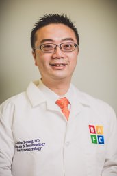 John Leung合作论文数Department of Gastroenterology, Tufts Medical Center12




