The King's College London (KCL) first X-ray microprobe (MKI) and the third generation microfocus X-ray sources (MKIII) are intended to be used for various applications including the study of physical and biological interactions at the atomic and molecular scales. The microfocus ultra-soft X-ray sources (MKI and MKIII) with interchangeable targets will provide a superior spatial resolution (a focal spot a few hundreds of nanometres in diameter can be achieved) and the control of the dose delivered to irradiated cells. This will require characterization of the spectra and intensities of the source, measurements of the focus intensities and spot sizes of suitable X-ray optics such as zone plates, grazing incidence microstructured optical arrays and multilayer mirrors.
The aim of this work is to design and build a source for a range of applications, with optimized multilayer structures in order to use the source output as efficiently as possible. The source is built around a Nd:YAG laser with fundamental wavelength 1064 nm, frequency doubled 532 nm (green) and tripled 355 nm, with a pulse length of about 800 ps and a repetition rate up to 50 Hz. The target material is Mylar (C10H8O4) tape, which is cheap, readily available and has many benefits as explained in this article. A versatile cubic target chamber and a set of computer controlled stage motors are used to allow positioning of the X-ray emission point. A range of measures is used to protect delicate components and optics, including a glass slide between the focusing lens and the target to prevent the lens being coated with debris. A low pressure gas (typically 3-6 mbar) is used inside the chamber as collision of atomic size debris particles with gas molecules reduces their kinetic energy and consequently their adhesion to the surrounding surfaces. The gas used is typically helium or nitrogen, the latter also acting as a spectral filter. Finally, the chamber is continually pumped to ensure that more than 70% of the debris particles are pumped out of the chamber.
The Smart X-Ray Optics (SXO) project comprises a UK-based consortium developing active/adaptive micro-structured optical arrays (MOAs). MOA devices are designed to focus X-rays using grazing incidence reflection through consecutive aligned arrays of microscopic channels. Adaptability is achieved using a combination of piezoelectric actuators, which bend the edges of the silicon chip, and a spider structure, which forms a series of levers connecting the edges of the chip with the active area at the centre, effectively amplifying the bend radius. The spider actuation concept, in combination with deep silicon etching stopped close to the surface, can also be used to create deformable mirrors where the curvature and tip/tilt angles of the mirror can be controlled. Finite Element Analysis (FEA) modelling, carried out for the optimization of the spider MOA device, indicates that deformable mirrors with curvature varying from flat to 5cm ROC and control over the tip/tilt angles of the mirror of +/-3mrad could be achieved. Test spider structures, manufactured using a Viscous Plastic Processing Process for the PZT piezoelectric actuators and a single wet etch step using <111> planes in a (110) silicon wafer for both the silicon channels and the spider structure, have been bent to a radius of curvature smaller than 5 cm. This paper evaluates the spider MOA's concept as a means to achieve deformable mirrors with controllable ROC and control over the tip/tilt angles. FEA modelling results are compared with obtained characterization data of prototype structures. Finally, manufacturing and integration methods and design characteristics of the device, such its scalability, are also discussed.
X-ray spectroscopy is a powerful tool for diagnosing the emission characteristics of X-ray sources. It may also be used in characterizing the elemental and chemical states present in compound materials, including the spatial distribution of these states – spectromicroscopy. This paper describes the appropriate spectroscopic techniques along with examples of their uses – the characterization of laser-plasma sources and the study of chemical state distributions in medium density fibreboard. The possibility of using laboratory-scale sources for spectromicroscopy, as opposed to synchrotrons, are discussed, taking into account the signal to noise ratios that are required to provide the necessary precision.
Characterization methods for grazing-incidence reflecting channel arrays are discussed. Characterization of single-reflection, unactuated micro-structured optical arrays is required to evaluate their performance as focusing elements. Numerical simulations allow the contribution of the x-rays reflected by the channel walls to be distinguished from the overall transmitted signal, and are applied to axial sources. Experimental results are also shown to support the simulations by translation of the channel structure parallel to a detector plane, allowing separation of reflected and transmitted x-rays through the array on the detector.
Transmission electron microscopy images and hard x-ray reflectivity curves are used to obtain information on the growth characteristics of x-ray multilayer mirrors. A multi-resolution approach based on wavelet analysis is used to decompose the interface profiles obtained from transmission electron microscopy images into a number of different spatial frequency ranges. The replication factor (vertical) and the characteristic length (lateral) of the interfacial roughness are determined in these spatial frequency ranges. By changing the upper or lower limits of the wavelet transform of the x-ray reflectivity curves, the structure uniformity (lateral) and evolution of the surface damage (vertical) are determined. These results are significant for the study of the growth characteristics of ultrathin x-ray multilayer mirrors and enable more effective methods to determine their imperfections.
Piezoelectric actuators are widely utilised in adaptive optics to enable mirrors having an actively controlled reflective surface for the purpose of the wavefront correction by reducing the effects of rapidly changing optical distortion. Two new prototype adaptive X-ray optical systems are under development with the aim of approaching the fundamental diffraction limit. One proposed technology is microstructured optical arrays (MOAs) involving two or four piezoelectric strips bonded to a silicon wafer to produce a micro-focused X-ray source for biological applications, and which uses grazing incidence reflection through consecutive aligned arrays of channels obtained using deep silicon etching. Another technology is large scale optics which uses a thin shell mirror bonded with 20–40 piezoelectric actuators for the next generation of X-ray telescopes with an aim to achieve a resolution greater than that currently available by Chandra (0.5"). PZT-based piezoelectric actuators are being developed in this programme according to the design and implementation of the proposed mirror and array structures. Viscous plastic processing is chosen for the preparation of the materials system, which is subsequently formed and shaped into the suitable configurations. Precise controls on the thickness, surface finish and the curvature are the key factors to delivering satisfactory actuators. Unimorph type piezoelectric actuators have been proposed for the applications and results are presented regarding the fabrication and characterisation of such piezo-actuators, as well as the related design concepts and comparison to modelling work.
The structures of ultrathin sputtered Cr/C multilayers were determined by high-resolution transmission electron microscopy. The evolution of layer thickness errors, interdiffusion and interfacial roughness were simulated using time series models. The results show that with increasing of interdiffusion and roughness the multilayer thickness ratio changes, thereby influencing the optical performance. All structural parameters show good correlation with and influence adjacent layers. The system errors of the deposition equipment can also be evaluated by the models.
Boron carbide (B4C) thin films were prepared by magnetron sputtering and residual gas impurities in the films were analyzed by X-ray photoelectron spectroscopy. The impurities, mainly oxygen, decrease with improving vacuum. By using argon ion beam etching of the films, the atomic concentration was measured as a function of etching depth. The binding energy spectra were analyzed using wavelet transform and curve fitting, showing that most of the oxygen impurity is in the form of boron oxides, and that the impurities are physically trapped among columnar structures in the film. In order to improve the base vacuum before coating the film, a range of methods were used, including argon gas filling on the target surface and titanium pre-sputtering. The experimental results show that the latter is an efficient and feasible method. Based on the titanium pre-sputtering technology, the optical performance of W/B4C multilayer was improved so much. (C) 2011 Elsevier B.V. All rights reserved.
Soft x‐ray microscopy is an attractive tool for the study of biological samples in‐vitro, due to the penetrating nature of x‐rays and the natural contrast which can be achieved in hydrated samples. There has been a roadblock to the commercialisation and rollout of small, laboratory scale, x‐ray microscopes for use in the wider community, as high resolution x‐ray microscopy requires tuneable, high brightness x‐ray sources. NANO‐UV has engaged in a product development programme to introduce the first affordable stand‐alone compact soft x‐ray microscope for in‐vitro studies, known as McXI. The initial specification of McXI is to provide 100 nm resolution on in‐vitro specimens, with a unique wavelength selection mechanism in the 2.3–4.4 nm region.
Introduction to XAFS. A Practical Guide to X-ray Absorption Fine Structure Spectroscopy, by Grant Bunker, Cambridge, Cambridge University Press, 2010, 268 pp., £55.00 (hardback), ISBN 9780521767750...
The UK Smart X-Ray Optics (SXO) programme is developing active/adaptive optics for terrestrial applications. One of the technologies proposed is microstructured optical arrays (MOAs), which focus X-rays using grazing incidence reflection through consecutive aligned arrays of microscopic channels. Although such arrays are similar in concept to polycapillary and microchannel plate optics, they can be bent and adjusted using piezoelectric actuators providing control over the focusing and inherent aberrations. Custom configurations can be designed, using ray tracing and finite element analysis, for applications from sub-keV to several-keV X-rays, and the channels of appropriate aspect ratios can be made using deep silicon etching. An exemplar application will be in the microprobing of biological cells and tissue samples using Ti Kα radiation (4.5 keV) in studies related to radiation-induced cancers. This paper discusses the optical design, modelling, and manufacture of such optics.
The Smart X-Ray Optics (SXO) project comprises a U.K.-based consortium developing active/adaptive micro-structured optical arrays (MOAs). These devices are designed to focus X-rays using grazing incidence reflection through consecutive aligned arrays of microscopic channels etched in silicon. Adaptability is achieved using a combination of piezoelectric actuators, which bend the edges of the silicon chip, and a spider structure, which forms a series of levers connecting the edges of the chip with the active area at the centre, effectively amplifying the bend radius. Test spider structures, have been bent to a radius of curvature smaller than 5 cm, indicating that in complete devices a suitable focal length using a tandem pair configuration could be achieved. Finite Element Analysis (FEA) modelling has been carried out for the optimization of the spider MOA device design. Prototype devices have been manufactured using a Viscous Plastic Processing technique for the PZT piezoelectric actuators, and a single wet etch step using {111} planes in a (110) silicon wafer for both the silicon channels and the spider structure. A surface roughness of 1.2 nm was achieved on the silicon channel walls. Characterisation techniques have been developed in order to evaluate the device performance in terms of the bending of the MOA channels produced by the actuators. This paper evaluates the progress to date on the development of spider MOA's comparing FEA modelling with the results obtained for prototype structures.
Interactions of Photons and Neutrons with Matter, by S.-H. Chen and M. Kotlarchyk, Singapore, World Scientific, 2007, xvi + 441 pp., £38.00 (hardback), ISBN 9789810242145. Scope: textbook. Level: b...
Narrowband multilayer monochromator mirrors have been designed for a soft x-ray microfocus source, selecting a narrow band around the chromium K-alpha line. The reflected beam from the mirror can be focused by a zone plate in a microprobe to study radiation effects in cell and tissue samples. The design, taking into account fabrication limitations, was based on theoretical analysis and a local optimal algorithm. The results show that Cr/B4C and Ni/B4C multilayer mirrors working in first or second order can provide high reflectivity and energy resolution to satisfy the requirement of the zone plate. Potential problems in fabrication are also discussed briefly.
The Smart X-Ray Optics (SXO) project comprises a U. K.-based consortium developing active/adaptive micro-structured optical arrays (MOAs). These devices are designed to focus X-rays using grazing incidence reflection through consecutive aligned arrays of microscopic channels etched in silicon. The silicon channels have been produced both by dry and wet etching, the latter providing smoother channel walls. Adaptability is achieved using piezoelectric actuators, which bend the device and therefore change its focal distance. We aim to achieve a 5 cm radius of curvature which can provide a suitable focal length using a tandem pair MOA configuration.Finite Element Analysis (FEA) modelling has been carried out for the optimization of the MOA device design, consider different types of actuators (unimorph, bimorph and active fibre composites), and different Si/piezoelectric absolute and relative thicknesses. Prototype devices have been manufactured using a Viscous Plastic Processing Process for the piezoelectric actuators and dry etched silicon channels, bonded together using a low shrinkage adhesive. Characterisation techniques have been developed in order to evaluate the device performance in terms of the bending of the MOA channels produced by the actuators. This paper evaluates the progress to date on the actuation of the MOAs, comparing FEA modelling with the results obtained for different prototype structures.
The Smart X-ray Optics (SXO) programme is developing advanced active-adaptive optics for X-rays. There are two main themes: large optics for applications in astronomy and small scale optics for micro-probing of biological cells and tissue samples using Ti or Cr K-alpha radiation (4.5keV and 5.4keV, respectively) in studies related to radiation induced cancers. For the latter objective, microstructured optical arrays (MOAs) have been proposed. These consist of an array of channels deep etched in silicon. They use grazing incidence reflection to focus the X-rays through consecutive aligned arrays of channels, ideally reflecting once off a channel wall in each array. Bending the arrays allows variable focal length. The adaptivity is achieved by flexing the arrays using PZT (Lead Zirconate Titanate)-based piezo actuators.The array bending has been modelled using finite element analysis (FEA) and the results showed that for reasonable efficiency, the wall roughness of the channels should not exceed 2nm.This paper describes two techniques of fabrication the MOAs: dry etching and wet etching. The first method requires a special equipment called "inductively coupled plasma" (ICP) using Bosch processes that are designed to produce features with a high aspect ratio with vertical walls. The second method involves using an alkaline solution for etching < 110 > silicon wafers. This type of wafer was selected because of the large wet etch ratio between the (111) and (100) planes that leads to smooth vertical walls. For our application tetra-methyl-ammonium hydroxide (TMAH) was used as it is fully compatible with CMOS integrated circuit processes.
Understanding the structural properties of wood fibre composites, widely used in building and other applications, is an important area of research in devising robust new materials for a range of environmental conditions, including those encountered in developing countries. Wood fibre composites are prepared by mixing fibres with prescribed amounts of glue, but exactly where the glue distributes in relation to the fibre components is unclear. Characterisation of the bonding conditions has largely been a matter of trial and error, which is time consuming and imprecise. In soft x-ray spectromicroscopy experiments carried out at the NSLS, a range of wood fibre composites, with different glues and glue loadings, was studied in order to attempt to quantify the relative glue content in different components of the composite.
 Chris Buckley合作论文数Ge Healthcare UK Limited;GE Healthcare20
 P. J. Duke合作论文数14P. Charalambous合作论文数14R. E. Burge合作论文数13M. T. Browne合作论文数13Melvyn Folkard合作论文数13Graeme Morrison合作论文数12
 Boris Vojnovic合作论文数Department of Oncology, Medical Sciences Division, University of Oxford;Gray Institute for Radiation Oncology and Biology, University of Oxford12a m rogoyski合作论文数10Philip Prewett合作论文数10
 Kevin M. Prise合作论文数School of Medicine, Dentistry & Biomedical Sciences, Queen's University Belfast10
 Daniel Rodriguez Sanmartin合作论文数10
 Tim W. Button合作论文数10Sławka J. Pfauntsch合作论文数10
 Camelia Dunare合作论文数9
 Richard Willingale合作论文数School of Physics and Astronomy, University of Leicester9
 j tom m stevenson合作论文数8Charlotte H. Feldman合作论文数8Shahin Sahraei合作论文数8
 Giuseppe Schettino合作论文数National Physical Laboratory7
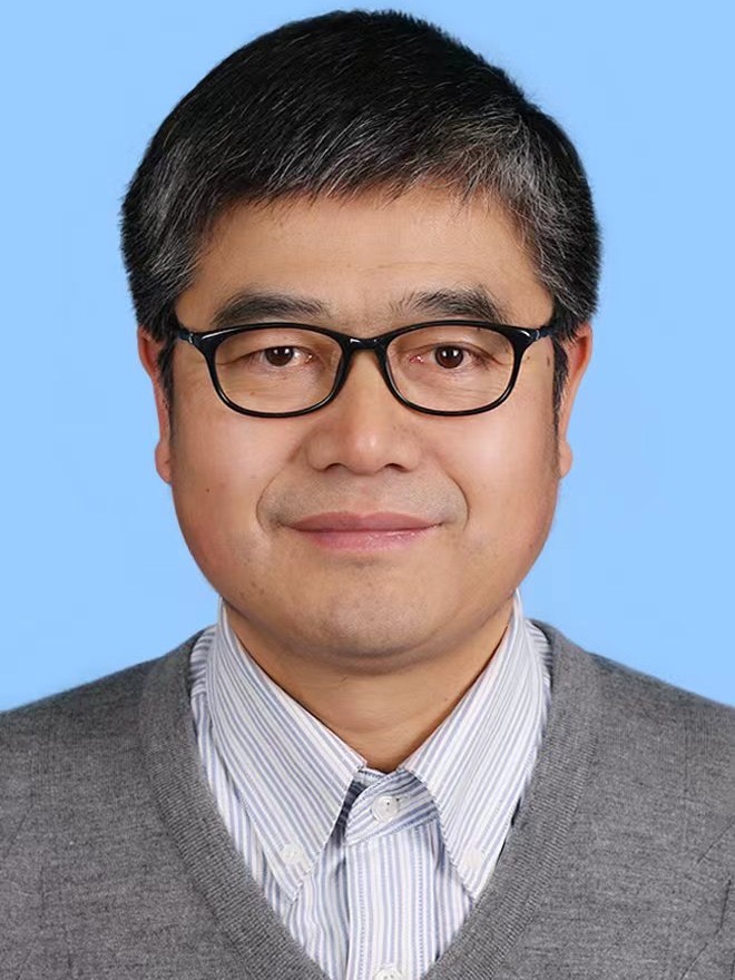 Zhanshan Wang (王占山)合作论文数Institute of Precision Optical Engineering, Tongji University;School of Physics Science and Engineering, Tongji University7
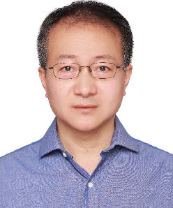 Dou Zhang (张斗)合作论文数Power Metallurgy Research Institute, Central South University;State Key Laboratory of Powder Metallurgy, Central South University7Annie K Powell合作论文数7I. C. E. Turcu合作论文数7N Khaleque合作论文数7r c cave合作论文数6Re Burge合作论文数6B.P. Hills合作论文数6Ady James合作论文数6J M Kenney合作论文数5M. J. Simpson合作论文数5
 Carolyn Atkins合作论文数Department of Physics and Astronomy, University College London5Matthew T. Shand合作论文数5
 Peter Doel合作论文数Physics and Astronomy Department, Faculty of Mathematical & Physical Sciences, University College London5A. R. Hare合作论文数5g dermody合作论文数4g f foster合作论文数4Chris McFaul合作论文数4
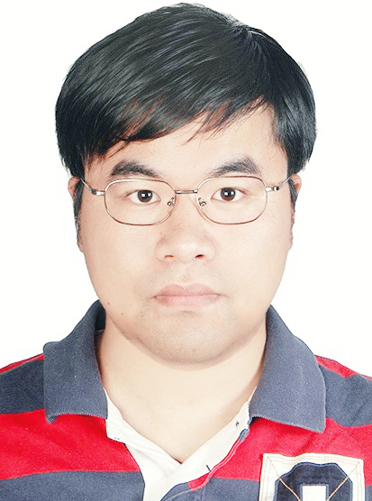 Hui Jiang (蒋晖)合作论文数University of Chinese Academy of Sciences;Shanghai Institute of Applied Physics, Chinese Academy of Sciences4t taguchi合作论文数4
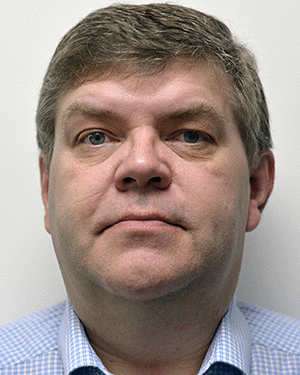 Meggs Carl合作论文数4
 Wang Hongchang合作论文数Diamond Light Source, Harwell Science and Innovation Campus4
 William Parkes合作论文数4
 Kirk Atkinson合作论文数Nuclear Department, Defence Academy College of Management and Technology4Stuart Gilchrist合作论文数4
 Jingtao Zhu (朱京涛)合作论文数School of Physical Science and Engineering, Tongji University4Barry D. Michael合作论文数Mount Vernon Hospital, Gray Cancer Institute4




