Background Pancreatic ductal adenocarcinoma (PDAC) is characterized by frequent SMAD4 inactivation and extensive lipid metabolic rewiring, yet the mechanistic crosstalk between these processes remains poorly elucidated. Methods Multi-omic profiling involving CUT&Tag, transcriptomic, and spatial metabolomic analyses reveals a novel regulatory mechanism of arachidonic acid metabolism governed by SMAD4 that determines PDAC radioresistance. Results Spatial metabolomics revealed that SMAD4 deficiency decreases arachidonic acid metabolism in PDAC tissues. Mechanistically, SMAD4 binds to the promoters of SLC27A3 and FADS2, facilitating the enrichment of the linoleic acid-arachidonic acid axis. Conversely, SMAD4 deficiency leads to a decrease in the linoleic acid-arachidonic acid axis, which may inhibit ferroptosis and promote radioresistance in PDAC tumors. Conclusions Collectively, our study identifies that SMAD4 acts as a transcriptional activator of SLC27A3 and FADS2, driving linoleic acid uptake and its conversion to arachidonic acid, which may subsequently trigger ferroptosis and enhance radiosensitivity.
WTX.AP peptide demonstrates potent antitumor activity in both in vitro and in vivo models
The mammalian STE20-like kinase 4 (MST4) has been implicated as a potential oncogene in various cancers, including pancreatic cancer (PC). However, the precise mechanisms through which MST4 promotes PC malignancy remain incompletely understood. A Biotin identification (BioID)-based proximity labeling approach was employed to identify key MST4-interacting molecules in human PANC-1 pancreatic cancer (PC) cells, followed by cell migration assay to validate the synergistic promotional effect of the MST4-interacting complex on PC cell migration. Direct binding between MST4 and its interacting partners was confirmed via microscale thermophoresis (MST) and isothermal titration calorimetry (ITC), with further validation through X-ray crystallographic structural analysis. The efficacy of rationally designed peptides targeting this interaction was evaluated in both in vitro cell models and in vivo xenograft mouse models. In this study, we demonstrate that MST4 forms a phosphorylation-dependent complex with 14-3-3ζ, leading to Yes-associated protein (YAP) activation and synergistic enhancement of PC cell migration. We observed significant upregulation of both MST4 and 14-3-3ζ in PC patient samples, which correlated strongly with YAP activation and poor prognosis. Structural analysis revealed the detailed interface of the MST4–14-3-3ζ complex, facilitating the rational design of peptide inhibitors that disrupt this interaction. These peptides effectively suppressed YAP activation and exhibited potent anti-tumor effects both in vitro and in vivo. Our findings establish the MST4–14-3-3ζ complex as a critical regulator of YAP signaling that synergistically promotes PC cell migration. Furthermore, rationally designed peptides targeting this MST4–14-3-3ζ interaction represent a promising therapeutic strategy for pancreatic cancer and warrant further clinical exploration.
Video footage obtained from a blue-light activation experiment using a 488-nm laser to stimulate WTX-CCD2.mCherry.Cry2
ABSTRACT Pancreatic ductal adenocarcinoma (PDAC) is characterized by frequent SMAD4 inactivation and profound lipid metabolic rewiring, yet how these processes intersect especially in SMAD4+ PDAC remains elusive. Here, we identify palmitoylation as a previously unrecognized post‐translational modification of SMAD4. Combining biochemical labeling, mutagenesis, and functional assays, we demonstrate that SMAD4 is palmitoylated at cysteine 363 by the acyltransferase ZDHHC22 and depalmitoylated by APT2. Mechanistically, palmitoylation enhances SMAD4 protein stability, facilitates the interaction of SMAD4 with importins, and amplifies subsequent transcriptional output, leading to direct upregulation of the key fatty acid biosynthetic enzyme FASN. Consequently, elevated palmitic acid levels in turn reinforce SMAD4 palmitoylation, establishing a self‐amplifying SMAD4 palmitoylation–FASN–palmitic acid positive feedback loop that drives PDAC tumor growth. Intriguingly, SMAD4 palmitoylation sensitizes PDAC cells to radiotherapy both in vitro and in vivo, revealing a dual role between tumor progression and treatment responses. Notably, clinically relevant SMAD4 mutants (R361C and R361H) exhibit enhanced palmitoylation, underscoring the pathological relevance of this mechanism for tumorigenesis. Collectively, these findings unveil a metabolic‐transcriptional circuit wherein palmitoylation bridges lipid metabolism with SMAD4‐driven oncogenesis, and posit SMAD4 palmitoylation as a therapeutic vulnerability in pancreatic cancer.
ABSTRACT Gastric cancer (GC) remains a formidable global health challenge, characterized by pronounced molecular heterogeneity, late‐stage diagnosis, and limited durable responses to existing therapies. This review synthesizes recent advances in GC research through an integrated, multidisciplinary lens, spanning tumor biology, microenvironmental dynamics, and therapeutic innovation. We first consolidate updated histopathological and molecular classification systems, highlighting oncogenic programs that underpin GC development, including Hippo‐YAP signaling and emerging neural–stem cell interactions. We then examine the immunosuppressive tumor microenvironment, emphasizing the dynamic crosstalk among tumor‐associated macrophages, regulatory T cells, tertiary lymphoid structures, and cancer‐associated fibroblasts that collectively drive metastatic dissemination and therapeutic resistance. Emerging biomarker‐guided strategies, including CLDN18.2‐targeted therapies, dual immune checkpoint blockade, and engineered cellular therapies, are critically discussed alongside rational combination approaches designed to overcome resistance. Beyond canonical paradigms, we highlight transformative frontiers, such as cancer neuroscience, microbiome‐driven immune modulation, and spatially resolved multiomics technologies, that enable high‐resolution mapping of cellular interactions. Finally, we critically assess translational barriers, including organ‐specific metastatic tropism and resistance evolution, and propose that the convergence of deep molecular profiling, neural‐immune modulation, and AI‐enabled computational oncology will be central to advancing precision medicine for GC. This integrated framework aims to accelerate the development of mechanism‐based combination therapies.
Nuclear paraspeckles play critical roles in orchestrating gene transcription to support tumor development and progression. Elucidating regulators of their dynamic compositions and formation could provide potential targets for treating cancer. In this study, we discovered that the short isoform of Wilms tumor gene on the X chromosome (WTX-S) specifically disrupted paraspeckle stability. WTX-S selectively interacted with and sequestered NONO (also as p54nrb) from paraspeckles. Mechanistically, interaction between WTX-S and NONO induced formation of nuclear bodies via liquid-liquid phase separation (WTX-NB). The coiled-coil domain 2 directly interacted with NONO-coiled-coil domain to compartmentalize paraspeckle proteins and subsequently disrupt paraspeckle stability. Functionally, the paraspeckle disintegration induced by WTX-NBs resulted in enhanced chemotherapy sensitivity and favorable prognosis in gastric cancer. Importantly, the cell-penetrating synthetic peptide WTXAP, based on the WTX-NONO interface, mimicked WTX-NB-mediated paraspeckle disintegration and exhibited potent antitumor activity. Likewise, treatment with KPT-330, an FDA-approved XPO1 inhibitor, significantly increased the extent of WTX-S-mediated paraspeckle disintegration, sensitizing WTXhigh gastric cancer to chemotherapy. Collectively, these findings characterize the function and potential clinical significance of WTX-NBs, offering a strategy for sensitizing gastric cancer to chemotherapy.Significance: WTX binding to NONO regulates the balance of biomolecular condensates, providing an axis that can be harnessed to perturb paraspeckles and improve chemotherapy sensitivity in gastric cancer.
WTX.AP and WTX.SP peptides treated orthotopic xenograft GC tumor formation and liver metastasis analysis
The serotonin axis, encompassing the biosynthesis, transport, receptor signaling and metabolism of serotonin, has emerged as a context-dependent regulatory network implicated in cancer biology. Beyond its classical neurotransmitter functions, serotonin modulates tumor progression through receptor-dependent signaling pathways and non-canonical mechanisms such as histone serotonylation, which directly links cellular metabolism to epigenetic regulation. Available evidence suggests that these processes can influence tumor cell proliferation, metabolic adaptation, plasticity and therapeutic drug resistance. Furthermore, the serotonin axis plays a context-dependent role in shaping the tumor microenvironment by regulating the functions of T cells, macrophages and other stromal components, thereby influencing responses to cancer immunotherapy. These diverse regulatory mechanisms have prompted the development of therapeutic strategies targeting serotonin biosynthesis and metabolism, serotonin transport, receptor signaling, and serotonylation, although their efficacy remains highly context-dependent. We further discuss emerging mechanistic insights, unresolved questions, and the opportunities and challenges for developing context-dependent therapeutic strategies targeting this axis.
Protein monoaminylation represents a new layer of neural–cancer regulation, but its role in gastric tumorigenesis is not understood. Using untargeted plasma metabolomics, we revealed that the level of serotonin (5-HT) is significantly elevated in gastric cancer (GC) patients. Functionally, 5-HT treatment dramatically promoted GC cell proliferation and tumor growth in a dose-dependent manner. Importantly, this oncogenic effect was abrogated by the inhibition of transglutaminase 2 (TGM2), indicating a crucial role for protein serotonylation via a receptor-independent mechanism. Using a 5-HT-based chemoproteomic probe, we identified a broad spectrum of serotonylation targets, including key ferroptosis-related proteins such as glutathione peroxidase 4 (GPX4). Specifically, we found that GPX4 is serotonylated by TGM2 at residues Gln55 and Gln77, which increases GPX4 protein stability by attenuating its ubiquitin-mediated degradation, thereby conferring resistance to ferroptosis and facilitating tumor growth. Clinically, TGM2 levels were positively correlated with tumoral GPX4 expression in GC patient specimens. Collectively, our results establish TGM2-mediated GPX4 serotonylation as a key mechanism driving GC progression through ferroptosis resistance, highlighting its potential as both a diagnostic biomarker and a therapeutic target within the neural–tumor axis.
Acidic CCD1 domain potently enhances CCD2-driven phase separation of WTX, amplifying its propensity to form dynamic biomolecular condensates
Gastric cancer (GC) remains a major global health challenge, with chemotherapy resistance significantly hindering treatment efficacy. A significant proportion of chemotherapeutics impact DNA replication, yet the mechanisms by which tumors evade this lethality remain incompletely understood. Notably, minichromosome maintenance 10 replication initiation factor (MCM10) is pivotal in initiating DNA replication, holding promise in mediating acquired chemotherapy resistance. This work aims to elucidate the driving roles of MCM10 GC pathogenesis and chemotherapeutic resistance. The expression pattern of MCM10 and its clinical relevance in GC patients were investigated by adopting single-cell RNA-seq data and in-house GC tissue microarray. Functional roles were evaluated through bioinformatic analyses and experimental assays, including in vivo xenograft formation assay and patient-derived organoid (PDO) models. The transcriptional regulation of MCM10 by the YAP1-TEAD4 complex was examined via Yap1−/−;Taz−/− transgenic mice models and functional rescue assays. Candidates for targeting MCM10 were predicted by virtual screening and further validated by cellular thermal shift assay (CETSA). MCM10 was the most upregulated MCM family member in GC cell lines, and its elevated levels correlated with poor patient prognosis. Bioinformatic analysis linked MCM10 to DNA replication and DNA damage repair, a finding confirmed by functional assays showing that MCM10 depletion induced DNA damage accumulation and impaired DNA replication. MCM10 was further proven to promote GC cell malignancy and tumorigenesis by activating Wnt/β-catenin signaling in GC cell lines, clinical samples, and xenograft models. Critically, MCM10 conferred resistance to chemotherapeutic agents by enhancing cancer cell stemness acquisition and DNA damage response. Mechanistically, YAP1/TEAD4 was identified as the transcriptional activator of MCM10, as TEAD4 silencing downregulated MCM10. TEAD4 overexpression failed to rescue the tumor-suppressing effects in MCM10-depleted cells. Furthermore, Momordin Ic was identified as a promising MCM10-targeted inhibitor, which effectively attenuated GC cell malignancy and chemoresistance. MCM10 drives gastric tumorigenesis by enhancing DNA replication and maintaining cancer stemness, positioning it as a key mediator of YAP1-TEAD4 oncogenic signaling. These findings establish MCM10 as a promising therapeutic target to overcome chemotherapy resistance in GC.
The tumor-associated microbiome has emerged as a novel hallmark of cancer. Gastric cancer (GC) serves as a paradigm for microbiome-associated carcinogenesis, with well-established roles for specific pathogens such as Helicobacter pylori and Epstein–Barr virus. However, the detailed molecular and cellular mechanisms by which microbial communities drive gastric tumorigenesis remain incompletely understood. Recent technological advancements have unveiled a broader spectrum of microorganisms implicated in GC, highlighting the emerging roles of the mycobiome (fungal microbiome) and bacterial-fungal interactions in shaping the tumor microenvironment. This review comprehensively summarizes recent progress in GC-associated pathogens, with a particular emphasis on how bacteria and fungi contribute to GC through direct carcinogenic effects, modulation of host immune responses, and influencing the efficacy of immune checkpoint inhibitors. We critically analyze the “double-edged sword” nature of the microbiome in GC and discuss the potential of multi-omics approaches to unravel complex microbe-host interactions. Finally, we explore the frontier of leveraging synthetic biology to develop engineered microbes (including bacteria and oncolytic viruses) as novel anticancer therapeutics, while addressing the challenges in translating these strategies into clinical practice. A deeper understanding of the gastric microbiome, particularly the multi-kingdom microbial network, is poised to open new avenues for microbiome-based precision diagnosis and innovative therapies for GC management.
To the editor: Pancreatic ductal adenocarcinoma(PDAC)represents the most prevalent form of malignancy affecting the pancreas and is asso-ciated with a very grim prognosis1.PDAC is often diagnosed at advanced stages due to the lack of early symptoms and bio-markers1.
Serine/threonine kinase 3 (STK3) is recognized as a key regulator in Hippo pathway and a tumor-suppressing gene in various cancer types. However, its non-canonical role has been gradually revealed in cancer development. Our objective is to elucidate the upregulation pattern and molecular mechanisms of STK3 in advancing gastric cancer (GC) progression. The regulation of YAP1 on STK3 was assessed through a combination of bulk and single-cell RNA-sequencing, Western blot, ChIP-qPCR, gene knockout mouse models, and functional rescue assays. The oncogenic roles of STK3 were confirmed through subcutaneous xenograft formation models and functional assays including spheroid formation and organoid growth. The phosphorylated target of STK3 was revealed by co-immunoprecipitation and in vitro kinase assays. STK3-targeted drugs were screened out by molecular docking and cellular thermal shift assay (CETSA). Reduction of YAP1 significantly impaired STK3 expression at both mRNA and protein levels, and deletion of STK3 partially attenuated the oncogenic activity of YAP1. Notably, MNNG-induced tumors in Yap1−/−Taz−/− mice exhibited decreased STK3 expression. Knockdown of STK3 led to reduced expression of stemness markers and xenograft growth, while sensitizing GC organoids and xenografts to 5-fluorouracil treatment. Mechanistically, the direct interaction between STK3 and GSK-3β promoted GSK-3β phosphorylation and β-catenin nuclear accumulation, and thus the activation of Wnt signaling. Furthermore, aminopterin demonstrates as a promising STK3-targeted small molecule with remarkable effectiveness in inhibiting GC cell malignance and xenograft growth. STK3 was identified as a transcriptional target of YAP1, leading to enhanced DNA repair ability and stemness acquisition during GC progression by activating Wnt/β-catenin activity through GSK-3β degradation. Moreover, STK3-targeted therapy offered a novel approach to concur acquired chemo-resistance in GC patients.
Oxidative stress is a "double-edged sword" in mediating cellular activities. Here, we report that Mist1+ cells are resistant to oxidative-stress-induced cell death and that persistent oxidative stress and Kras mutation act in Knudson's "two-hit" paradigm to promote gastric cancer initiation. Reactive oxygen species (ROS) accumulation causes metaplastic lineage expansion of Mist1+ cells, licensing them as a cellular origin of gastric cancer. Mechanistically, the transcription factor Mist1 upregulates its downstream target genes Bnip3 and Tmed6, enhancing ROS resistance. Persistent ROS induce R-loop accumulation and activate YAP signaling in Mist1+ cells for proliferation and tumorigenesis. Importantly, ROS and Kras mutation synergistically drive the malignant transformation of Mist1+ cells, while ROS or Kras mutation alone could not do so. Collectively, our study offers insights into the two-hit theory by demonstrating that oxidative stress and oncogenic mutation cooperatively drive Mist1+ cell expansion and transcriptional reprogramming-critical events during early tumor initiation.
Kappa opioid receptor (KOR) signaling is involved in joint development and inflammation in Osteoarthritis (OA), while the biochemical mechanism remains unclarified. This study aims to investigate downstream molecular events of KOR activation, to provide novel perspectives in OA pathology. U50,488H, a selective KOR agonist, was intra-articularly injected in mice upon destabilization of the medial meniscus (DMM) as OA models, with PBS injection as control. The behavioral and histological evaluation was assessed by hot plate test and red solid green staining, respectively. Alterations in mRNA and protein expression were assessed by RNA-seq, RT-qPCR, immunohistochemistry and western blotting (WB) in chondrocytes treated with TNF-α or TNF-α + U50,488H. Proteins interacted with KOR were explored using proximity labeling followed by mass spectrometry and then testified by co-immunoprecipitation (Co-IP) assay and immunofluorescence (IF). OA-induced pain was reduced and cartilage degeneration was alleviated upon KOR activation in DMM mice. In chondrocytes, activation of KOR reversed the upregulation of MMPs, IL-6, IL-1β and phosphorylated(p-) STAT3, stimulated by TNF-α, while the expression of NF-κB, MAPKs and AKT signaling weren't reversed. RNA-seq and IF results presented that KOR activation evidently reduced STAT3 nuclear translocation in chondrocytes upon TNF-α stimuli. The reduction may be resulted from the binding of KOR and STAT3 in the plasma membrane, revealed by proximity labeling and Co-IP results. KOR activation protects cartilage from OA, and this protective effect is mainly exerted via sequestering STAT3 on the plasma membrane, resulting in inactivation of STAT3-dependent immune responses which otherwise contributes to OA.
AbstractTranscriptional factors (TFs) act as key determinants of cell death and survival by differentially modulating gene expression. Here, we identified many TFs, including TEAD4, that form condensates in stressed cells. In contrast to YAP-induced transcription-activating condensates of TEAD4, we found that co-factors such as VGLL4 and RFXANK alternatively induced repressive TEAD4 condensates to trigger cell death upon glucose starvation. Focusing on VGLL4, we demonstrated that heterotypic interactions between TEAD4 and VGLL4 favor the oligomerization and assembly of large TEAD4 condensates with a nonclassical inhibitory function, i.e., causing DNA/chromatin to be aggregated and entangled, which eventually impede gene expression. Based on these findings, we engineered a peptide derived from the TEAD4-binding motif of VGLL4 to selectively induce TEAD4 repressive condensation. This “glue” peptide displayed a strong antitumor effect in genetic and xenograft mouse models of gastric cancer via inhibition of TEAD4-related gene transcription. This new type of repressive TF phase separation exemplifies how cofactors can orchestrate opposite functions of a given TF, and offers potential new antitumor strategies via artificial induction of repressive condensation.
Targeting tumor-infiltrating regulatory T (TI-Treg) cells is a potential strategy for cancer therapy. The ATPase p97 in complex with cofactors (such as Npl4) has been investigated as an antitumor drug target; however, it is unclear whether p97 has a function in immune cells or immunotherapy. Here we show that thonzonium bromide is an inhibitor of the interaction of p97 and Npl4 and that this p97-Npl4 complex has a critical function in TI-Treg cells. Thonzonium bromide boosts antitumor immunity without affecting peripheral Treg cell homeostasis. The p97-Npl4 complex bridges Stat3 with E3 ligases PDLIM2 and PDLIM5, thereby promoting Stat3 degradation and enabling TI-Treg cell development. Collectively, this work shows an important role for the p97-Npl4 complex in controlling Treg-TH17 cell balance in tumors and identifies possible targets for immunotherapy.
 Zhaocai Zhou (周兆才)合作论文数School of Life Sciences, Fudan University30
 Shi Jiao (焦石)合作论文数School of Life Sciences, Fudan University25
 Yi Han合作论文数16
 Wenjia Wang合作论文数15
 Hui Zhang合作论文数13Zhifa Cao合作论文数12Pingping Nie合作论文数11Yan Meng合作论文数10
 Yang Tang合作论文数9
 Yuanzhi Xu (徐远志)合作论文数School of Medicine, Tongji University8HuanHu Zhang (张桓虎)合作论文数7Ju Junyi合作论文数7
 Yun Zhao (赵允)合作论文数Center for Excellence in Molecular Cell Science, Chinese Academy of Sciences6
 Michael Shing Yan Huen (禤承恩)合作论文数School of Biomedical Sciences, Li Ka Shing Faculty of Medicine, The University of Hong Kong6FengHua Guo合作论文数6Zhenzhu Tong合作论文数5Yangwei Xu合作论文数Department of Pathology, School of Basic Medical Sciences, Southern Medical University, Guangzhou, 510515, China.5Yanyan Li合作论文数5Feng Wang合作论文数5Yi-Chuan Li合作论文数5Chao Dong (董超)合作论文数5
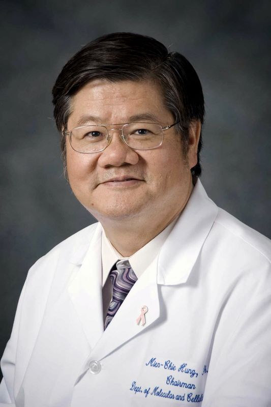 Mien-Chie Hung (洪明奇)合作论文数China Medical University;China Medical University Hospital5
 Kang Wei合作论文数Prince of Wales Hospital, The Chinese University of Hong Kong5
 Yingqun Zhou (周莹群)合作论文数School of Medicine, Tongji University;Tenth People's Hospital of Tongji University5
 Qingling Zhang (张庆玲)合作论文数南方医科大学基础医学院病理学系5Weihong Zhang合作论文数4Yang Wang合作论文数4Ruixian Yu合作论文数4Chen Fan合作论文数4
 Xulin Chen (陈绪林)合作论文数Institute of Medical Microbiology, Jinan University4
 Lixin Wei (卫立辛)合作论文数第二军医大学东方肝胆外科医院肿瘤免疫与基因治疗中心4
 Ka-fai To (杜家輝)合作论文数Department of Anatomical and Cellular Pathology, Faculty of Medicine, Chinese University of Hong Kong;State Key Laboratory in Oncology, Chinese University of Hong Kong4
 Tianyu Yu合作论文数4
 Huixiong Xu (徐辉雄)合作论文数School of Forensic Medicine and Science, Fudan University4
 Moubin Lin (林谋斌)合作论文数Yangpu Hospital, Tongji University;School of Medicine, Tongji University4Wenqi Bai合作论文数3Yan Sang合作论文数3Shilong Wang合作论文数3LiJie Tan (谭黎杰)合作论文数3Fuping Wen (文富平)合作论文数中国原子能科学研究院辐射安全研究所监测与评价研究室3
 Jie Chen合作论文数The University of Hong Kong, The University of Hong Kong3
 Gemin Fang (方葛敏)合作论文数Institutes of Physical Science and Information Technology, Anhui University3
 Yiming Zhang (张以鸣)合作论文数School of Medicine, Tongji University;Shanghai Tenth People's Hospital3Yiran Song (宋亦然)合作论文数2Fan Chen合作论文数2Yazhi He合作论文数2Hoi-Man Ng合作论文数2
 Zaisheng Ye (叶再生)合作论文数Interdisciplinary Institute for Medical Engineering, Fuzhou University;Fujian Cancer Hospital2Kangmin Zhuang合作论文数2




