Amyotrophic lateral sclerosis (ALS) and frontotemporal dementia (FTD) are devastating neurodegenerative diseases linked by similar pathological mechanisms, which, in some familial forms, may be associated with the same genetic alterations. Among them, the most common is the C9ORF72 (C9) mutation. The C9 mutation consists in an aberrant expansion of the hexanucleotide repeat (G4C2)n that leads to the production and accumulation of toxic dipeptide repeat proteins (DPRs). Some of these C9-DPRs contribute to neuronal dysfunction and degeneration through different mechanisms. One of these involves alterations in the protein quality control (PQC) system, specifically in the autophagy-lysosomal pathway. Valosin-containing protein (VCP) is a critical component of the PQC system, assisting the degradation of misfolded proteins and damaged organelles and the maintenance of cellular homeostasis. In this study, we investigated the role of VCP in modulating pathological features associated with C9 mutation. Using neuronal cell models, we demonstrated that VCP overexpression significantly reduced C9-DPRs levels. This reduction is mediated by mechanisms involving both the ubiquitin-proteasome system (UPS) and autophagy. Additionally, we also observed that C9-DPRs induce lysosomal damage, which is counteracted by VCP overexpression, as indicated by decreased galectin-3 puncta and restored lysosomal pH. We then pharmacologically activated VCP-mediated clearance through SMER28, increasing the clearance of the most toxic DPR, the polyPR. We also determined that in this model, SMER28 activity is mediated by the UPS and is associated with the mitigation of DPR-induced lysosome damage. Additionally, using motor neurons derived from induced pluripotent stem cells (iPSC-MNs) from C9-ALS mutation carriers, we demonstrated that SMER28 treatment significantly decreased polyGA levels, a marker for C9-DPR accumulation. Moreover, SMER28 rescued C9-MNs commitment to differentiation and the alteration in the expression of autophagy-related genes. Taken together, our findings strongly support VCP as a modulator of C9 pathology and highlight its potential as a therapeutic target.
Spinal and bulbar muscular atrophy (SBMA) is a slowly progressive X-linked neuromuscular disorder for which disease-modifying therapies are under investigation. SBMA Functional Rating Scale (SBMAFRS), its subscale (mSBMAFRS), and Six-Minute Walk Test (6MWT) are commonly used trial endpoints, but thresholds for clinically meaningful change remain undefined. Minimal clinically important difference (MCID) estimates are needed to interpret longitudinal outcomes and inform trial design. We retrospectively analysed ambulatory, genetically confirmed SBMA patients. Eighty consecutive visit pairs from 44 patients included concurrent Global Rating of Change (GRC) assessments, and 47 visit pairs from 30 patients included concurrent 6MWT data. At follow-up, patients rated overall change since the previous visit on a 3‑level GRC (unchanged, slightly worse, much worse). Anchor-based MCIDs for worsening were derived from differences in change scores between GRC categories and compared with distribution-based estimates (0.5 baseline standard deviation). Sensitivity analyses and Monte Carlo resampling assessed robustness. Anchor‑based MCID estimates for worsening were −1.13 and −1.46 points for the SBMAFRS total score, −0.53 and −1.09 points for the mSBMAFRS, and −34.5 and −32.4 m for the 6MWT. The mSBMAFRS showed the most consistent gradient across GRC categories and remained significant in sensitivity analyses. Distribution‑based MCIDs (2.30, 1.42 points and 61.85 m, respectively) were consistently larger than anchor‑based values. Age, disease duration, and CAG repeat length did not predict perceived worsening. These data provide the first patient‑anchored MCID estimates for SBMA outcome measures, support use of the mSBMAFRS, and offer thresholds for responder definitions and sample-size calculations in future SBMA trials.
Breast cancer (BC) is a widespread and heterogeneous disease in which autophagy plays an essential role in tumor development and progression. It has been suggested that autophagy activation may prevent tumor development in the initial stages of the disease, while in more advanced stages, autophagy might activate survival mechanisms for cancer cells. Moreover, autophagy may be involved in developing therapies resistance and in forming metastases. Some Heat Shock Proteins (HSPs) play an important role in autophagy. The small HSPB8 draws attention because it is generally highly expressed in Estrogen Receptor positive (ER+) BC and its over-expression increases autophagic flux, proliferation, migration and survival of BC cells under stress conditions. HSPB8 mediates the autophagic degradation of client proteins via the chaperone- assisted selective autophagy (CASA) complex in which it binds the BAG cochaperone 3 (BAG3), the HSP70 and the E3-ubiquitin ligase STUB1. Similarly to HSPB8, BAG3 is also highly expressed in BC cells, and its unregulated expression is linked to a poor prognosis. In our previous studies, we showed that HSPB8 and BAG3 silencing reduces proliferation and migratory capacities of hormone-sensitive MCF-7 BC cells. Here, we analyzed the signal transduction mechanisms involved in HSPB8- and BAG3-mediated regulation of metastatic potential. We evaluated the function of protein tyrosine kinase 2 (PTK2 also known as FAK), known to activate several downstream signals controlling PI3K, AKT and MAP kinases. We demonstrated that HSPB8 and BAG3 downregulation correlated with a significant reduction in the phosphorylated and active form of FAK which, in a cascade mechanism, induces a decrease in the proliferative, migratory, and adhesive capacity of MCF-7 and T47D ER+ human BC cell lines. Furthermore, co-immunoprecipitation studies demonstrated a co-localization between BAG3 and FAK, also confirmed by Immunofluorescence staining in BC specimen sections. Based on our results, we suggest that BAG3 associates with FAK and, together with HSPB8, modulates the signal transduction mechanisms controlling the survival of the hormone-sensitive human ER+, PR positive (PR+), HER2 negative (HER2−) BC cell lines MCF-7 and T47D.
Cytoskeletal alterations and axonal transport deficits are key factors in many neurodegenerative disorders. The neuronal kinesin family member 5A (KIF5A) is a microtubule-based motor protein critical for anterograde transport of RNA granules, organelles, and neurofilaments along axons and dendrites. Heterozygous missense and nonsense mutations in the N-terminal motor and stalk domains are associated with hereditary spastic paraplegia 10 (SPG10) and Charcot-Marie-Tooth disease type 2 (CMT2), while frameshift mutations in KIF5A C-terminal cargo-binding domain are linked to amyotrophic lateral sclerosis (ALS). We recently reprogrammed an iPSC line from a SPG10 patient carrying the novel missense variant c.50G>A (p.R17Q) in the KIF5A motor domain, classified as variant of unknown significance (VUS) and predicted to affect ATP binding. Here we gene-edited this mutant iPSC line by CRISPR-Cas9 to obtain an isogenic wild-type (WT) KIF5A cell line. We next examined functionally the impact of the p.R17Q VUS on KIF5A protein sub-cellular distribution and on axonal transport of mitochondria and lysosomes in differentiated iPSC-motoneurons (MNs). The presence of neurofilament-positive axonal swellings and an increased distribution of KIF5A protein in distal neurites was observed in the mutant p.R17Q compared to the WT KIF5A iPSC-MNs, indicating a likely defective axonal transport. The anterograde velocity and distance travelled by mitochondria and lysosomes along neurites was indeed significantly reduced in the mutant KIF5A iPSC-MNs compared to the WT ones. These findings demonstrate that the p.R17Q VUS is pathogenic, thereby extending the spectrum of KIF5A mutations causing SPG10 and support the use of patient-derived iPSC-MNs to functionally validate KIF5A-associated VUS.
Chaperone-assisted selective autophagy (CASA) is a crucial process aimed at maintaining proteostasis in several neurodegenerative diseases associated with protein misfolding, including polyglutamine (polyQ) diseases. Autophagy is a critical lysosome-mediated degradation pathway, particularly essential in neurons, which are highly susceptible to proteotoxic stress due to their post-mitotic nature. Selective autophagy pathways, including CASA, ensure the targeted removal of misfolded proteins and damaged organelles, thereby preserving cellular homeostasis. CASA is based on the intersection of chaperones and autophagy, where HSPB8 and BAG3 interact with HSPA and STUB1 forming a complex that identifies, ubiquitinates, and directs aberrant proteins toward autophagosomes for subsequent lysosomal degradation. In polyQ diseases, such as spinal and bul muscular atrophy (SBMA) and Huntington's disease (HD), mutant proteins accumulate, overwhelming the protein quality control systems. The CASA components are upregulated as a compensatory response, promoting toxic aggregates clearance and cellular damage mitigation. However, chronic proteotoxic stress and progressive impairment of autophagic and lysosomal pathways eventually limit CASA efficiency, contributing to disease progression. The review highlights how CASA exerts its protective activities in polyQ diseases and reports therapeutic strategies aimed at enhancing CASA activity, including pharmacological inducers and combinatorial approaches targeting autophagy and the ubiquitin-proteasome system. Overall, CASA emerges as a crucial adaptive mechanism and a promising therapeutic target in polyQ-related neurodegeneration.
INTRODUCTION:Spinal and bulbar muscular atrophy (SBMA) is an X-linked neuromuscular disease caused by a CAG-repeat expansion in the androgen receptor (AR) gene, translated into an elongated polyglutamine (polyQ) tract in the protein. Androgens trigger ARpolyQ toxicity, thus most potential therapeutic approaches involve androgen reduction or AR negative modulation, with severe endocrine side effects. OBJECTIVE:A start codon (I-AUG) controls AR translation, while a second one, (II-AUG) downstream to I-AUG and the CAG repeat, drives translation of the shorter AR isoform (AR-A) lacking the polyQ tract, but preserves all relevant AR functional domains. Here, we studied AR-A behaviour and its effect on ARpolyQ toxicity in SBMA. METHODS:We took advantage of cellular, mice and Drosophila melanogaster SBMA models to asses AR-A behaviour and effect on ARpolyQ aggregation and functions. RESULTS:Comparative expression analyses of AR isoforms revealed that AR-A is predominantly located in the mouse brainstem and spinal cord of the central nervous system, but not in muscle. AR-A retains partial androgenic activity, but does not aggregate. Since ARpolyQ and AR-A can heterodimerize, we tested the effect of AR-A on ARpolyQ behaviour, showing that AR-A has a pro-solubilizing effect on ARpolyQ aggregates and that the heterodimers retain a partial transcriptional activity. Finally, we tested the effect of AR-A in a fly model of SBMA. Flies expressing AR-A alone showed no signs of external eye degeneration, in contrast to those expressing expanded ARpolyQ. Notably, co-expression of AR-A with ARpolyQ significantly reduced ARpolyQ aggregation and eye degeneration, supporting our hypothesis that AR-A enhances ARpolyQ solubility and mitigates its toxicity in vivo. CONCLUSIONS:Altogether, our results demonstrate that an increased expression of AR-A may have a role in protecting against ARpolyQ aggregation and toxicity. These findings suggest that AR-A could represent a promising avenue for developing an alternative therapeutic strategy that warrants further investigation.
Abstract TAR DNA-binding protein 43 (TDP-43) aggregation is the defining pathological hallmark of nearly all cases of amyotrophic lateral sclerosis (ALS), yet physiologically relevant human models that faithfully recapitulate disease-associated TDP-43 proteinopathy and dysfunction remain limited. To cover this gap, we generated a novel human-based model of cortical neurons carrying the endogenous ALS-linked TDP-43 A315T mutation together with an in-frame Dendra2 fluorescent reporter, enabling temporal and spatial monitoring of the protein. Neurons expressing TDP-43 A315T exhibited progressive neurite degeneration, altered neuronal activity, and impaired mitochondrial respiration, recapitulating several ALS-associated phenotypes. Our model also displays autophagy-dependent accumulation of cytoplasmic aggregates of mutant TDP-43 without overt loss of nuclear function, maintaining normal processing of canonical cryptic exon targets. In contrast, experimental induction of TDP-43 nuclear exclusion readily triggered cryptic exon incorporation, demonstrating that the model faithfully reproduces loss-of-function phenotypes under stress conditions. In line with perturbed protein solubility, mutant TDP-43 neurons show increased stress granule (SG) formation at baseline and under an oxidative stress condition. Finally, treatment with the RNA chaperone Clip34 significantly reduced TDP-43 aggregation under both basal and oxidative stress conditions as well as its localization to SGs. Taken together, these findings establish a physiologically relevant human model that separates early TDP-43 toxic gain-of-function from basal loss-of-function while providing a robust platform for investigating TDP-43 biology and accelerating therapeutic discovery in ALS.
Heat shock protein family B member 8 (HSPB8) is a chaperone involved in the chaperone-assisted selective autophagy (CASA) complex. HSPB8 in conjunction with cochaperone BAG3, promotes autophagy-mediated removal of misfolded proteins associated with various neurodegenerative diseases. Mutations in HSPB8 , previously associated with Charcot Marie Tooth disease type 2L, have recently been linked to an autosomal dominant rimmed vacuolar myopathy (MFM13), and is considered a multisystem proteinopathy. Patients have distal and proximal limb girdle myopathy with muscle biopsy showing fatty replacement, endomysial fibrosis, and rimmed vacuoles leading to muscle atrophy and early demise. We have demonstrated reduced expression of HSPB8, altered autophagy and TDP-43 accumulation in patient fibroblasts. Using CRISPR technology, we generated a knock-in Hspb 8 mouse model of the c.515dupC hot spot frameshift variant to study disease pathology. Overexpressed murine Hspb8 frameshift mutant (c.515dupC, fs) displays insolubility and aggregation propensity in Murine Neuroblastoma X Spinal Cord 34 (NSC-34) cells. Mutant Hspb8 mice developed late-onset muscle weakness beginning at 15 months. Muscle biochemical analyses revealed reduced HSPB8 levels, increased TDP-43, and altered autophagy markers, partially recapitulating the human phenotype. Fiber type analysis, neuromuscular junction integrity, and motor neurons show mild myopathy without neurodegeneration. Given the lack of available treatments, we evaluated trehalose, a natural disaccharide that induces HSPB8 and enhances autophagy. Administration of 2% trehalose in drinking water improves motor performance, restores HSPB8 expression, and ameliorates autophagic and TDP-43 pathology in mutant mice. These findings support the value of our preclinical models for translational studies, and autophagy enhancement as a potential therapeutic strategy for HSPB8-related myopathy.
HSPB8 belongs to the small heat shock protein family, which comprises ten chaperones with molecular weights below 29 kDa. HSPB8 is broadly expressed across human tissues, with the highest levels in skeletal muscles, the cardiac muscle, and the nervous system. In muscles, HSPB8 plays a crucial role in chaperone-assisted selective autophagy (CASA), contributing to protein quality control and maintaining proteostasis. The most extensively studied mutations affecting the HSPB8 K141 codon are associated with autosomal dominant neuromuscular disorders such as Charcot-Marie-Tooth disease type 2L and distal hereditary motor neuropathy type 2 (dHMN2). Of note, recent findings have identified Myofibrillar Myopathy type 13 (MFM13) with Rimmed Vacuoles as a distinct disorder caused by frameshift (fs) mutations in the carboxy-terminus of HSPB8. This review focuses on the known HSPB8-fs mutations leading to MFM13, their associated clinical phenotypes and histological findings, and highlights the need to further understand the underlying etiologies and mechanisms.
Voltage-gated potassium channels, Kv3.1, Kv3.2, Kv3.3, and Kv3.4, facilitate rapid repolarization and shape action potentials, which are crucial to maintaining high-frequency firing. Little is known about the expression and function of Kv3 channels in skeletal muscle. We show that these channels are expressed in type IIa/IIx fibers, and their transcript levels progressively increase from postnatal age to adulthood in physiological context. In mature myofibers, the Kv3.1 and Kv3.4 channels are enriched in the muscle triads. The expression of the Kv3 channel is lost upon acute motor unit damage, in mouse models of amyotrophic lateral sclerosis (ALS) and spinal and bulbar muscular atrophy (SBMA), and the skeletal muscle of patients with sporadic ALS. Early treatment of ALS and SBMA mice with AUT00201, a positive allosteric modulator of Kv3 channels, improved the phenotype of ALS mice specifically, suggesting that positive modulation of Kv3 channels is a novel therapeutic option for patients with ALS.
Kinesin family member 5 A (KIF5A) is a neuron-specific molecular motor involved in anterograde transport. KIF5A mediates a wide range of trafficking processes that are only partially shared with the other members of the KIF5 family. Since 2002, several disease-causing mutations have been found in the KIF5A gene and a link between the specific domain in the encoded protein affected by mutations and the associated phenotype has become evident. Point mutations targeting KIF5A motor and stalk domains, that are expected to impair KIF5A motility, mainly associate with spastic paraplegia type 10 (SPG10) and axonal Charcot-Marie-Tooth (CMT) disease. Oppositely, translational frameshifts causing the elongation of KIF5A tail enhance KIF5A migration towards cell periphery, induce kinesin aggregation, and are linked to amyotrophic lateral sclerosis (ALS) or neonatal intractable myoclonus (NEIMY). This review correlates KIF5A structure and roles in neuronal trafficking with its involvement in the above-mentioned neurodegenerative and neurodevelopmental conditions.
Conformational changes of proteins can occur due to mutations or stress conditions, altering their functionality through loss of physiological or gain of pathological function. A Protein Quality Control (PQC) system exists in cells to deal with the accumulation of misfolded proteins and aggregates, comprising a network of chaperones and degradative pathways to refold or remove the aberrant proteins. Protein misfolding and PQC system impairment lead to a broad range of diseases, including neurodegenerative and neuromuscular disorders, among them spinal and bulbar muscular atrophy (SBMA). SBMA is a neuromuscular disorder caused by a polyglutamine expansion (polyQ) in the androgen receptor (AR) protein. Expanded AR (ARexp) is highly prone to misfolding and aggregation, leading to its accumulation in affected tissues. Here, we summarise the dynamics that control AR protein stability and its degradation in physiological conditions. Next, we recapitulate the current knowledge of the molecular mechanisms of SBMA pathogenesis involving the PQC system. Finally, we provide an overview of promising approaches to SBMA intervention involving the modulation of PQC system functions to reduce ARexp accumulation and its toxic effects in affected cells.
Amyotrophic lateral sclerosis (ALS) is a fatal neurodegenerative non-cell-autonomous disease with no cure, thus research is intensely focused on identifying pharmacological targets. Several studies aimed to clarify the pathogenic mechanisms and involvement in various cell types. A crucial factor in ALS is autophagy, which plays a key role in degrading intracellular protein aggregates. The connection between ALS and autophagy is reinforced by the fact that several genes mutated in ALS are linked to fundamental aspects of autophagy. The blockage of the autophagic flux was observed in ALS motor neurons, where it occurs earlier than in glia. However, the inconsistent effects of autophagy modulators in preclinical and clinical studies indicate the need for a deeper understanding of the role of autophagy in other cell types, such as astrocytes, microglia, and oligodendrocytes. Astrocytes and microglia are significantly impacted by autophagy dysregulation, contributing to neurodegeneration in both mouse and human-derived models. Autophagy is overactivated early in the disease, even before symptoms appear. This overactivation is influenced by the timing and specific tissue involved. It can alter cells' immunophenotype, favouring proinflammatory responses and affecting the cellular environment and autophagy in the surrounding cells. In contrast, oligodendrocytes show mild autophagic alterations. Additionally, sex hormones may affect proper autophagy function and ALS progression. The lack of information on how sex influences autophagy in glia highlights the need for more nuanced investigation into this mechanism. Future research should focus on these aspects, paving the way for personalised pharmacological approaches that consider the roles of cell types, time of intervention, and sex.
Introduction Cachexia is a lethal syndrome with massive muscle wasting that occurs in 60 % of colon cancer patients. Several studies in humans and mice show that males are more prone than females to muscle atrophy caused by colorectal cancer. Understanding whether muscle atrophy precedes or follows other organ alterations may unravel the sex-specific drivers of cachexia. Objectives In two mouse models of colon cancer, we explored when cachexia affects multiple organs in both sexes and their sex development, if/how sex hormone may affect in vitro the inflammatory state of colon adenocarcinoma C26, HCT116 and human primary colorectal cancer cells and in vivo the progression of cachexia in C26-carriers. Methods We compared tumor growth and cachexia-related responses in C26 males and females and C57BL/6J-ApcMin/+ mice of both sexes. C26, HCT116 and human primary colorectal cancer cells exposed to 17β-estradiol or the antagonist fulvestrant were analysed for Il-6 expression and secretion. β-estradiol 3-benzoate was given to C26 males. Results In both models, cancer-bearing males display more/earlier muscle wasting than females. Muscle proteasome activity is enhanced only in cachectic males and only when they are sexually mature. During cachexia hypogonadism appears earlier in males than females of both models. Tumor-bearing females as long as they are “cycling” are more preserved from cachexia than males. Circulating levels of IL-6 increased more in C26 males than C26 females and spleens were bigger in C26 males also displaying more atrophic and inflamed muscles. 17β-estradiol halved the expression of Il-6 and abrogated the secretion of IL-6 from C26 cells, while fulvestrant surprisingly highly enhanced Il-6 expression, supporting an anti-inflammatory effect of estrogens directly on C26 cells. Similar data were obtained from human (primary) colorectal cancer cells. In vivo β-estradiol 3-benzoate prevented some signs of cachexia in C26 carriers. Conclusion Overall, we herein report a novel direct role of 17β-estradiol on colon cancer cells explaining why multiple tissues from males display more signs of cachexia than females.
Expanded G4C2 repeats derived from mutations of the C9orf72 gene are causative factors in amyotrophic lateral sclerosis (ALS) and frontotemporal dementia (FTD) patients, leading to multiple pathological events. Bis thiophene para dinicotinimidamide 2a was reported to preferentially stabilize G-quadruplex G4C2 RNA structures at sub-micromolar concentrations. We replaced its amidine groups with BBB-compliant guanyl hydrazones, and carried out scaffold variations to improve water solubility. An eight-membered array was built around bis-thiophene- (4b-6a), bis-oxazole- (7b), diphenylurea diamide- (8b) and phenyldioxy ditriazolephenyl scaffolds (9a,b). Biological profiling of the array identified 4b as a promising, drug-like hit, active in cellular assays on ALS patient-derived cells.
Huntington disease (HD) is a neurodegenerative disease caused by a polyglutamine expansion (polyQ) in the Huntingtin protein (muHTT), which makes it prone to misfolding and aggregation. muHTT aggregates sequester a wide variety of proteins essential for cell homeostasis, including chaperones and transcription factors, and their depletion may contribute to HD pathogenesis. Lysosomes are the main hubs for degradative and signaling activities in cells, and their functionality is crucial for cell homeostasis, especially for neurons. Different forms of cellular stresses, including proteotoxic stresses, can alter lysosome integrity and induce lysosomal membrane permeabilization (LMP). Damaged lysosomes are recognized by galectins, in particular galectin-3 (LGALS3) with activation of the lysosome quality control (LQC) system responsible for repairing, degrading, or replacing leaky lysosomes. The system is transcriptionally regulated by the transcription factors EB and E3 (TFEB and TFE3, respectively). Using HD mouse and cell models, we demonstrated that TFEB and TFE3 are sequestered in muHTT aggregates, and muHTT proteins associates with LMP triggering the translocation of LGALS3 to the lumen of lysosomes, with a close relation between polyQ size and severity of these events. Moreover, we demonstrated that TFEB and TFE3 silencing or overexpression modulate muHTT aggregation. TFEB and TFE3 knockdown worsens muHTT aggregation, while their overexpression reduces muHTT inclusions and concurrently reduces LGALS3 accumulation via lysophagy and lysosome replacement. Our findings suggest that both TFEB and TFE3 are implicated in HD, and their sequestration in muHTT inclusions increase the vulnerability of neurons to lysosome injury, altering LQC and contributing to disease pathogenesis.
Heat shock protein family B (small) member 8 (HSPB8) promotes chaperone-assisted selective autophagy (CASA), which assures proteostasis in muscles and neurons. HSPB8 frameshift mutations found in neuromyopathies are translated on the same frame, generating the same C-terminal extension, which causes HSPB8 aggregation and proteostasis defects. Here, we describe three novel HSPB8 frameshift variants, translated to protein using the third alternative frame to stop codons downstream to the canonical one and to the one used by other known HSPB8 frameshift mutants. Therefore, these variants are predicted to encode a C-terminal extension that is different in length and amino acids. HSPB8 c.562delC and c.520_523delTACT were identified in two unrelated sporadic patients, while c.515delC, in a familial case of early-onset myopathy. Patients may differentially exhibit additional pathological features, such as neuropathy, respiratory insufficiency, and, remarkably, severe cardiomyopathy. Skeletal muscle biopsies revealed variations in fiber size, atrophy, multiple vacuoles, fat infiltration, and eosinophilic inclusions. In a reconstituted cell model of disease the expression of one representative novel HSPB8 mutant results in i) aggregation of the HSPB8 mutant, ii) sequestration of both the HSPB8 wild-type and CASA complex members, as well as iii) the autophagy receptor sequestosome-1 (SQSTM1/p62), iv) accumulation of ubiquitinated substrates, and v) defects in CASA-mediated degradation. Our results prove that the last exon of the HSPB8 gene is highly susceptible to pathogenic mutations, resulting in a wider phenotypic spectrum associated with HSPB8 frameshift variants. Our studies suggest the importance of HSPB8 genetic testing not only for neuropathy and myopathy but also for cardiomyopathy.
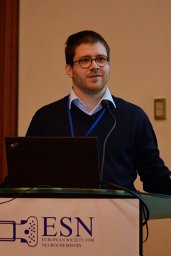 Cristofani Riccardo合作论文数113
 V. Crippa合作论文数94P. Rusmini合作论文数86Valeria Crippa合作论文数80
 Paola Rusmini合作论文数73Barbara Tedesco合作论文数72
 Cicardi Maria Elena合作论文数Centre of Excellence on Neurodegenerative Diseases, Università degli Studi di Milano67Veronica Ferrari合作论文数67M. Galbiati合作论文数56
 Mariarita Galbiati合作论文数56Margherita Piccolella合作论文数55Elena Casarotto合作论文数44Marta Chierichetti合作论文数43
 Serena Carra合作论文数43
 Luciano Martini合作论文数Department of Endocrinology, University of Milan42Elio Messi合作论文数35
 Giorgetti Elisa合作论文数33Caterina Bendotti合作论文数26M. Meroni合作论文数25
 Fabio Celotti合作论文数25Elisa Onesto合作论文数23Marta Cozzi合作论文数22Alessandra Boncoraglio合作论文数20
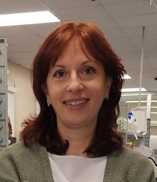 Maria Pennuto合作论文数Centro Acidi Nucleici CNR, Università La Sapienza20Paola Pramaggiore合作论文数19P Negri-Cesi合作论文数19E. Onesto合作论文数17
 Cosimo Melcangi合作论文数Department of Pharmacological and Biomolecular Sciences, Università degli Studi di Milano15F. Simonini合作论文数14
 Adriana Maggi合作论文数Centro di Eccellenza Per Le Malattie Neurodegenerative, l’Università Degli Studi di Milano;Centro di Biotecnologie Farmacologiche, l’Università Degli Studi di Milano;Dipartimento di Scienze Farmacologiche, l’Università Degli Studi di Milano14
 Cristina Cereda合作论文数ASST Fatebenefratelli Sacco;Università Degli Studi di Pavia14
 Alessandro Provenzani合作论文数Fiorgen Foundation;Laboratory of Metabolomics and Systems Biology;Magnetic Resonance Center ( CERM );University of Florence;Magnetic Resonance Center ( CERM ), University of Florence14Daniela Sau合作论文数13Elena Bolzoni合作论文数13Marco Meroni合作论文数12
 Giuseppe Vezzoli合作论文数Division of Nephrology;Laboratory of Pediatric Endocrinology;Istituto di Ricovero e Cura a Carattere Scientifico;San Raffaele Hospital;Laboratory of Pediatric Endocrinology, San Raffaele Hospital12M. Motta合作论文数11Patrizia Limonta合作论文数11
 Paolo Ciana合作论文数Institute of Pharmacological Sciences;University of Milan;Institute of Pharmacological Sciences, University of Milan11Francesco Mina合作论文数10Paolo Bonaldo合作论文数Department of Histology, Microbiology and Medical Biotechnologies, University of Padova10E. Bolzoni合作论文数9
 Nancy L. Weigel合作论文数Baylor College of Medicine9
 Isabella Palazzolo合作论文数9S. De Biasi合作论文数9M. Piccolella合作论文数8D. Dondi合作论文数8




