3D conventional photoacoustic (PA) imaging often suffers from visibility artifacts caused by the limited bandwidth and constrained viewing angles of ultrasound transducers, as well as the use of sparse arrays. PA fluctuation imaging (PAFI), which leverages signal variations due to blood flow, compensates for these visibility artifacts at the cost of temporal resolution. Deep learning (DL)-based PA image enhancement has previously demonstrated strong potential for improved reconstruction at a high temporal resolution. However, generating an experimental training dataset remains problematic. Herein, we propose creating an experimental training dataset based on single-shot 3D PA images (input) and corresponding PAFI images (ground truth) of chicken embryo vasculature, which is used to train a 3D ResU-Net neural network. The trained DL-PAFI network predictions on new experimental test images reveal effective improvement in visibility and contrast. We observe, however, that the output image resolution is lower than that of PAFI. Importantly, incorporating only experimental data into training already yields a good performance, while pre-training with simulated examples improves the overall accuracy. Additionally, we demonstrate the feasibility of real-time rendering and present preliminary in vivo predictions in mice, generated by the network trained exclusively on chicken embryo vasculature. These findings suggest the potential for achieving real-time, artifact-free 3D PA imaging with sparse arrays, adaptable to various in vivo applications.
Quantitative photoacoustic imaging involves an optical inverse problem asking for priors. We propose to add Ultrasound Power Doppler information to: 1) measure the fluence in blood vessels, 2) solve the inverse problem for (mu(a), mu(s)'). (c) 2025 The Author(s)
Objective. Quantitative photoacoustic (PA) imaging aims at recovering optical properties in optically turbid media. Here, we aim to recover uniqueness of the solution in absorption and scattering coefficient in a single illumination and wavelength setting. Approach. By considering a numerical tissue model composed of optically contrasted regions surrounded by blood vessels, we combine PA imaging with ultrasound power Doppler (UPD). We show that quantitative absorption coefficients inside the blood vessels can be derived from UPD, serving as a prior for a radiance Monte Carlo inverse model. Main results. We show that using this vascular imaging prior mitigates the non-uniqueness issue, providing quantitative reconstructions of absorption and scattering even in the absence of background or blood absorption prior information. We generalize to unresolved capillary beds by degrading the resolution of UPD and PA. Significance. Since dual PA-ultrasound systems are already widespread, this approach could become an essential brick of quantitative PA imaging.
Conventional photoacoustic (PA) imaging suffers from visibility artefacts due to limitations in ultrasound transducer bandwidth, viewing angles, and the use of sparse arrays. PA fluctuation imaging (PAFI), exploiting the signal changes due to blood flow, compensates for these artefacts, at the cost of temporal resolution. Our study addresses this limitation employing a deep learning approach in which PAFI images serve as ground truths for training a 3D neural network to obtain real-time single-shot artefact-free images. Following a pre-training with simulated examples, a 3D-ResUnet network was trained with 90 PA chicken embryo vasculature volumes as input and corresponding PAFI as ground truths. Notably, inclusion of experimental data significantly improves predictions over simulation-only training, even accounting for transducer angular filtering. Furthermore, applying the same network exclusively trained in-ovo to predict the femoral artery in mice demonstrates the potential of this method for real-time, full-visibility multispectral PA imaging in vivo using sparse arrays.
Spatial light distribution prediction is highly useful but challenging; no imaging method is currently capable of measuring it at different depths. This study introduces a novel technique for fluence quantification within blood vessels through the ratio of photoacoustic fluctuation imaging (PAFI) and ultrasound power Doppler (USPD). However, their direct coupling fails in accurately estimating the fluence due to differences in the Point Spread Functions (PSFs), leading to varying image resolution and amplitude dependence over vessel sizes. To address this, we propose a model-based matrix approach to apply a non-stationary PSF filter to USPD. Validation through 3D simulations and experiments with tissue-mimicking phantoms demonstrates accurate fluence recovery. Results indicate a robust correlation with the Monte Carlo-simulated ground truth, even in unresolved vessels. This direct imaging technique uniquely offers precise measurement of light distribution in ubiquitous blood vessels, showing great potential for clinical applications and quantitative photoacoustic inverse problems.
Objectives. Numerous optical biomedical imaging or therapeutic modalities suffer from unknown light fluence distribution at depths. Photoacoustic (PA) imaging, which enables imaging blood vessels at the acoustic resolution, probes the product between the fluence and effective optical absorption that depends on the size or density of blood vessels. In the case of unresolved vessels, fluence and absorption can not be decoupled using PA imaging alone without the use of inverse problems. Thus, we propose combining two modalities that are sensitive to blood vessels to directly image fluence maps within vascularized areas, including in unresolved vessels. Approach. To achieve fluence imaging, the combination of photoacoustic fluctuation (PAFI) and Ultrasound Power Doppler (UPD) images is considered. After exposing a new theoretical expression of the UPD image, we establish a fluence imaging method giving quantitative fluence in blood vessels. Fluence imaging involves resolution compensation with a PSF filter that is compared to alternative simpler corrections. Main results. This method universally applies to arbitrary hematocrit and multi-scale vessel imaging. Using a spherical sparse array, we demonstrate 3D fluence imaging within blood vessels in simulation and experiments which is not possible with PAFI alone. Significance. Overall, we show that combining PAFI and UPD has the potential for real-time light dosimetry or could enhance quantitative inverse problems in PA imaging.
We present a method and setup that provide complementary three-dimensional (3D) images of blood oxygenation (via quantitative photoacoustic imaging) and blood flow dynamics (via ultrasound Doppler). The proposed approach is label-free and exploits blood-induced fluctuations, and is implemented on a sparse array with only 256 elements, driven with a commercially available ultrasound electronics. We first implement 3D photoacoustic fluctuation imaging (PAFI) to image chicken embryo, and obtain full-visibility images of the vascular morphology. We obtain simultaneously 3D ultrasound power Doppler with a comparable image quality. We then introduce multispectral photoacoustic fluctuation imaging (MS-PAFI), and demonstrate that it can provide quantitative measurements of the absorbed optical energy density with full visibility and enhanced contrast, as compared to conventional delay-and-sum multispectral photoacoustic imaging. We finally showcase the synergy and complementarity between MS-PAFI, which provides 3D quantitative oxygenation (SO2) imaging, and 3D ultrasound Doppler, which provides quantitative information on blood flow dynamics. MS-PAFI represents a promising alternative to model-based inversions with the advantage of resolving all the visibility artefacts without prior and regularization, by use of a straightforward processing scheme.
Photoacoustic (PA) imaging (PAI) images blood vessels through the hemoglobin contrast. However, conventional images are affected by visibility artefacts which prevents seeing all the blood vessels morphology. We introduced PA fluctuation imaging (PAFI) exploiting the absorption fluctuations due to blood flow. Here, we demonstrate how PAFI enhances the image quality and elaborate if it can be used for quantitative multispectral (MS) imaging for 3D blood oxygenation (SO2) imaging. A spherical array (256 elements, 8 MHz, Imasonic) connected to a Verasonics Vantage scanner, was coupled to the laser (Innolas Spitlight) through a fiber bundle (Ceramoptek). The PAFI sequence consisted in acquiring 1000 frames at 100 Hz repetition rate while scanning continuously the wavelength from 680 nm to 880 nm. Image processing resulted in PAFI image per wavelength. The compensation of predictable effects of pertubations provided quantitative SO2 images with visibility and contrast enhancement. The technique was validated in glass capillaries and in the chicken embryo model by comparing the SO2 values with the one obtained in the visible structures using conventional imaging (resp. 3.5% and 9% errors). Thus, MS-PAFI can provide quantitative SO2 full-visibility 3D imaging with an intrinsic specificity to blood which simplifies spectral unmixing.
Photoacoustic imaging (PAI) provides optical contrast at depth beyond the optical transport mean free path. From the generation of ultrasound by light absorption, images can be reconstructed at the acoustic resolution ( 100 μm) with a penetration of a few cm. The design of imaging systems often leads to limited view artifacts, where a part of the information needed for a complete reconstruction of the objects is missing. We theoretically show that a dynamic approach based on the analysis of fluctuations induced by blood flow can suppress visibility artefacts. We demonstrate the performance of 3D Photoacoustic Fluctuation imaging (PAFI) using a spherical array with limited number of channels (256 elements, 8 MHz) in the chicken embryo model. Due to the low number of channels, standard PAI reconstructions additionally suffer from a poor contrast, which is enhanced by 2 to 3-fold using PAFI. We present an implementation of simultaneous PAFI and Ultrasound Power Doppler and present some results with coupled flow direction evaluations and optical contrast. Photoacoustic fluctuation imaging overcomes many limitations of conventional imaging and will be further evaluated for in-vivo imaging.
In conventional photoacoustics (PA) imaging, the finite size and limited-bandwidth of ultrasound transducers often lead to visibility artifacts resulting in a degraded image quality. We propose a reconstruction algorithm based on deep learning to address theses issues. An in vitro vasculature mimicking model has been used in order to show the capability of a conventional neural network to remove these artefacts in an experimental configuration. The deep learning algorithm is trained using couples of PA images and ground truth photographs. The uncertainty of the model prediction is estimated through the Monte carlo dropout method allowing the display of a pixel-wise degree of confidence. Finally, the interest of using simulation data through transfer learning in order to reduce the size of the experimental dataset is investigated. (C) 2021 The Author(s)
It has previously been demonstrated that model-based reconstruction methods relying on a priori knowledge of the imaging point spread function (PSF) coupled to sparsity priors on the object to image can provide super-resolution in photoacoustic (PA) or in ultrasound (US) imaging. Here, we experimentally show that such reconstruction also leads to super-resolution in both PA and US imaging with arrays having much less elements than used conventionally (sparse arrays). As a proof of concept, we obtained super-resolution PA and US cross-sectional images of microfluidic channels with only 8 elements of a 128-elements linear array using a reconstruction approach based on a linear propagation forward model and assuming sparsity of the imaged structure. Although the microchannels appear indistinguishable in the conventional delay-and-sum images obtained with all the 128 transducer elements, the applied sparsity-constrained model-based reconstruction provides super-resolution with down to only 8 elements. We also report simulation results showing that the minimal number of transducer elements required to obtain a correct reconstruction is fundamentally limited by the signal-to-noise ratio. The proposed method can be straigthforwardly applied to any transducer geometry, including 2D sparse arrays for 3D super-resolution PA and US imaging.
We investigate the use of a 256-channel spherically focused sparse array for volumic real-time ultrasound (US) and photoacoustic (PA) imaging of chicken embryo in vivo. Reconstructions were performed offline and signal processing techniques exploiting spatial and temporal dynamics of the blood flow were applied to visualize the vasculature. The resulting reduction of the clutter enhances the contrast by up to a factor of 2, providing an enhanced visualization of vascular networks. This methodology has a potential for in vivo 3D real time visualization of the vasculature and other features using complementary information provided by US and PA imaging.
Photoacoustic fluctuation imaging, which exploits randomness in photoacoustic generation, provides enhanced images in terms of resolution and visibility, as compared to conventional photoacoustic images. While a few experimental demonstrations of photoacoustic fluctuation imaging have been reported, it has to date not been described theoretically. In the first part of this work, we propose a theory relevant to fluctuations induced either by random illumination patterns or by random distributions of absorbing particles. The theoretical predictions are validated by Monte Carlo finite-difference time-domain simulations of photoacoustic generation in random particle media. We provide a physical insight into why visibility artefacts are absent from second-order fluctuation images. In the second part, we demonstrate experimentally that harnessing randomness induced by the flow of red blood cells produce photoacoustic fluctuation images free of visibility artefacts. As a first proof of concept, we obtain two-dimensional images of blood vessel phantoms. Photoacoustic fluctuation imaging is finally applied in vivo to obtain 3D images of the vascularization in a chicken embryo.
Conventional photoacoustic imaging may suffer from the limited view and bandwidth of ultrasound transducers. A deep learning approach is proposed to handle these problems and is demonstrated both in simulations and in experiments on a multi-scale model of leaf skeleton. We employed an experimental approach to build the training and the test sets using photographs of the samples as ground truth images. Reconstructions produced by the neural network show a greatly improved image quality as compared to conventional approaches. In addition, this work aimed at quantifying the reliability of the neural network predictions. To achieve this, the dropout Monte-Carlo procedure is applied to estimate a pixel-wise degree of confidence on each predicted picture. Last, we address the possibility to use transfer learning with simulated data in order to drastically limit the size of the experimental dataset.
Limited-view and bandwidth-limited acquisition in acoustic–resolution photoacoustic imaging lead to known imaging artifacts. To eliminate these artifacts, it has first been proposed to use fluctuations induced by multiple speckle illuminations to recover otherwise invisible features. However, a very small size of the optical speckle grain at depth in tissue against the acoustic resolution makes this approach unrealistic in practice. Here, we demonstrate experimentally in vitro that fluctuations induced by blood flow at physiological concentration may be exploited to improve visibility in photoacoustic imaging. We first illustrate how our method reveals features otherwise invisible due to the source directivity: a bended capillary tube (inner diameter 100µm) filled with blood flowing at 1.7cm/s was illuminated by 5ns laser pulses (=532nm, fluence=3.0mJ/cm2, PRF=100Hz) and imaged with a linear ultrasound array (128 elements, pitch=0.1mm, fc=15MHz) connected to an acquisition device. Being partially invisible in the conventional image, the whole capillary is reconstructed by means of a second-order analysis of photoacoustic images. Second, we illustrate how our approach allows for visualization of the inside of large objects otherwise invisible due to highpass filtering: we performed a second-order analysis on photoacoustic data resulting from illumination (=800nm, fluence=9.0mJ/cm2, PRF=10Hz/100Hz) of a glass tube (inner diameter 1mm) with blood flowing at 1cm/s. Whether the tube is perpendicular to the imaging plane or is lying inside the imaging plane parallel to the probe, the whole blood volume is visible on the fluctuation-based image, whereas conventional imaging only reveals the blood stream boundaries.
It has been shown that sub-diffraction structures can be resolved in acoustic resolution photoacoustic imaging thanks to norm-based iterative reconstruction algorithms exploiting prior knowledge of the point spread function (PSF) of the imaging system. Here, we demonstrate that super-resolution is still achievable when the receiving ultrasonic probe has much fewer elements than used conventionally (8 against 128). To this end, a proof-of-concept experiment was conducted. A microfluidic circuit containing five parallel microchannels (channel’s width 40μm, center-to center distance 180μm) filled with dye was exposed to 5ns laser pulses (=532nm, fluence=3.0mJ/cm2, PRF=100Hz). Photoacoustic signals generated by the sample were captured by a linear ultrasonic array (128 elements, pitch=0.1mm, fc=15MHz) connected to an acquisition device. The forward problem is modelled in a matrix form Y=AX, where Y are the measured photoacoustic signals and X is the object to reconstruct. The matrix A contained the PSFs at all points of the reconstruction grid, and was derived from a single PSF acquired experimentally for a 10-μm wide microchannel. For the reconstruction, we used a sparsity-based minimization algorithm. While the conventional image obtained by beamforming the signals measured with all the 128 elements of the probe cannot resolve the individual microchannels, our sparsity-based reconstruction leads to super-resolved images with only 8 elements of the probe (regularly spaced over the full probe aperture), with an image quality comparable to that obtained with all the 128 elements. These results pave the way towards super-resolution in 3D photoacoustic imaging with sparse transducers arrays.
The resolution of photoacoustic imaging (PAI) is limited at depths by the diffraction limit. Several ways have been introduced to achieve super-resolution. In the context of imaging the vasculature, the presence of flow can be exploited in two regimes, distinct by the concentration of flowing absorbing particles. In the high concentration regime, we proposed to exploit the absorption fluctuation caused by flowing absorbers by analyzing nth-order statistics of temporal signal fluctuations. In the low concentration regime, when absorbers appear one-by-one in each acoustic resolution spots, the localization microscopy technique can be adapted to our problem. While these two methods improve the resolution greatly, their cost is to reduce temporal resolution, because of the need to record thousands of images. Supposing the knowledge of the PSF (point spread function) of the imaging system, it is possible to the regain temporal resolution. After the simulation of the forward model, the imaged object can be recovered by solving a minimization problem. We will show that adding a sparsity constraint to this problem can enhance the resolution. These techniques have been investigated in both simulations and experiments in microfluidic channels. Such super-resolution approaches bring the optical contrast at depth closer to the cellular level.
 Pernot Mathieu合作论文数Institute Physics for Medicine Paris, Inserm U1273, ESPCI Paris, PSL University, CNRS UMR 806335
 Tanter Mickael合作论文数Institute Physics for Medicine Paris, ESPCI Paris29
 Emmanuel Bossy合作论文数Laboratoire d’Imagerie Paramétrique, Université Paris VI24
 Matthew O’Donnell合作论文数Department of Bioengineering, College of Engineering & UM Medicine, University of Washington23
 Thu-Mai Nguyen合作论文数Dept. of Bioeng., Univ. of Washington;c;Dept. of Bioeng., Univ. of Washington16
 Sergey Vilov合作论文数Univ Grenoble Alpes, CNRS, LIPhy, F-38000 Grenoble, France15
 Ivan M. Pelivanov合作论文数Department of Bioengineering, College of Engineering & UW Medicine, University of Washington15
 Jinjun Xia合作论文数10Guillaume Godefroy合作论文数CNRS, Univ Grenoble Alpes9
 Shaozhen Song合作论文数9Wong, E.Y.合作论文数9
 Zhihong Huang合作论文数7
 Justine Robin合作论文数ESPCI Paris, PSL Res Univ6
 Olivier Villemain合作论文数Department of Medical Biophysics, Temerty Faculty of Medicine, University of Toronto6
 Camilo Perez合作论文数6Michael Lombardo合作论文数6
 Wojciech Kwiecinski合作论文数Laboratoire de recherche en imagerie, Institut Langevin6Alain Bel合作论文数6
 Mathias Fink合作论文数Langevin Institute, École Supérieure de Physique et de Chimie Industrielles6
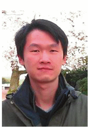 Chen-Wei Wei合作论文数Dept. of Bioeng., Univ. of Washington;c;Dept. of Bioeng., Univ. of Washington5Emmanuel Messas合作论文数5
 Ori Katz合作论文数Applied Physics Department, Hebrew University of Jerusalem5J Robin合作论文数4
 Ricky Wang合作论文数College of Engineering, University of Washington4
 Thomas Chaigne合作论文数4
 Thomas J. Matula合作论文数Department of Bioengineering, University of Washington4
 Guillaume Goudot合作论文数3
 Tristan Mirault合作论文数3Danilo Pozzo合作论文数3Li Junwei合作论文数3
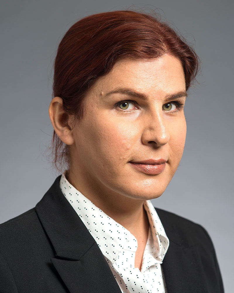 Lilo Pozzo合作论文数Department of Chemical Engineering, University of Washington3Patrick Bruneval合作论文数Laboratoire d'Anatomie Pathologique INSERM U 430, Hopital Broussais3Catherine Boisson合作论文数3Bernard F. Le Bonniec合作论文数3
 Xiaohu Gao (高虓虎)合作论文数Department of Bioengineering, College of Engineering, University of Washington3
 Mathieu Remond合作论文数3Eliel Hojman合作论文数2
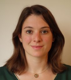 Charlotte Constans合作论文数Univ Paris, PSL Res Univ2
 Wei-Ning Lee (李維寧)合作论文数Department of Electrical and Electronic Engineering, Faculty of Engineering, The University of Hong Kong2
 Soon Joon Yoon合作论文数2Bao-Yu Hsieh合作论文数2
 Yonina Eldar合作论文数Signal Acquisition Modeling Processing and Learning Lab, Department of Computer Science and Applied Mathematics, Faculty of Mathematics and Computer Science, Weizmann Nstitute of Science2Yak-Nam Wang合作论文数1Nicolas Tsapis合作论文数1Vera Khokhlova合作论文数1Georgios Z. Papadakis合作论文数1
 Bjørn Atle J. Angelsen合作论文数Department of Circulation and Medical Imaging, Norwegian University of Science and Technology;Institutt for Sirkulasjon og Bildediagnostikk, Norwegian University of Science and Technology1




