Attention deficit hyperactivity disorder (ADHD) is a common neurodevelopmental disorder that exhibits significant symptom overlap with concussion. While ADHD diagnosis is often considered a confounding factor in concussion evaluation, its impact on performance on standard assessment tools is not well understood. This cross-sectional analysis examined 45 participants with a self-reported doctors’ diagnosis of ADHD and 45 matched controls. Performance on the King-Devick (KD) test, SCAT6 symptoms, Vestibular Oculomotor Screening (VOMS), and 3D-MOT was first compared between ADHD and control participants in independent baseline and post-injury cohorts, and secondarily, between independent baseline and post-injury cohorts within ADHD and control groups. The Mann-Whitney U test and effect size analysis revealed higher symptoms as measured by SCAT6 and VOMS in post-injury cohorts compared to baseline cohorts in both ADHD and control groups. No differences were seen between the ADHD and control groups in the baseline or post-injury cohort on any assessment tool. A forward stepwise regression indicated ADHD diagnosis did not significantly predict overall concussion symptom severity as measured by SCAT6 symptom severity. The findings suggest that ADHD diagnoses do not appear to confound performance on concussion assessment tools when analysed in a heterogenous sample. Post-injury cohorts show higher concussion symptoms compared to control cohorts, regardless of ADHD status. The wide variability in symptom presentation and lack of detected differences between ADHD and control groups in this heterogenous sample suggest that factors beyond ADHD, such as age, sex, and injury history, may play a more prominent role in assessment outcomes.
Major depressive disorder (MDD) has traditionally been linked to deficient serotonergic neurotransmission, chronic stress, and heightened inflammation. Compelling evidence implicates the kynurenine pathway (KP), activated by inflammatory cytokines and stress-related signals, as a critical mediator connecting these factors. The KP degrades tryptophan, the metabolic precursor of serotonin, into neuroactive metabolites called kynurenines, such as quinolinic acid and kynurenic acid. Patients with MDD exhibit KP dysregulation, often marked by an overproduction of quinolinic acid, an N-Methyl-D-aspartic acid receptor (NMDAR) agonist that drives excitotoxicity, alongside reduced production of kynurenic acid, an NMDAR antagonist that protects from excitotoxicity and has anti-inflammatory effects. This review examines dysregulation of the KP in MDD, emphasizing KP metabolites – particularly quinolinic acid and kynurenic acid – as biomarkers and mediators of excitotoxicity, neuroinflammation, and oxidative stress, and discusses the therapeutic efficacy of antidepressants that modulate this pathway. Understanding KP dysregulation could inform the development of targeted interventions that address the underlying biological drivers of MDD, offering new hope for patients who do not respond to conventional treatments.
Traumatic brain injury (TBI) is a leading cause of long-term disability, with limited effective treatment options. A key factor of TBI pathophysiology is neuroinflammation, which can involve the activation of the nucleotide-binding domain leucine-rich repeat protein 3 (NLRP3) inflammasome. Aberrant inflammation following injury has the ability to reduce the capacity to induce long-term changes in synaptic plasticity, a leading mechanism for the development of learning and memory deficits following injury. This study investigated the potential of a novel NLRP3 inhibitor, AMS-17, to mitigate synaptic plasticity deficits following mild TBI (mTBI) in mice. Adult C57Bl/6 mice were subjected to mTBI or a sham injury, and hippocampal slices were then prepared for field electrophysiological recordings in the medial perforant pathway of the dentate gyrus. We found that mTBI induced deficits in long-term potentiation that were not immediate at 2 h post-injury but developed by 3 days post-injury. We next incubated slices in AMS-17 or a control solution prior to electrophysiological recordings. Here we found that incubation with AMS-17 rescued these LTP deficits, bringing them to levels observed in sham-injured controls. Importantly, AMS-17 did not affect the capacity to induce LTP in sham-injured mice. These findings suggest that targeting the NLRP3 inflammasome may offer a promising therapeutic strategy to reduce learning and memory impairments following mTBI. Further studies are needed to determine the optimal therapeutic window and long-term efficacy of AMS-17 in mTBI.
Traumatic brain injury (TBI) causes persistent neurobehavioral deficits and increases the risk of psychiatric disorders, including depression, anxiety, and cognitive dysfunction linked to disrupted neuroplasticity, neuroinflammation, and serotonergic (5-HT) signaling. No effective pharmacotherapies exist for chronic TBI. Psilocybin, a psychedelic 5-HT2A receptor agonist, shows promise due to its neuroplasticity-enhancing, anti-inflammatory, and antidepressant effects. Here, male rats received fluid-percussion or sham injury, followed one year later by a single psilocybin (1 mg/kg) or saline injection. Behavioral testing began 24 h later, and positron emission tomography assessed 5-HT2A binding after two weeks. TBI produced persistent sensorimotor, learning and memory, and affective deficits; reduced 5-HT2A binding; and microglial alterations in the medial prefrontal cortex characterized by decreased process branching and enlarged soma size. Psilocybin treatment could improve sensorimotor function, restore 5-HT2A binding, and reduce microglial cell counts. These findings highlight psilocybin's therapeutic potential in chronic TBI and support further investigation of psychedelic treatments.
Exercise evokes many physiological changes, including the release of hormones and growth factors that are known to improve cognition via unknown mechanisms. Here, we have compared the ability of two physiologically relevant factors, corticosterone (CORT) and brain-derived neurotrophic factor (BDNF), to affect long-term potentiation (LTP) in the hippocampus. Using a compressed theta-burst stimulation (cTBS) protocol, we found that CORT has no effect on LTP, BDNF enhances LTP and combined CORT + BDNF treatment results in significantly greater LTP. We also find that CORT + BDNF, but not either compound alone, results in phosphorylation of protein kinase A (PKA). These findings show that BDNF and CORT act synergistically to enhance LTP at these synapses, potentially via a PKA-dependent mechanism. Such a synergistic action could underlie the positive cognitive effects of exercise.
Introduction Although as many as 92% of survivors of physical intimate partner violence (IPV) report impacts to the head and/or non-fatal strangulation (NFS) that raise clinical suspicion of brain injury (BI), there are no evidence-based methods to document and characterise BI in this vulnerable population, limited clinical practice guidelines and insufficient understanding about long-term risks for conditions including Alzheimer’s Disease and Related Dementias (ADRD). This leaves most survivors of IPV-caused BI (IPV-BI), overwhelmingly women, without adequate access to medical care and support, safe housing, back-to-school/work accommodations or follow-up care for long-term neurocognitive health. Although traumatic brain injury (TBI) is an established ADRD risk factor, little is known about the attributable risk of ADRD due to IPV-BI, particularly in women.Methods of analysis Our overarching objectives are to (1) use plasma biomarkers as novel tools to assist clinicians to improve diagnosis of IPV-BI at the acute, subacute and chronic stages in a manner sensitive to the needs of this vulnerable population and (2) raise awareness of the importance of considering IPV-BI as a potential ADRD risk factor. A prospective observational study funded by the US Department of Defense (HT9425-24-1-0462), Brain Canada (6200) and the Canadian Institutes of Health Research (523320-NWT-CAAA-37499) leverages collaborative research at multiple clinical sites in British Columbia to maximise equity, diversity and inclusion among participants, with a target enrolment of n=600 participants.The Advocates, Academics, Survivors and Clinicians to END Intimate Partner Violence Biomarkers study, which is predicated on pre-specified research questions, represents one of the most significant community-based studies on plasma biomarkers affected by an IPV-BI incident. Of particular significance is the fact our study uses robust biomarker approaches being applied in the TBI and ADRD fields to determine how the biomarker profile after IPV-BI compares to typical TBI and the early stage of neurodegenerative disorders.Ethics and dissemination This study was approved by the University of British Columbia Clinical Research Ethics Board (H24-01990, H22-02241 and H16-02792) and the Island Health Research Ethics Board (H22-03510). Upon publication of primary papers, de-identified data and biospecimens will be made widely available, including the US Federal Interagency Traumatic Brain Injury Research (FITBIR) federated database. Our data and integrated knowledge translation activities with persons with lived experience of IPV-BI and those working in the healthcare sector will be synthesised into co-designed and implemented knowledge tools to improve outcomes for survivors of IPV-BI.
Traumatic brain injury (TBI) is a significant global health challenge, with limited effective treatments for its acute and chronic consequences. TBI is characterized by neuroinflammation, oxidative stress, impaired neuroplasticity, imbalances in neurotransmission, and cell death-factors that contribute to the development of neurological and psychiatric disorders. Emerging evidence suggests that serotonergic psychedelics psilocybin and 5-methoxy-N,N-dimethyltryptamine (5-MeO-DMT) may hold promise as treatments for TBI. These compounds promote neuroplasticity, exert anti-inflammatory and neuroprotective effects, and have shown efficacy in treating psychiatric conditions that share pathophysiological features with TBI. 5-HT1A and 5-HT2A receptors are implicated in their effects, but psilocybin also targets neurotrophic TrkB receptors, whereas 5-MeO-DMT targets sigma-1 receptors, known to have neuroprotective properties. This review integrates current preclinical and clinical research, highlighting both the shared and distinct mechanistic pathways through which psilocybin and 5-MeO-DMT may alleviate TBI-related impairments, such as cognitive and affective dysfunction and neuroinflammation. Additionally, the safety profiles, dosing paradigms, and clinical challenges of these psychedelics are critically examined. By bridging insights from psychedelic science and neurotrauma research, this review underscores the innovative potential of psilocybin and 5-MeO-DMT as adjunctive treatments for TBI, paving the way for novel interventions in neurorehabilitation.
Mild traumatic brain injuries (mTBIs) are caused by biomechanical forces being transmitted to the brain, causing neuronal connections to be subjected to sheering forces. The injury severity can be affected by a number of factors that include age and sex, however, there remains a paucity of data on how repeated mTBI (r-mTBI) impacts the female brain. In these studies, male and female juvenile rats [postnatal day (PND) 25-26] were administered a total of eight mTBIs over a 2-day period. Following each mTBI, rats were immediately assessed for acute neurological impairment. After eight mTBIs were completed, the Barnes maze was used to assess spatial learning and memory. Axonal injury was assessed using silver stain histological analyses. We found that injured females exhibited less acute neurological impairment than males. Three days after the final r-mTBI, no significant differences were observed in spatial learning and memory, with all animals showing similar times to locate the escape platform on the reversal trial, additionally there was no main effect of sex in the Barnes maze. Silver stain uptake was significantly increased in the optic tract, corpus callosum, and cortex compared with sham animals at seven days postinjury in a sex-specific manner. Females showed significant increase in all three regions following r-mTBI, whereas males only showed a significant increase in staining in the optic tract. Overall, these findings show that females may be more susceptible to axonal damage than males, and that cognitive deficits were not evident in this population following r-mTBI. These results indicate that there may be benefits in examining biomarkers that reflect axonal injury and the therapies that target reducing axonal degradation. NEW & NOTEWORTHY Diffuse axonal injury is a hallmark feature of all severities of traumatic brain injury (TBI) yet, in preclinical mild (m)TBI research no studies have yet investigated axonal damage with silver stain immunohistochemistry in female animals. This is a critical gap in the literature as recent studies suggest that females experience mTBI more frequently than males. We found that repeated mTBI (r-mTBI) caused significant diffuse axonal injury that was more pronounced in females compared with males.
Adolescent binge drinking has lasting behavioral consequences by disrupting the endocannabinoid system (ECS) and depleting brain omega-3. The natural accumulation of omega-3 fatty acids in cell membranes is crucial for maintaining the membrane structure, supporting interactions with the ECS, and restoring synaptic plasticity and cognition impaired by prenatal ethanol (EtOH) exposure. However, it remains unclear whether omega-3 supplementation can mitigate the long-term effects on the ECS, endocannabinoid-dependent synaptic plasticity, and cognition following adolescent binge drinking. Here, we demonstrated that omega-3 supplementation during EtOH withdrawal increases CB1 receptors in hippocampal presynaptic terminals of male mice, along with the recovery of receptor-stimulated [35S]GTPγS binding to Gαi/o proteins. These changes are associated with long-term potentiation (LTP) at excitatory medial perforant path (MPP) synapses in the dentate gyrus (DG), which depends on anandamide (AEA), transient receptor potential vanilloid 1 (TRPV1), and N-methyl-D-aspartate (NMDA) receptors. Finally, omega-3 intake following binge drinking reduced the time and number of errors required to locate the escape box in the Barnes maze test. Collectively, these findings suggest that omega-3 supplementation restores Barnes maze performance to levels comparable to those of control mice after adolescent binge drinking. This recovery is likely mediated by modulation of the hippocampal ECS, enhancing endocannabinoid-dependent excitatory synaptic plasticity.
The expanding legalization of cannabis raises significant public health concerns about its use during pregnancy, particularly due to the limited understanding of its impact on neurodevelopment. Existing research suggests that perinatal cannabis or cannabinoid exposure may impair learning and memory; however, variations in study design hinder the ability to draw generalizable conclusions. Clinical studies are limited in their observational nature and the lack of insight into neural or cellular mechanisms underlying cognitive changes, underscoring the importance of preclinical studies to explore the effects of perinatal cannabinoids in greater detail. The objective of this systematic review is to consolidate findings from existing preclinical research that investigates the effects of perinatal cannabinoid exposure on learning and memory and the putative mechanism of learning and memory, hippocampal synaptic plasticity, in rodents. This review summarizes studies on hippocampal synaptic plasticity (n = 2), spatial/visual memory (n = 13), working memory (n = 6), recognition memory (n = 12), and associative memory (n = 7). Perinatal cannabinoid-induced impairments were reported in the two synaptic plasticity studies, and in 24 out of 30 studies that examined learning and memory, with spatial memory tasks showing the most consistent deficits. While the existing evidence converges on the notion that perinatal cannabinoid exposure negatively impacts hippocampal physiology and associated memory functions, further research is needed to disentangle the influence of various methodological factors, including offspring sex and age, cannabinoid type, time of gestational exposure, and method of administration.
Stroke is a leading cause of death and disability, with ischemic stroke representing most cases. Age is the most significant nonmodifiable risk factor for stroke, and with an aging population, there is an urgent need for effective prevention and treatment strategies. Physical inactivity is a strong risk factor for stroke, and exercise has long been held as a promising approach to improve poststroke outcomes. During exercise, the myokine irisin is released as a product of a type 1 membrane protein cleavage that is encoded by the fibronectin type III domain containing 5 (FNDC5) gene. This review summarizes recent literature on irisin's role in ischemic stroke, examining central effects, stroke risk, poststroke functional outcomes, and exogenous administration. Irisin has value as a prognostic marker for risk stratification. Low levels of irisin correlate with worse outcomes and higher mortality in patients with ischemic stroke. Irisin may also be a key to the benefits of exercise, particularly for high-intensity resistance training, which significantly increases irisin levels. Beyond exercise, exogenous irisin is neuroprotective in murine models, reducing brain edema, inflammation, and apoptosis, and increasing blood-brain barrier integrity and brain-derived neurotrophic factor levels. This underscores irisin's potential to mitigate ischemic damage and promote recovery. Human trials are necessary to validate these findings and explore the feasibility of irisin-based interventions in acute stroke care. This review lays a foundation for future research to clarify irisin's therapeutic benefits, establish optimal exercise protocols, and explore exogenous irisin as a novel intervention for ischemic stroke.
There is growing interest in exploring the therapeutic potential and mechanisms of action of psilocybin on stress-related neuropsychiatric disorders, including depression, generalized anxiety disorder (GAD), post-traumatic stress disorder (PTSD), obsessive-compulsive disorder (OCD), addiction, and disordered eating. Despite promising progressions in preclinical and clinical research, the neurobiological and physiological mechanisms underlying the therapeutic effects of psilocybin remain complex, involving multiple systems with numerous homeostatic feedback signaling pathways throughout the body. This review paper explores how psilocybin mechanistically interacts with the gut microbiota, enteric nervous system, hypothalamic-pituitary axis, and how psilocybin influences the bidirectional communication between peripheral and neuronal systems. Shifting towards a more integrated paradigm to unravel the mechanisms through which psilocybin affects the bidirectional gut-brain axis holds the promise of significantly advancing our understanding of psilocybin-based therapies from preparation of treatment, administration, to proceeding long-term integration. Such an understanding can extend beyond the treatment of psychiatric disorders, further encompassing a broader spectrum of inflammatory-related disorders.
Fragile X syndrome (FXS) is the most common inherited cause of intellectual disability and is the leading known single-gene cause of autism spectrum disorder. Patients with FXS display varied behavioural deficits that include mild to severe cognitive impairments in addition to mood disorders. Currently, there is no cure for this condition; however, there is an emerging focus on therapies that inhibit mechanistic target of rapamycin (mTOR)-dependent protein synthesis owing to the clinical effectiveness of metformin for alleviating some behavioural symptoms in FXS. Adiponectin (APN) is a neurohormone that is released by adipocytes and provides an alternative means to inhibit mTOR activation in the brain. In these studies, we show that Fmr1 knockout mice, like patients with FXS, show reduced levels of circulating APN and that both long-term potentiation (LTP) and long-term depression (LTD) in the dentate gyrus (DG) are impaired. Brief (20 min) incubation of hippocampal slices in APN (50 nM) was able to rescue both LTP and LTD in the DG and increased both the surface expression and phosphorylation of GluA1 receptors. These results provide evidence for reduced APN levels in FXS playing a role in decreasing bidirectional synaptic plasticity and show that therapies which enhance APN levels may have therapeutic potential for this and related conditions. This article is part of a discussion meeting issue ‘Long-term potentiation: 50 years on’.
BACKGROUND:Cannabis is increasingly being legalized and socially accepted around the world and is often used with alcohol in social settings. We recently showed that in utero exposure to both substances can alter the density of parvalbumin-expressing interneurons in the hippocampus. Here we investigate the effects of in utero alcohol and cannabis exposure, alone or in combination, on somatostatin- and neuropeptide Y-positive (NPY) interneurons. These are separate classes of interneurons important for network synchrony and inhibition in the hippocampus. METHODS:A 2 (Ethanol, Air) × 2 (tetrahydrocannabinol [THC], Vehicle) design was used to expose pregnant Sprague-Dawley rats to either ethanol or air, in addition to either THC or the inhalant vehicle solution, during gestational days 5-20. Immunohistochemistry for somatostatin- and NPY-positive interneurons was performed in 50 μm tissue sections obtained at postnatal day 70. RESULTS:Exposure to THC in utero had region-specific and sex-specific effects on the density of somatostatin-positive interneurons in the adult rat hippocampus. A female-specific decrease in NPY interneuron cell density was observed in the CA1 region following THC exposure. Combined exposure to alcohol and THC reduced NPY neurons selectively in the ventral dentate gyrus hippocampal subfield. However, overall, co-exposure to alcohol and cannabis had neither additive nor synergistic effects on interneuron populations in other areas of the hippocampus. CONCLUSIONS:These results illustrate how alcohol and cannabis exposure in utero may affect hippocampal function by altering inhibitory processes in a sex-specific manner.
 Joana Gil-Mohapel合作论文数Div Med Sci, Univ Victoria37
 Anna Patten合作论文数Island Medical Program, University of Victoria26
 Christine J Fontaine合作论文数Division of Medical Sciences, University of Victoria21
 Patricia Souza Brocardo合作论文数Center of Biological Sciences, Federal University of Santa Catarina20
 Hannah Reid合作论文数Ctr Behav Teratol, San Diego State Univ19
 Taylor Snowden-Richardson合作论文数15
 Sandy Shultz合作论文数Department of Neuroscience, Central Clinical School, Monash University15
 Titterness Andrea K合作论文数Graduate Program in Neuroscience, University of British Columbia14
 Patrick C Nahirney合作论文数National Institute of Arthritis and Musculoskeletal and Skin Diseases, National Institutes of Health Muscle Proteomics and Nanotechnology Section14
 Luis E B Bettio合作论文数Div Med Sci, Univ Victoria13
 Sonata Suk-Yu Yau合作论文数12
 Alicia Meconi合作论文数Island Medical Program, University of British Columbia11Wortman Ryan C合作论文数11
 Timal S. Kannangara合作论文数Division of Medical Sciences and Department of Biology, University of Victoria11
 Cristina Pinar合作论文数10E. L. Grafe合作论文数Div Med Sci, Univ Victoria10Eric Eyolfson合作论文数Alberta Children's Hospital Research Institute (ACHRI), Calgary, AB, Canada10
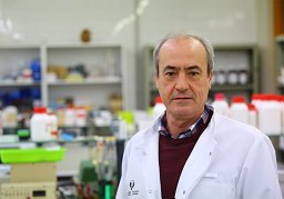 Pedro Grandes合作论文数Faculty of Medicine and Dentistry, Basque Country University9B D Eadie合作论文数8Jd Thomas合作论文数8C. Rodriguez合作论文数Dept Psychol, San Diego State Univ8Gawryluk Jodie R合作论文数Department of Psychology, Dalhousie University8
 Daniel Johnston合作论文数Center for Learning and Memory, College of Natural Sciences, The University of Texas at Austin;Department of Neuroscience, College of Natural Sciences, The University of Texas at Austin8
 Crystal Bostrom合作论文数8
 Stuart McDonald合作论文数Department of Neuroscience, Central Clinical School, Monash University8Wickliffe C. Abraham合作论文数8Justin Brand合作论文数7
 Katie Neale合作论文数7Irene Shkolnikov合作论文数7Jamie Morrison合作论文数7
 Juan Sebastian Trivino Paredes合作论文数Univ Victoria7Carl Ernst合作论文数Department of Psychiatry, Faculty of Medicine and Health Sciences, McGill University7Jennifer L. Helfer合作论文数Psychology Department, University of Delaware7Andrea K. Olson合作论文数7
 K.F. So (蘇國輝)合作论文数Department of Ophthalmology, School of Clinical Medicine, The University of Hong Kong;Department of Psychology, Faculty of Social Sciences, The University of Hong Kong;Jinan University7Brennan D. Eadie合作论文数6Itziar Bonilla-Del Rio合作论文数6
 Kristen R. Breit合作论文数Department of Psychology, West Chester University6
 Terrence Sejnowski合作论文数Department of Neurobiology, School of Biological Sciences, University of California, San Diego;Howard Hughes Medical Institute;The Salk Institute for Biological Studies6
 Van Redila合作论文数5Jessica M Simpson合作论文数5
 Mariana A. Vetrici合作论文数5
 Henriette Van Praag合作论文数Department of Biomedical Science, Charles E. Schmidt College of Medicine, Florida Atlantic University;Brain Institute, Florida Atlantic University5Ramos Almudena合作论文数5
 Aimin Xu (徐愛民)合作论文数Department of Medicine, School of Clinical Medicine, The University of Hong Kong;Department of Pharmacology and Pharmacy, Li Ka Shing Faculty of Medicine, The University of Hong Kong5Jennifer D. Thomas合作论文数Center for Behavioral Teratology, San Diego State University5
 Nagore Puente合作论文数5Helle M. Sickmann合作论文数5
 Mohamed Ghilan合作论文数4




