INTRODUCTION:Adjuvant chemotherapy can improve recurrence-free survival (RFS) in gastrointestinal malignancies. Previous review of phase III randomized controlled trials (RCTs) for colorectal cancer observed that RFS improvements were driven by early divergences during active chemotherapy; late recurrences were not influenced by adjuvant therapy. The broader applicability of this finding is unknown. METHODS:PubMed, Cochrane Library (CENTRAL), Embase, Scopus, and Web of Science were queried from database inception to December 31, 2025, for phase III RCTs including pancreatic, gastroesophageal, hepatocellular, and biliary tract malignancies. Trials where significant differences in RFS were observed between experimental (adjuvant chemotherapy) and control (resection alone) arms were included. Summary data were extracted from Kaplan-Meier curves using DigitizeIT, and absolute differences in RFS were compared at matched intervals using Wilcoxon matched-pairs signed rank tests. RESULTS:A total of 14 RCTs were identified, investigating periampullary (n = 4), gastroesophageal (n = 5), hepatocellular (n = 4), and biliary tract (n = 1) carcinomas. Across pooled RCTs, the highest rates of recurrence were observed in the first year following resection. Median RFS event rate was significantly higher with resection alone (0-0.5 y: resection alone 44.9 [IQR 14.2-84.1] versus adjuvant chemotherapy 26.4 [IQR 7.7-41.9], P < 0.001; 0.5-1 y: resection alone 33.2 [22.7-42.1] versus adjuvant chemotherapy 25.0 [13.8-44.5]; P = 0.007). No difference was observed during later intervals from postoperative randomization (1-2 y: P = 0.952; 2-3 y: P = 0.191; 3-4 y: P = 0.999; 4-5 y: P = 0.110). For the subset of trials where ≤6 mo of adjuvant chemotherapy was used (n = 10), improvements in RFS event rates were observed only during and immediately following the treatment interval (0-6 mo: P = 0.001; 6-12 mo: P = 0.037). CONCLUSIONS:Across multiple gastrointestinal malignancies, improvements in RFS associated with active adjuvant chemotherapy regimens are driven by early changes in recurrence dynamics. These data may provide in vivo understanding of residual tumor cell populations after curative-intent resection and how chemotherapy can be best used to prevent recurrence.
To review the current data on preoperative workup, operative approach, and multi-modality therapy for cholangiocarcinoma. Essential preoperative work-up starts with a comprehensive history and physical exam, assessing the patient’s performance status. Tumor markers and cross-sectional imaging should be obtained including computed tomography (CT) of the chest, multi-phase thin-slice CT of the abdomen and pelvis and magnetic resonance cholangiopancreatography (MRCP). Additional imaging and technical considerations vary widely based on anatomic location (intrahepatic, perihilar, distal). General operative principles include resection to negative margins and performing a regional lymphadenectomy, both of which are associated with improved outcomes. After resection, most patients receive multimodality treatment with adjuvant chemotherapy. Thorough preoperative planning is needed to determine resectability for patients with cholangiocarcinoma. Multi-modality therapy should be considered, especially in the adjuvant setting, with future studies needed to evaluate the role and efficacy of neoadjuvant therapy.
BACKGROUND:The optimal treatment strategy for patients undergoing curative-intent resection of colorectal liver metastases remains uncertain. While perioperative and adjuvant chemotherapy are widely used, randomized trials have not demonstrated a consistent overall survival benefit, and guidelines continue to endorse approaches ranging from surgery alone to multimodal therapy. Randomized studies evaluating perioperative or adjuvant chemotherapy-along with trials incorporating biologic therapy-have shown improvements in disease control but inconsistent overall survival benefit. We evaluated national trends in perioperative chemotherapy use and factors associated with receipt over 2 decades. METHODS:Using Surveillance, Epidemiology, and End Results-Medicare (2000-2020), we identified patients who underwent hepatectomy for liver-only colorectal liver metastases. Perioperative chemotherapy was defined as systemic therapy within 6 months before surgery, 4 months after, or both. Temporal trends were assessed, multivariable logistic regression identified predictors of treatment, and overall survival was evaluated with Kaplan-Meier and adjusted Cox models. RESULTS:Among 42,235 patients with liver-only metastases, 3,752 (9%) underwent resection; 54% of these received perioperative chemotherapy (14% preoperative only, 22% postoperative only, and 18% both). Use declined modestly over time (odds ratio per year, 0.97; 95% confidence interval, 0.95-0.98; P < .001). Chemotherapy was more common with rectal primaries (odds ratio, 1.42; 95% confidence interval, 1.05-1.92; P = .02) and synchronous disease (odds ratio, 1.57; 95% confidence interval, 1.30-1.92; P < .001) but less likely in patients ≥75, <65, and with overlapping or unspecified primaries. Perioperative chemotherapy was independently associated with improved overall survival, greatest with both preoperative and postoperative regimens (hazard ratio, 0.54; 95% confidence interval, 0.47-0.60; P < .001). CONCLUSION:In this national cohort of adults, perioperative chemotherapy was used in half of patients undergoing hepatectomy for colorectal liver metastases with minimal change over time and was associated with improved survival, supporting continued multimodal management.
Rates of metastatic cancer are increasing, but our knowledge of the accuracy of metastatic diagnoses within real world data is limited. In this brief report, to evaluate the accuracy of various data sources, we assessed concordance between cancer registry diagnoses and electronic health record (EHR) International Classification of Diseases diagnoses for the presence of colorectal liver metastases (CRLM) for patients within a single academic health care system. 1025 patients diagnosed with CRLM were evaluated. Concordance between cancer registry data and EHR data for the diagnosis of synchronous CRLM was low (k = 0.66). Additional data quality issues identified included (1) a 9 % rate of false positives in cancer registry data for the diagnosis of CRLM and (2) discordant data between the metastatic diagnosis and cancer registry stage affecting 16 % of cases. These results highlight important limitations of real world databases in the identification and study of patients with metastatic cancer.
Background This study aimed to identify the incidence of clinical understaging in early gastric cancer (GC) and to identify variables associated with increased risk of a patient being understaged. Methods Clinical stage I (T1/2 N0) GC patients undergoing oncologic resection without neoadjuvant therapy from 2013 to 2020 were identified in the National Cancer Database (NCDB). Patients found to have a higher stage of cancer on final pathologic review were considered understaged. Clinicopathologic predictors of understaging were identified using multivariable logistic regression. The relationship between identified variables and understaging was modeled using logistic regression with natural cubic splines to allow for a flexible, nonlinear analysis. Cox proportional hazards analysis and Kaplan-Meier curves were used to evaluate survival outcomes. Results The study identified 4370 clinical stage I GC patients, 36% of whom were initially understaged on clinical staging. Tumor size per millimeter increase (OR, 1.05; 95% confidence interval CI 1.04-1.05; p < 0.001), higher grade (moderate: OR, 2.76; 95% CI 1.99-3.84; poor/anaplastic status: OR, 5.99; 95% CI 1.99-3.84, p < 0.001), and non-academic treatment facilities (OR, 1.19; 95% CI 1.03-1.38; p = 0.017) were associated with increased risk of understaging. Spline analysis showed increased risk of understaging based on tumor size. This was compounded when tumor differentiation was considered, such that a tumor measuring 2.5 cm was associated with 16.9%, 31.8%, or 51.6% likelihood of clinical understaging if the tumor was well, moderately, or poorly differentiated, respectively. Understaging was associated with increased mortality (OR, 2.31; 95% CI 2.10-2.95; p < 0.001). Conclusions More than one third of clinical stage I GC patients are understaged. Tumor grade and size should be considered at preoperative evaluation of early GC to improve identification of patients at risk for understaging.
Postoperative pancreatic fistula (POPF) remains the defining complication of pancreatectomy, leading to significant morbidity, mortality, and increased healthcare costs. Multiple studied interventions have failed to meaningfully reduce the incidence of POPF, which is estimated to affect 25–30
Objectives Pancreatic ductal adenocarcinoma (PDAC) exhibits ill-defined appearance on imaging during neoadjuvant therapy (NAT), which may limit consistency of dimensional measurements for objective response. This study evaluates inter-reader variability of current clinical measures of tumor size and interval response between baseline and restaging CT, and compares reader reliability with and without image registration assistance. Methods Two senior and two junior radiologists retrospectively reviewed imaging of resectable PDAC patients enrolled in an IRB-approved Phase II clinical trial. Readers segmented lesion volumes from baseline and restaging CT and measured axial plane diameters to evaluate therapeutic response according to RECIST v1.1 and WHO criteria. Reliability of criteria index measures were compared with a computer-assisted image registration approach. Inter-reader agreements were quantified via generalized conformity index (GCI), concordance correlation coefficient (CCC), and Fleiss’ Kappa (k). Statistical analyses were performed with significance level α = 0.05. Results Baseline and restaging imaging of 30 patients (67 ± 10y, 50% female) were evaluated. Inter-reader agreements of segmented tumor volumes at baseline/restaging were low (junior: GCI = 0.42/0.35, CCC = 0.36/0.68; senior: GCI = 0.43/0.39, CCC = 0.62/0.96; all readers: GCI = 0.43/0.38, CCC = 0.66/0.78). Agreements in percent change in tumor size differed by experience level (longest diameter: CCC = 0.26/0.77/0.59, p < 0.01; diameter product: CCC = 0.32/0.86/0.66, p < 0.001) and produced low kappa agreements (RECIST: k = 0.0/0.37/0.23, p < 0.01; WHO: k = 0.0/0.31/0.18, p = 0.01 [junior/senior/all readers]). Image registration-assisted tumor response produced higher agreements across all levels of reader experience (k = 0.82/0.80/0.81, p < 0.01; CCC = 0.83/0.95/0.91, p < 0.05 [junior/senior/all readers]). Conclusion Substantial measurement variability exists in CT assessment of PDAC tumor response during NAT. Longitudinal image registration improves reproducibility and diminishes experience gap in radiologist assessments of objective response.
Purpose: Pancreatic ductal adenocarcinoma (PDAC) often presents with poorly defined lesions and low imaging contrast, which impedes accurate assessment of therapeutic response. Current non-invasive clinical markers are limited to RECIST and blood-based biomarkers CA19-9 and CEA, which are widely regarded to be insufficient descriptors of PDAC response to therapy. We propose a contrast-independent and modality-independent biomechanical image registration workflow for characterizing therapeutic response by leveraging the Eshelby inclusion problem to estimate the transformation strain of the tumor from whole pancreas segmentations across the treatment interval. Methods: An Eshelby inclusion model was constructed to parameterize pancreatic mass effect that arises due to transformation strain of the tumor. This model was integrated into a biomechanical image registration framework to simultaneously resolve changes in tumor and parenchymal shape between baseline and restaging imaging. In 25 patients with measured pathologic response to a standardized neoadjuvant chemotherapy regimen, Eshelby transformation strain was compared to RECIST v1.1 score and changes in serum CA19-9 and CEA as indicators for therapeutic response. Correlations between each variable and pathologic response were assessed using Spearman's., and prognostic value towards stratifying pathologic response was evaluated via Wilcoxon rank sum tests at a significance level of alpha=0.05. Results: Tumor transformation strain was significantly associated with pathologic response (rho=0.55, p=0.008), whereas the change in CA19-9 (rho=0.29, p=0.19), RECIST score (rho=0.09, p=0.71), and change in CEA (rho=0.07, p=0.75) did not exhibit significant correlation. Current clinical markers for therapeutic response were not able to stratify pathologic response based on above- versus below-median values of each marker (RECIST: p=0.49;.Delta A19-9: p=0.24;Delta CEA: p=0.54), whereas the Eshelby transformation strain effectively distinguished patients who achieved more favorable pathologic response (p=0.009). Conclusions: The Eshelby biomechanical model of tumor transformation strain offers a modality- and intensity-independent mechanism for characterizing therapeutic response that surpasses current clinical markers for PDAC.
BACKGROUND:The benefit of adjuvant chemotherapy (AC) for ampullary adenocarcinoma is unclear. The Hidden Genome model classifies prognostic subtypes with greater accuracy than standard histologic classification (intestinal [INT] vs pancreatobiliary [PB]), but its predictive capacity to guide the use of AC remains unstudied. METHODS:We applied the Hidden Genome model to an international cohort of 183 patients with resected ampullary adenocarcinoma who underwent genomic sequencing. The model quantified the predicted cell of origin (colorectal vs pancreas/distal bile duct) in all specimens. Overall survival (OS) was compared using Kaplan-Meier estimates, stratified by AC use versus surgery alone (SA). RESULTS:Most patients (n=128; 69.9%) received AC, which was not associated with a significant improvement in OS (median, 50.9 months [95% CI, 36.5-76.9] vs 53.8 months [95% CI, 32.4-119.0]; P=.816). Histologic subtype was neither associated with prognosis (P=.241) nor predictive of chemotherapy efficacy for INT-subtype (P=.379) or PB-subtype (P=.544) tumors. When stratified by genomic subtype, the colorectal group had a favorable prognosis regardless of AC use (median OS, 74.4 months [95% CI, 33.8-97.8] for AC vs 98.7 months [95% CI, 32.4-140.9] for SA; P=.889). Among patients with pancreas/distal bile duct tumors, those who received AC had longer survival compared with those who underwent SA (78.2 months [9.8-not reached] vs 22.7 months [2.3-not reached], respectively; hazard ratio, 0.17 [95% CI, 0.04-0.80]; P=.024). CONCLUSIONS:AC regimens were not associated with improved survival in histologically defined INT- or PB-subtype ampullary adenocarcinoma. However, genomic classification better stratified risk groups and identified patients more likely to benefit from AC.
OBJECTIVE:To evaluate the role of SMAD4 mutational status on clinical outcomes in patients with localized pancreatic ductal adenocarcinoma (PDAC) treated with neoadjuvant chemotherapy (NAC). BACKGROUND:NAC is increasingly utilized in the management of localized PDAC. In biologically unselected patients, FOLFIRINOX (FFX) and gemcitabine plus nab-paclitaxel (gem/nab-p) demonstrate equivalent oncologic outcomes. SMAD4 has been identified as a potential biomarker of resistance to FFX in single institution studies but validation is needed. METHODS:A multicenter, retrospective cohort study was conducted of patients with localized PDAC who received neoadjuvant FFX or gem/nab-p. Alterations in SMAD4 were assessed by targeted next-generation sequencing. The association of SMAD4 alterations with rates of metastatic progression and surgical resection were assessed by multivariable logistic regression accounting for resectability status and tumor location. RESULTS:The rate of SMAD4 alterations was 27.3% (85/311). A total of 168 (54.0%) patients underwent surgical resection; there was no difference in rates of surgical resection between FFX vs. gem/nab-p treatment groups (52.0% vs. 59.3%; P =0.248). When stratified by SMAD4 mutational status, SMAD4 alterations were associated with increased likelihood of metastatic progression (OR 1.89, 95% CI 1.01-3.55; P =0.047) and failure to complete surgical resection (OR 0.49, 95% CI 0.26-0.91; P =0.024) uniquely among patients who received FFX. SMAD4 alterations were not associated with metastatic progression ( P =0.804) or surgical resection ( P =0.689) for gem/nab-p treated patients. CONCLUSION:Alterations in SMAD4 were predictive of treatment failure for patients receiving neoadjuvant FFX. These data warrant prospective evaluation and support future trial designs incorporating SMAD4 as a predictive genomic biomarker.
Biliary tract cancers include cholangiocarcinoma, gallbladder cancer, and ampullary cancer. Although overall rare, the incidence is increasing globally, particularly the subset of intrahepatic cholangiocarcinoma. Surgery is currently considered to be the only curative treatment approach; however, survival outcomes after surgery remain poor. Moreover, many patients already have advanced-stage, unresectable disease at the time of diagnosis. Herein, we will review the role of adjuvant radiotherapy to improve local control after surgery, the role of neoadjuvant radiotherapy to increase the proportion of patients able to undergo surgery, and the use of definitive/palliative radiotherapy to provide local control/symptom relief for patients who have inoperable disease. Most studies observed a survival benefit associated with radiotherapy, with the strongest evidence for those with high-risk disease features (e.g., positive surgical margins, lymph node involvement). However, due to the low incidence of biliary tract cancers, most existing studies are retrospective; there is very limited randomized data and prospective studies tend to have small sample sizes, underscoring the need for more high-quality research on radiotherapy for biliary tract cancers. As some studies show evidence of a dose-dependent response, further investigation into the delivery of dose-escalated radiotherapy with modern techniques such as proton therapy is warranted.
BACKGROUND:Access to liver surgeons for assessing colorectal cancer liver metastases reflects a critical resource influencing disparities in oncologic care. We analyzed the geographic distribution of liver surgeons treating Medicare patients with colorectal cancer liver metastases and its impact on survival. METHODS:Liver surgeons were identified from hepatectomy billing claims for a cohort of patients aged 66-85 years with colorectal cancer liver metastases and no extrahepatic disease in the paired Surveillance, Epidemiology, and End Results-Medicare database (2008-2019) linked to the Medicare Data on Provider Practice and Specialty file. Surgeon distribution, clinic evaluation, and treatment rates were analyzed at the level of the health service area and the contributing state registry. Survival outcomes were assessed using multivariable Cox regression. RESULTS:A total of 421 surgeons performed 1,774 hepatic resections for 16,686 patients (resection rate 10.6%). Surgeon distribution varied, with 1 liver surgeon for every 143,000-534,000 people per contributing state. Many patients (42.4%) lived in a health service area where no liver surgery (for the study population) was performed. Living in a health service area with a liver surgeon significantly increased the odds of a clinic evaluation (odds ratio 1.22, 95% confidence interval 1.11-1.34). States with the fewest population-adjusted liver surgeons demonstrated lower odds of clinic evaluation (odds ratio 0.83, 95% confidence interval 0.74-0.93). Evaluation by a liver surgeon was associated with a decreased risk of death (hazards ratio 0.72, 95% confidence interval 0.67-0.78). CONCLUSIONS:Geographic disparities in liver surgeon distribution impacted rates of clinic evaluation and survival of patients with colorectal cancer liver metastases identified in Surveillance, Epidemiology, and End Results-Medicare. Future research should focus on strategies for improving access to liver surgeons.
 Charles M. Vollmer合作论文数Surgery Faculty, Perelman School of Medicine, University of Pennsylvania36
 Jeffrey A. Drebin合作论文数Memorial Sloan-Kettering Cancer Center32
 Fane Mitchell合作论文数29
 Ashani T. Weeraratna合作论文数Biochemistry and Molecular Biology, Bloomberg School of Public Health, Johns Hopkins University28
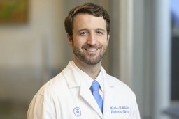 Matthew Tate McMillan合作论文数the Department of Surgery, From;Division of Hematology/Oncology, Department of Medicine;and Department of Radiation Oncology, University of Pennsylvania Perelman School of Medicine;Department of Medicine; and Department of Radiation Oncology, University of Pennsylvania Perelman School of Medicine27
 Gretchen Alicea合作论文数27
 Douglass Stephen合作论文数Institute of Cellular Medicine, Newcastle University27
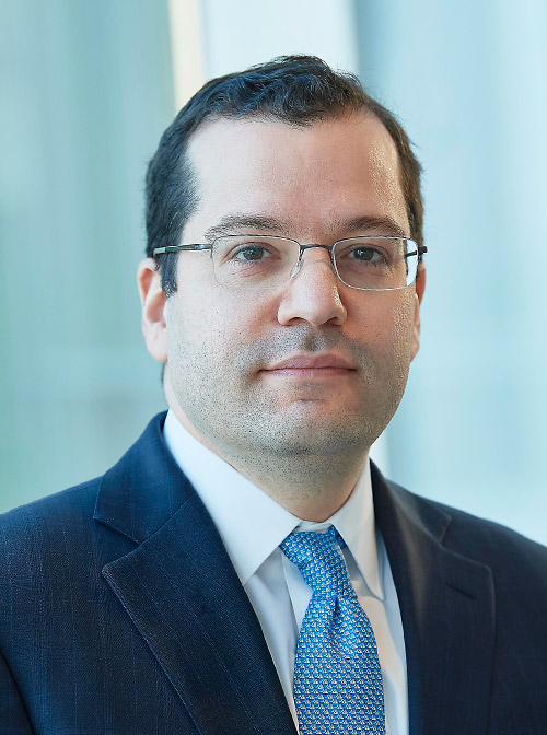 Robert E. Roses合作论文数Surgery Faculty, Perelman School of Medicine, University of Pennsylvania26
 Giorgos C. Karakousis合作论文数Surgery Faculty, Perelman School of Medicine, University of Pennsylvania23
 Douglas L. Fraker合作论文数Division of Surgical Oncology, University of Pennsylvania;Laboratory of Surgical Oncology, University of Pennsylvania;Department of Surgery, University of Pennsylvania Perelman School of Medicine22Langan Russell C合作论文数22
 Laura Maggino合作论文数21Mariam F Eskander合作论文数20
 Marie R Webster合作论文数The Wistar Institute20Kugel Curtis H合作论文数20
 Henry A. Pitt合作论文数Rutgers Cancer Institute of New Jersey;Robert Wood Johnson University Hospital;Rutgers Robert Wood Johnson Medical School, Rutgers University19
 Xiaowei (George) Xu合作论文数Department of Pathology and Laboratory Medicine, School of Medicine, University of Pennsylvania Perelman18Behera Reeti合作论文数17Qin Liu合作论文数17
 Dmitry Gabrilovich合作论文数Institute for Immunology and Immune Health, Perelman School of Medicine, University of Pennsylvania;AstraZeneca16
 Timothy E Kennedy合作论文数Department of Neurology and Neurosurgery, Faculty of Medicine and Health Sciences, McGill University;Division of Neuroscience, McGill University;Montreal Neurological Institute, McGill University15
 Michael I. D'Angelica合作论文数Memorial Sloan Kettering Cancer Center15Soares Kevin合作论文数15
 Amanpreet Kaur合作论文数14
 Jashodeep Datta合作论文数Department of Surgery, University of Pennsylvania Health System14
 William R. Jarnagin合作论文数Memorial Sloan Kettering Cancer Center14Yash Chhabra合作论文数Institute for Molecular Bioscience (IMB),, The University of Queensland14Xiangfan Yin合作论文数The Wistar Institute14T. Peter Kingham合作论文数Department of Surgery, Memorial Sloan Kettering Cancer Center14
 Meenhard Herlyn合作论文数Ellen and Ronald Caplan Cancer Center, The Wistar Institute of Anatomy and Biology;The Wistar Institute Melanoma Research Center, The Wistar Institute of Anatomy and Biology13
 Alice Wei合作论文数David M. Rubenstein Center for Pancreatic Cancer Research, Memorial Sloan Kettering Monmouth;Weill-Cornell School of Medicine13Miral S. Grandhi合作论文数12Jason C. Maggi合作论文数12
 Mithat Gönen合作论文数Memorial Sloan Kettering Cancer Center12Balachandran Vinod P合作论文数Department of Surgery, Memorial Sloan Kettering Cancer Center11
 Vinit Kumar合作论文数11
 Giuseppe Malleo合作论文数Manchester Royal Infirmary11
 Haejin In合作论文数10Lee Major K合作论文数10
 David W. Speicher合作论文数The Speicher laboratory, The Wistar Institute;Biochemistry and Molecular Biophysics Graduate Group, University of Pennsylvania10
 Claudio Bassi合作论文数Department of General and Pancreatic Surgery, Verona University10Sinnamon Andrew J合作论文数10Evgenii Tcyganov合作论文数9
 Emilio Sanseviero合作论文数Wistar Inst Anat & Biol, 3601 Spruce St, Philadelphia, PA 19104 USA9
 Gloria Marino合作论文数Johns Hopkins Sch Med, Sidney Kimmel Canc Ctr9
 Rejji Kuruvilla合作论文数Department of Biology, Krieger School of Arts & Sciences, Johns Hopkins University;The Solomon H. Snyder Department of Neuroscience, School of Medicine, Johns Hopkins University9Kelz Rachel R合作论文数Dept Surg, Univ Penn9
 Vito Rebecca合作论文数Department of Biochemistry and Molecular Biology, Bloomberg School of Public Health, The Johns Hopkins University9Kossenkov Andrew V合作论文数9




