Immune cells in the tumor microenvironment are not only powerful regulators of immunosuppression and tumorigenesis but also a dominant cell population, with tumor-associated macrophages (TAMs) comprising up to 50% solid tumor mass. Immunotherapies such as immune checkpoint inhibitors derive efficacy from this cancer-immune interface; however, immune-related adverse events from systemic blockade remain a major challenge. To address this need for potent, tumor-specific immunotherapies, we developed tumor immune cell targeting chimeras (TICTACs) that selectively deplete immune checkpoint receptors such as SIRPα from TAM surfaces. These chimeras consist of a synthetic ligand targeting CD206, a TAM marker, conjugated to a nonblocking antibody that binds to the checkpoint receptor without inhibiting it. By engaging CD206, which constitutively recycles between the plasma membrane and early endosomes, TICTACs drive robust checkpoint degradation in CD206high macrophages, with no effect on CD206low cells. This decoupling of antibody selectivity from blocking function presents a new paradigm for tumor-specific immunotherapies.
Accurate prognostic assessment of prostate cancer (PCa) requires an integrated understanding of tissue morphology-encompassing cell structure, glandular architecture, and tissue organization-and the immune environment. We present Prostate-TriMod, a novel tri-modal histology dataset designed to integrate high-resolution visual morphology with spatial tissue maps, immune infiltration patterns, and clinical outcomes. This dataset, generated from the Cell DIVE™ multiplexed imaging platform, consists of three synchronized modalities: (1) multiscale virtual H&E tiles (224px, 256px, 512px, and 2040px) providing visual morphological context, (2) spatial tissue maps identifying cancerous/non-cancerous epithelial cells, stroma and immune cell populations (via TOPAZ and CAT models), and (3) text captions generated from single-cell data and patterns. The dataset includes comprehensive clinical annotations, including Grade Groups and biochemical recurrence (BCR) status. By providing high-fidelity alignment between visual features, spatial tissue maps, and textual descriptions, Prostate-TriMod empowers the development of advanced multimodal AI frameworks. We expect this resource to support reuse in multimodal representation learning, spatial analysis, and benchmarking studies that link histology morphology and immune context to clinical outcomes in prostate cancer.
Background Benign prostatic hyperplasia (BPH) leads to prostate enlargement and lower urinary tract symptoms that can resist treatment. A histologic hallmark of BPH is glandular epithelial hyperplasia with new ductal branching morphogenesis. The stromal inductive factors driving tissue morphogenesis may provide new therapeutic targets, but are incompletely known due in part to a paucity of cell culture model systems. We thus sought to create a reliable cell culture assay for human prostatic branching morphogenesis.Methods Various combinations of input cells, cell densities and cell culture medium were trialed. A robust solution comprised embedding BHPrE1 immortalized human prostatic epithelial cells in Matrigel, together with an inductive source of BPH stromal fibroblasts or their conditioned medium. Resultant spheroids were measured and budding/branching morphogenesis evaluated by brightfield microscopy and optionally immunofluorescence. Candidate paracrine signaling pathways were interrogated using small molecule inhibitors and neutralizing antibodies.Results We have detailed a simple, robust cell culture assay for human prostatic epithelial branching morphogenesis. Preliminary application of the assay to investigate signaling pathways previously implicated in developmental budding/branching morphogenesis identified functions of epidermal growth factor (EGF), insulin-like growth factors (IGFs), and bone morphogenetic proteins (BMPs) in stimulating prostatic spheroid growth. However, only BMP inhibition blocked prostatic epithelial budding/branching morphogenesis.Conclusions We have described a straightforward cell culture assay for human prostatic budding/branching morphogenesis that in particular spotlights IGFs and BMPs as candidate therapeutic targets in BPH. Additional preclinical testing is warranted.
Extracellular vesicles (EVs) are lipid nano-to-micro-sized vesicles increasingly studied for their role in intercellular communication and their potential as minimally invasive molecular indicators in various diseases. However, challenges remain in characterizing specific surface molecules on EVs due to cargo heterogeneity and the lack of convenient quantification methods. In this study, we show the isolation, characterization, detection, and quantification of Trop2-carrying EVs (EV-Trop2) in serum of prostate cancer patients. This work combines the unique advantages of our EV isolation method with ELISA to enable surface-protein-specific EV analysis directly from serum. This is, to our knowledge, the first demonstration to isolate and quantify EV-Trop2 from prostate cancer patient serum to study its expression patterns in relation to prostate cancer status. Analysis of serum samples from three patient groups: high-risk prostate cancer (n = 22), low-risk prostate cancer (n = 23), and cancer-free groups (n = 21), revealed significantly different levels of EV-Trop2 expression between the high-risk and low-risk patient groups (p = 0.0015) and between high-risk patient and cancer-free groups (p < 0.0001). Multivariate modeling further showed that EV-Trop2 contributed to improved classifier metrics across the three sample groups. These findings highlight a strategy for probing EV-associated surface targets and suggest broader applicability of this approach across multiple cancers.
Glutamine reprogramming plays a crucial role in the growth and survival of clear cell renal cell carcinoma (ccRCC), although the mechanisms governing its regulation are still not fully understood. We demonstrate that the RNA demethylase fat mass and obesity-associated gene (FTO) drives glutamine reprogramming to support ccRCC growth and survival. Genetic and pharmacologic inhibition of FTO in ccRCC cells impaired glutamine-derived reductive carboxylation, depleted pyrimidines, and increased reactive oxygen species. This led to increased DNA damage and reduced survival, which could be rescued by pyrimidine nucleobases or the antioxidant N-acetylcysteine. Mechanistically, FTO demethylates the glutamine transporter solute carrier family 1 member 5 (SLC1A5) messenger RNA to promote its expression. Restoration of SLC1A5 expression in FTO-knockdown cells rescued metabolic and survival defects. FTO inhibition reduced ccRCC tumor xenograft and PDX growth under the renal capsule. Our findings indicate that FTO is an epitranscriptomic regulator of ccRCC glutamine reprogramming and highlight the therapeutic potential of targeting FTO for the treatment of ccRCC.
Supplementary Table 1 shows the summary of clinical characteristics of patients for sera TROP2 pull-down.
FXYD5 is involved in various biological processes, including inflammation, tumor progression, drug resistance, and hypertension. It has been suggested as a potential biomarker for several cancers, with elevated levels found in ovarian and colon cancer. However, the role of FXYD5 in prostate cancer has not yet been elucidated. Here, we found that the FXYD5 was expressed in tumor cells by sc-RNA sequencing of human metastatic prostate tumor tissues. The expression levels of FXYD5 were higher in androgen receptor (AR)-negative prostate cancer cells compared to AR-positive prostate cancer cells. Intrinsically, FXYD5 knockout promoted PC3 cell proliferation, migration, and invasion. In vivo studies showed that FXYD5 knockout promoted PC3 tumor growth, while overexpression of FXYD5 inhibited PC3 tumor progression. However, in AR-positive 22Rv1 cells, overexpression of FXYD5 promoted tumor progression. In vitro assays indicate that overexpression of FXYD5 in PC3, C42B, and 22Rv1 cells reduces mitochondrial membrane potential and reactive oxygen species. Additionally, FXYD5 overexpression reduces lactate dehydrogenase activity and lactate levels in PC3 cells but has no effect on these parameters in 22Rv1 cells. In PC3 cells, FXYD5 overexpression promotes the formation of tight cell-cell interactions, accompanied by downregulation of membrane markers and nuclear enlargement. In contrast, these effects are not observed in 22Rv1 or C42B cells. Further, FXYD5 is identified as a potential Siglec-7 ligand by CRISPRi screen. FXYD5 knockout decreased Siglec-7 Fc binding capacity and enhanced the NK-cell mediated cytotoxicity in prostate cancer cells. In a humanized mouse model, 22Rv1 tumor growth was inhibited with human immune cell modulation. However, when FXYD5 was overexpressed in 22Rv1 cells, tumor growth was not impacted by immune cell-based therapy. This was due to the interaction of FXYD5 on cancer cells with Siglec-7 on immune cells, thereby inducing immunosuppressive signals. These findings suggest potential therapeutic strategies targeting FXYD5-based mechanisms. Ru Wen, G Edward W. Marti, Neeladrisingha Das, Zhengyuan Qiu, Nathan Lam, Eric E. Peterson, Zenghua Fan, Aram Lyu, Fernando Jose Garcia Marques, Abel Bermudez, Hongjuan Zhao, Lawrence Fong, Guillem Pratx, Donna M. Peehl, Sharon J. Pitteri, James D. Brooks. FXYD5 plays diverse roles in immune evasion and tumor progression in prostate cancer [abstract]. In: Proceedings of the American Association for Cancer Research Annual Meeting 2025; Part 1 (Regular Abstracts); 2025 Apr 25-30; Chicago, IL. Philadelphia (PA): AACR; Cancer Res 2025;85(8_Suppl_1):Abstract nr 4325.
PURPOSE:No clear guidelines exist regarding MRI use after confirmatory biopsy during active surveillance. Our objective was to evaluate MRI performance after confirmatory biopsy in patients with vs without prior MRI-informed biopsy. MATERIALS AND METHODS:Patients in the Canary Prostate Active Surveillance Study with Gleason Grade Group (GG) 1 disease undergoing MRI-informed Biopsy 2, defined as second surveillance biopsy after diagnosis, were separated into prior vs no prior MRI-informed biopsy groups. Primary outcome was reclassification (≥GG2) at MRI-informed Biopsy 2. Reclassification rates and location (systematic cores, targeted cores, both) were compared between groups. Univariable and multivariable logistic regression identified predictors of reclassification. RESULTS:Patients with (n = 101) vs without (n = 103) prior MRI-informed biopsy had lower reclassification rates at Biopsy 2 (21% vs 36%, P = .017) and lower GG at reclassification (95% vs 73% of reclassifications to GG2, P = .039). In multivariable modeling, Prostate Imaging Reporting and Data System 4 or 5 lesion at MRI-informed Biopsy 2 was associated with increased odds of reclassification (odds ratio = 2.04, 95% CI [1.04-4.05]). The negative predictive value of MRI at Biopsy 2 was 87% (95% CI [78-96]) and 73% (95% CI [61-85]) in with vs without prior MRI groups. Reclassification location was identified by targeted cores only in 36% vs 19% of patients with vs without prior MRI (P = .4). Reclassification location was identified by systematic cores only in 36% vs 58% of patients with vs without prior MRI (P = .4). CONCLUSIONS:These results support MRI use at Biopsy 2 and suggest negative surveillance MRI should not replace Biopsy 2. Both targeted and systematic cores should be taken at Biopsy 2 in patients with and without prior MRI on active surveillance.
Siglecs (sialic acid-binding immunoglobulin-like lectins) are a family of cell surface proteins that are mainly expressed in immune cells. Recent studies show that Siglecs play important roles in immune evasion for tumors. We previously reported that Siglec-7 and Siglec-9 contribute to immune evasion by interacting with their ligands in prostate cancer, and blocking these interactions inhibits prostate cancer tumor progression. Further, Siglec-7 and Siglec-9 are highly expressed in macrophages in metastatic human prostate tumors. However, how macrophages mediate immune evasion via Siglec-dependent pathways remains unknown. Here, we have found that Siglec-7 and Siglec-9 are expressed in monocyte-differentiated macrophages derived from peripheral blood mononuclear (PBMC) cells. We will further profile Siglec expression in macrophages derived from PBMC and tumor tissues from patients with prostate cancer. The Siglecs on macrophages will be modulated, and their function will be evaluated through in vitro and in vivo models. Our study will provide insight into understanding macrophage-mediated immune evasion through Siglec-dependent mechanisms. Nathan S. Lam, Ru M. Wen, James D. Brooks. Determining the role of Siglecs in macrophage mediated immune evasion in prostate cancer [abstract]. In: Proceedings of the American Association for Cancer Research Annual Meeting 2025; Part 1 (Regular Abstracts); 2025 Apr 25-30; Chicago, IL. Philadelphia (PA): AACR; Cancer Res 2025;85(8_Suppl_1):Abstract nr 920.
Approximately 300,000 American men were diagnosed with prostate cancer in 2024. Existing screening approaches based on measuring levels of prostate-specific antigen in the blood lack specificity for prostate cancer. Studying the glycans attached to proteins has the potential to generate new biomarker candidates and/or increase the specificity of existing protein biomarkers, and studying protein glycosylation changes in prostate cancer could also add new information to our understanding of prostate cancer biology. Here, we present the analysis of N-glycoproteins in clinical prostate cancer tissue and patient-matched, non-cancerous adjacent tissue using LC-MS/MS-based intact N-linked glycopeptide analysis. This analysis allowed us to characterize protein N-linked glycosylation changes in prostate cancer at the glycoprotein, glycopeptide, and glycosite levels. Overall, 1894 unique N-glycosites on 7022 unique N-glycopeptides from 1354 unique glycoproteins were identified. Importantly, we observed an overall increase in glycoprotein, glycopeptide, and glycosite counts in prostate cancer tissue than non-cancerous tissue. We identified biological functions enriched in prostate cancer that relate to cancer development. Additionally, we characterized N-glycosite-specific changes in prostate cancer, demonstrating significant meta- and micro-heterogeneity in N-glycan composition in prostate cancer in comparison to non-cancerous tissue. Our findings support the idea that protein glycosylation is heavily impacted and aberrant in prostate cancer and provide examples of N-glycosite-specific changes that could be exploited for more specific markers of prostate cancer.
Prostate cancer is estimated to contribute to over 35,000 deaths of men residing in the United States, with the majority fatality due to metastatic disease. CDC7 is a kinase that regulates DNA replication and is found elevated during neuroendocrine transdifferentiation in lung and prostate cancer. In this study, we demonstrate that CDC7 is highly expressed in treatment-resistant prostate cancer, with even higher levels observed in treatment-resistant prostate cancer with neuroendocrine phenotype (NEPC). We further identify CDC7 as a critical regulator of prostate tumorigenesis. Downregulation of CDC7 significantly reduces prostate cancer cells growth and invasion in vitro and silencing CDC7 suppresses prostate tumor growth in vivo. Furthermore, we demonstrate that the inhibition of CDC7 using TAK-931, a selective CDC7 inhibitor, significantly reduces the proliferation, migration, and invasion of aggressive prostate cancer cells. TAK-931 treated prostate cancer cells exhibit an abnormal cell cycle profile, suggesting that CDC7 inhibition induces replication stress and promotes apoptosis. Collectively, our findings demonstrate that CDC7 is a regulator of tumor progression in prostate cancer and represents new therapeutic target in advanced prostate cancer.
Figure S2. Batiraxcept and docetaxel alone or in combination significantly inhibited bone tumor growth and metastasis in LuCaP mPCa AC PDX models. A-B, Representative images of Masson-Goldner staining and Ku70 IHC of PDX cells in the tibiae of LuCaP 147 (A), and LuCaP 35 (B). Human PCa cells marked by Ku70 were significantly decreased after 30 days of treatment by intraperitoneal injection with batiraxcept (20 mg/kg/QOD, n = 8–9, docetaxel (10 mg/kg/QW, n = 9) or the combination of batiraxcept (20 mg/kg/QOD, n = 9) and docetaxel (10 mg/kg/QW, n = 9) compared to vehicle controls (n = 8,10). C-D, Quantification of the IHC Ku70-positive areas in mouse tibia specimens treated with vehicle, batiraxcept, docetaxel, and batiraxcept + docetaxel in A-B. Fold change: combination batiraxcept and docetaxel (B+D) group set as 1. E, Inhibition of LuCaP 147CR tumor cells metastasis to mouse lung lobes by batiraxcept, docetaxel, and batiraxcept + docetaxel combination therapy. The arrowheads indicate Ku70-positive metastatic tumor nodules in the lung, which metastasized from the mice tibiae. F, Quantification of fold changes (docetaxel group set as 1) of human-specific GAPDH level normalized against universal GAPDH level in different treatment groups for LuCaP 147CR. *, p < 0.05; **, p < 0.01; ***, p < 0.001; NS, p > 0.05 not significant. All scale bar = 50 μm.
We discovered T-cell clonal expansions in benign prostatic hyperplasia, indicative of a specific adaptive immune response and with implications for disease pathogenesis and new treatments.
BACKGROUND AND OBJECTIVE:Prostate cancer (PC) and dementia may commonly co-occur; yet, prior evidence for bidirectional associations is inconsistent. This study aims to determine the associations between PC and dementia in large population-based studies, which may further inform clinical care. METHODS:To assess the dementia risk in men with PC, a national cohort study was conducted in 178 746 men diagnosed with PC in 1998-2017 and 1 787 460 age-matched control men in Sweden without prior dementia. Cox regression was used to estimate hazard ratios (HRs) for Alzheimer's disease (AD) and vascular dementia (VaD) through 2018. Subanalyses explored differences by PC treatment during 2005-2017. To assess the PC risk in men with dementia, case-control analyses were performed in 180 189 men with PC and 1 801 890 age-matched control men. Logistic regression was used to estimate odds ratios (ORs) for PC associated with prior AD or VaD. All analyses were adjusted for sociodemographic factors and health care utilization. RESULTS AND LIMITATIONS:Among men with high-risk PC, those treated with androgen deprivation therapy (ADT) only had a higher risk of AD (HR, 1.37; 95% confidence interval [CI], 1.19-1.58) and VaD (1.51; 1.29-1.78), but not those who received other treatments. Men with low- or intermediate-risk PC had little or no increased risk of AD (HR, 1.10; 95% CI, 1.03-1.18) or VaD (0.90; 0.83-0.98). Men with AD or VaD had lower odds of high-risk PC (OR, 0.39; 95% CI, 0.35-0.45, and 0.36; 0.30-0.42, respectively) and low- or intermediate-risk PC (0.30; 0.25-0.36, and 0.30; 0.24-0.38, respectively). This study was limited to Sweden and will need replication when feasible. CONCLUSIONS:In a large national study, men with high-risk PC treated with ADT had higher risks of AD and VaD. Such men should be monitored for timely detection and treatment of dementia. In contrast, men with AD or VaD had a lower subsequent risk of PC, possibly reflecting reduced screening in these subgroups.
Renal fibrosis (RF) is the main pathological feature and a potential therapeutic target of chronic kidney disease (CKD), a prevalent health problem causing a high economic burden to the health care system. Fat mass and obesity-associated (FTO) inhibition, either genetically or pharmacologically, significantly reduced collagen deposition, lipid peroxidation, and ferroptosis marker expression after unilateral ureteral obstruction (UUO) compared with sham-operated controls in mice. In murine and human kidney epithelial cells as well as in human embryonic stem cell-derived kidney organoids, FTO inhibition reduced erastin-induced ferroptosis by decreasing lipid peroxidation and reactive oxygen species production by downregulating the ferroptosis driver ACSL4. Moreover, FTO inhibition directly downregulated TGFBI, which was strongly associated with reduced M2 macrophage accumulation after UUO. Our results provide a strong rationale for targeting FTO to alleviate RF in patients subjected to obstruction-related kidney injury, thereby reducing the prevalence of CKD and associated treatment costs and improving the quality of life.
Background/Objectives: Clear-cell renal cell carcinoma (ccRCC) is a heterogenous disease that can be classified into multiple molecular subtypes with differential prognosis and sensitivities to treatments based on their genomic, transcriptomic, proteomic, and metabolic profiles. Patient-derived xenografts (PDXs) are high-fidelity cancer models because they maintain similar genotypes and immunohistologic phenotypes to the parental tumors and respond to standard-of-care therapies as expected. However, whether the molecular subtypes identified in ccRCC patient samples are preserved in PDX models is not clear. Our objective is to compare the transcriptional and proteomic profiles of our PDX models to those of ccRCC patients and identify both similarities and distinctions between molecular profiles of PDX subtypes and corresponding ccRCC patient subtypes, so that proper PDX subtypes can be used when investigating the corresponding ccRCC patient subtypes. Methods: To match PDXs to the human ccRCC molecular subtypes, we compared the transcriptomic and proteomic profiles of five ccRCC PDX models established in our lab to those of the human ccRCC molecular subtypes reported by our group, as well as other groups, using hierarchical analysis, Principal Component Analysis (PCA), and Permutation Correlation Analysis. The enrichment of key molecular pathways in PDXs and ccRCC subtypes was determined using Gene Set Enrichment Analysis. Results: We found that each PDX resembles one of the molecular subtypes closely at both transcript and protein levels. In addition, PDXs representing different molecular subtypes show unique metabolic characteristics. Moreover, molecular subtypes of PDXs correlated with ccRCC patient subtypes in key pathway activities implicated in ccRCC progression and therapy resistance. Conclusions: Our results suggest that PDX subtypes should be used when investigating the molecular mechanism of cancer progression and therapy resistance for corresponding ccRCC patient subtypes. This “matching” strategy will greatly facilitate the clinical translation of positive findings into the optimal management of ccRCC patients.
Metastasis is the main cause of prostate cancer-associated deaths, highlighting the urgent need to determine the mechanisms underlying prostate cancer progression. TROP2 (also known as tumor-associated calcium signal transducer 2) is an oncogenic transmembrane surface protein that is highly expressed in metastatic prostate cancer. Naturally occurring cleavage of TROP2 leads to a release of the TROP2 extracellular domain (TECD) into the extracellular environment. In this study, we identified an important functional role of TECD in prostate cancer metastasis. TECD was detectable in media from prostate cancer cells and serum from patients with clinically significant prostate cancer. Although shed TECD did not affect prostate cancer cell proliferation and tumor growth, it increased cell migration and invasion in vitro and promoted metastatic colonization and spontaneous metastasis in vivo. TECD interactome and proteomic studies revealed that TECD binds to EGFR and shed TECD modulates a set of proteins associated with invasion, migration, mTOR signaling, and epithelial-to-mesenchymal transition. Furthermore, elevated shed TECD increased EGFR phosphorylation, resulting in the activation of the EGFR-PI3K-AKT-mTOR pathway in prostate cancer. EGFR inhibitors suppressed the invasive ability of prostate cancer cells driven by TECD overexpression, further supporting the key role of EGFR in TECD-mediated prostate cancer progression. This study uncovers a function of TECD in driving prostate cancer progression and provides mechanistic insights into TECD signaling through EGFR.Significance: Shed extracellular domain of TROP2 binds to and activates EGFR and stimulates the PI3K-AKT-mTOR signaling cascade to promote prostate cancer metastasis, providing potential biomarkers and therapeutic targets.
 Daniel W. Lin合作论文数Department of Urology, University of Washington59
 Peter R. Carroll合作论文数Department of Urology, University of California, San Francisco58
 John Leppert合作论文数Departments of Medicine, School of Medicine, Stanford University;Department of Urology, School of Medicine, Stanford University;Stanford Cancer Institute, Stanford University57
 Lisa F. Newcomb合作论文数55HongJuan Zhao (赵红娟)合作论文数53
 Ian M. Thompson合作论文数Urology Division, The University of Texas Health Science Center at San Antonio50
 Peter Nelson合作论文数Stuart and Molly Sloan Precision Oncology Institute, Fred Hutch Cancer Center;Human Biology Division, Fred Hutch Cancer Center;University of Washington School of Medicine46
 Martin Gleave合作论文数Vancouver Prostate Centre, Faculty of Medicine, The University of British Columbia;Department of Urologic Sciences, The University of British Columbia42Jesse K. Mckenney合作论文数42Nolley Rosalie合作论文数40
 Tina Hernandez-Boussard合作论文数Boussard Lab, School of Medicine, Stanford University40
 Geoffrey Sonn合作论文数Radiology Department, School of Medicine, Stanford University;Department of Urology, School of Medicine, Stanford University37
 Sharon Pitteri合作论文数Department of Radiology, Stanford University School of Medicine;Cancer Early Detection-Canary Center, Stanford University36Yingye Zheng合作论文数Department of Biostatistics and Biomathematics, Fred Hutchinson Cancer Research Center36
 Christian Kunder合作论文数Stanford University;GynePath Laboratory, Inc34Michelle Ferrari合作论文数34
 Michael Andre Liss合作论文数32
 Benjamin I. Chung合作论文数Department of Urology, Stanford University32
 Dean A. Troyer合作论文数Pathology, Microbiology and Molecular Biology, Eastern Virginia Medical School;Department of Pathology, University of Texas Health Science Center32Ziding Feng合作论文数31
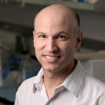 Jonathan Pollack合作论文数Department of Pathology, School of Medicine, Stanford University31
 Donna Peehl合作论文数Department of Radiology and Biomedical Imaging, School of Medicine, University of California, San Francisco31
 Richard E. Fan合作论文数Department of Urology, School of Medicine, Stanford University29Abel Bermudez合作论文数27
 Andrew A. Wagner合作论文数Department of Surgery, Harvard Medical School/Beth Israel Deaconess Medical Center27
 Atreya Dash合作论文数Department of Urology, University of Washington Medical Center;Department of Urology, Seattle Veterans Affair25Joseph C. Presti合作论文数24
 Todd Matthew Morgan合作论文数Division of Urologic Oncology, Department of Urology, University of Michigan;Visible Health;UrologyMatch.com24
 Lawrence D. True合作论文数Department of Laboratory Medicine & Pathology, School of Medicine, University of Washington;University of Washington Health Sciences Center23
 Douglas W. Blayney合作论文数Medicine Department, Stanford University23
 Tanya Stoyanova合作论文数Radiology Department, Stanford University23
 En-Chi Hsu合作论文数School of Life Science Pei-Tou, National Yang-Ming University Institute of Microbiology and Immunology23
 Matthew R. Cooperberg合作论文数Department of Urology, University of California San Francisco22
 John P. Higgins合作论文数Department of Internal Medicine, McGovern Medical School, University of Texas Health Science Center at Houston;Memorial Hermann Healthcare System;Lyndon Baines Johnson General Hospital, Harris Health System22
 Mirabela Rusu合作论文数Department of Biomedical Data Science, Stanford University22
 Jim E. Riviere合作论文数Institute of Computational Comparative Medicine, Kansas State University;North Carolina State University21
 Ladan Fazli合作论文数Vancouver Prostate Centre21
 Fernando Jose Garcia-Marques合作论文数20Thomas I-Chun合作论文数20
 John Thomas Wei合作论文数Department of Urology, University of Michigan20
 Pejman Ghanouni合作论文数Division of Body MRI, School of Medicine, Stanford University;Department of Radiology, Stanford University;Department of Urology, Stanford University20Shiqin Liu合作论文数Dewitt Daughtry Family Dept Surg, Univ Miami19
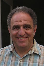 Rob Tibshirani合作论文数Department of Statistics, Stanford University;Department of Biomedical Data Science, Stanford University19Jeff P. Simko合作论文数19
 Sarah T. Hawley合作论文数Department of Health Management and Policy, School of Public Health, University of Michigan-Ann Arbor18Antonio Hurtado-Coll合作论文数18
 Jacques Lapointe合作论文数Division of Experimental Medicine, Department of Medicine, Faculty of Medicine and Health Sciences, McGill University17
 Martin G. Sanda合作论文数Department of Urology, Emory University School of Medicine;Winship Cancer Institute, Emory University17
 William J. Ellis合作论文数Medical Center, University of Washington;Fred Hutchinson Cancer Center, University of Washington17




