The belief that the moon disturbs sleep is widespread, but the factors associated with it remain poorly understood. I therefore examined how frequently poor sleep is attributed to moon phases, whether this varied across the lunar cycle, and which personal and environmental factors were associated with “moon blaming”. Data were derived from an ongoing online survey. At the time of analysis, 1815 participants had completed a 16-item questionnaire assessing sleep quality, sleep duration, sleep timing on workdays and free days, alarm clock use, environmental and personal sleep-disturbing factors, residential setting, age, gender, attention to lunar phases, and whether the moon was perceived as a cause of poor sleep. The primary outcome was endorsement of the moon as a sleep-disturbing factor. Logistic regression with stepwise Akaike information criterion selection was used to identify the strongest predictors of attributing the moon for poor sleep. Questionnaire timing was also examined across the lunar cycle. Among environmental factors, the moon was the most frequently endorsed cause of poor sleep (36%), followed by outdoor temperature (31%), indoor noise (26%), and bad weather (22%). Rumination was the most commonly reported personal factor (73%), but it did not predict moon attribution. Instead, the strongest correlates were weather-related sleep complaints, tracking lunar phases, age, and gender, with endorsement increasing with age and being more common among women. Moon-related complaints also peaked during the week after the full moon. These findings suggest that perceived lunar effects on sleep are shaped, at least in part, by attributional and expectation-related processes.
Thermoregulatory processes are closely linked to sleep initiation and maintenance throughout the circadian cycle, and may contribute to the increased tendency to nap in older adults. This cross-sectional study examined whether habitual napping in healthy older individuals is associated with altered skin temperature-derived heat-loss dynamics and their relationship with sleep onset. Thirty self-reported habitual nappers and 28 non-nappers (59-82 y) completed a 40-hour multiple-nap protocol under controlled laboratory conditions, with continuous polysomnography and distal-proximal skin temperature gradients (DPG) recordings. DPG was analyzed across scheduled wake episodes and at lights-off preceding each nap opportunity. Habitual nappers exhibited distinct changes in thermoregulatory dynamics compared to non-nappers. Overall, they had a lower DPG during scheduled wakefulness, particularly during the afternoon nap window (14:45-17:30). Their circadian organization of the DPG also differed markedly: they had a higher 24-hour DPG amplitude, a more pronounced 12-hour component, and an earlier DPG phase than the non-nappers. During nap opportunities, shorter sleep onset latency (SOL) was associated with a faster increase in DPG after lights-off in both groups. However, in habitual nappers, sleep onset occurred more rapidly despite a reduced dependence on pre-sleep DPG increase. Together, these findings indicate that habitual napping in older adults is accompanied by altered heat-loss rhythms across the circadian cycle and a reduced coupling between pre-sleep thermoregulatory dynamics and sleep initiation. Circadian-driven thermoregulatory changes may underlie the greater propensity to nap in older adults and differentiate habitual nappers from non-nappers. However, the causal direction of this relationship requires further investigation.
Circadian rhythms and inter-individual differences in sleep-wake preferences, or chronotypes, influence cognitive performance. The synchrony effect-the alignment of time of day with chronotype-modulates attention, memory, and executive control. This study examined behavioural and neural correlates of synchrony effects during an auditory oddball task performed in morning (1.5 h after wake-up) and evening (10.5 h after wake-up) sessions by extreme morning-type (n = 16) and evening-type (n = 15) young (age range: 22-32 y.o.) individuals. Behaviourally, no main, nor interaction effects of chronotype or time of day were observed for overall reaction time metrics (all p > 0.05). However, overall reaction times during the task were longer in the final third of the task in the evening compared to the morning session for morning but not evening types (p < 0.05). Functional magnetic resonance imaging (fMRI) revealed neural correlates underlying the synchrony effect: the inferior frontal cortex (IFC) showed higher task-related activation at optimal times (morning for morning types, evening for evening types, pcorr < 0.05), putatively reflecting enhanced top-down control. Conversely, a medial orbitofrontal region (mOFC), part of the default mode network, was more active at non-optimal times (pcorr < 0.05), consistent with reduced task engagement and internally directed processes. These findings suggest that synchrony effects interact with time-on-task-dependent effects with implications for sustained attention in real-world settings.
Light is the primary cue that synchronises the human circadian system to the 24-h day, advancing or delaying circadian rhythms depending on its timing. While it is known that morning light induces phase advances, most studies assess the timing of dim-light melatonin onset (DLMO) in the subsequent circadian cycle, over 24 h after the light intervention. However, it is unclear whether these phase advances occur within the same circadian cycle as the light intervention or the next one. This narrative review addresses the question of whether morning light can phase-advance human melatonin rhythms in less than 24 h. To answer this question, we review studies that use same-day or single-cycle protocols, in which light exposure and post-intervention DLMO assessment occur within the same 24-h period. To compare light interventions across studies, melanopic equivalent daylight illuminance (mEDI) values were estimated and related to the magnitude of the observed phase advance. The reviewed research suggests that modest phase advances of 10-30 min can be achieved within the same circadian cycle if light is delivered shortly after waking up in the morning. This is particularly effective if the light is bright, blue-enriched, or if it is delivered for a long time (over 1 h). There was a statistical trend (r = 0.51, p = 0.06) towards a positive association between the mEDI of the light intervention and the magnitude of the phase advance. Overall, same-day phase-advances seem possible but not well characterised, and more targeted work is needed to determine whether morning light can phase-advance human melatonin rhythms in less than 24 h. If this is confirmed, the length of circadian protocols could be reduced, accelerating the clinical use of treatments for circadian rhythms sleep-wake disorder.
Adolescents often experience chronic sleep restriction due to a biologically driven delay in bedtime and early school schedules. Caffeine is widely used to counteract resulting sleepiness, however, its effects on adolescents’ sleep under sleep restriction remain unclear. In this preregistered mixed-design polysomnographic study, 45 adolescents (14–17 years) underwent twice three nights of restricted (6h) or extended (9.5h) sleep opportunity. Three hours before bedtime of the third night, participants received caffeine (2mg/kg) or placebo (double-blinded) in quasi-randomized order. Bayesian analyses indicated that deep sleep intensity (indicated by EEG slow-wave energy) was reduced and sleep architecture disturbed after caffeine compared to placebo, particularly in the beginning of extended nights. Critically, caffeine reduced subjective sleepiness exclusively under sleep restriction and did not affect sleep evaluation in either sleep history. These findings suggest that caffeine's disruptive effects on adolescent sleep are more pronounced and unrecognised during nights of recommended duration than sleep-restricted nights.
During sleep, the human brain transitions to a "sentinel processing mode," enabling the continued processing of environmental stimuli despite the absence of consciousness. We employed advanced information-theoretic analyses, including mutual information (MI) and co-information (co-I), alongside event-related potential (ERP) and temporal generalization analyses (TGA), to characterize auditory prediction error processing across wakefulness and sleep. We hypothesized that a shared neural code would be present across sleep stages, with deeper sleep being associated with reduced information content and increased information redundancy. Twenty-nine participants (15 women) underwent an auditory "local-global" oddball paradigm during wakefulness and an 8 h sleep opportunity monitored via polysomnography. We focused on "local" mismatch responses to a deviating fifth tone after four standards. ERP analyses showed that prediction error processing continued throughout all sleep stages (N1-N3, REM). Mutual information analyses revealed a substantial reduction in encoded prediction error information particularly during N3 and REM, although ERP amplitudes increased with deeper NREM sleep. We also observed delayed information encoding during sleep, and co-information analyses showed neural dynamics became increasingly redundant with increasing sleep depth. TGA revealed a largely shared neural code between N2 and N3, though it differed between wakefulness and sleep. We demonstrate how the neural code of the "sentinel processing mode" changes from wake to light to deep sleep and REM, characterized by delayed processing, more redundant and less rich neural information in the human cortex as consciousness wanes. This altered stimulus processing reveals how neural information evolves with variations in consciousness across the night.
PurposeAdolescence is marked by changes in brain connectivity linked to risk decision-making, indexed by risk-taking propensity, optimal choices (safe, higher expected value), and irrational choices (risky, lower expected value). Insufficient sleep and caffeine consumption, common during adolescence, may independently and jointly influence risk decision-making in adolescents already predisposed to risk-taking. This study investigated whether sleep-restriction impairs risk decision-making, whether caffeine increases risk-taking propensity and optimal choices while decreasing irrational choices, and whether caffeine effects are enhanced under sleep-restriction.MethodsForty-one adolescents aged 14–17 years completed a between-subject, two-night sleep manipulation (sleep-restriction: 6 h time in bed, sleep-extension: 9.5 h time in bed), followed by a within-subject, double-blind, placebo-controlled caffeine manipulation. Risk decision-making was assessed using a modified Wheel of Fortune task, and Bayesian inference quantified sleep and caffeine effects.ResultsAcross all outcomes, posterior estimates for sleep condition, caffeine, and their interaction were small, with 95% highest density intervals spanning zero; region of practical equivalence overlap varied across predictors, indicating limited evidence for practically meaningful effects. Caffeine effects and sleep-caffeine interactions remained uncertain across outcomes.DiscussionAdolescent risk decision-making on the modified Wheel of Fortune task showed no clear evidence of practically meaningful average changes after two nights of sleep-restriction or acute caffeine. These results suggest limited sensitivity of this task to the present manipulations rather than evidence that sleep or caffeine is inconsequential. Findings provide quantitative effect size and uncertainty benchmarks for future experimental work.Clinical trial registrationhttps://www.humanforschung-schweiz.ch/en/trial-search/study-detail/58864/translate/en/, identifier SNCTP000004988.
Whether light exposure during the day reduces non-visual light effects later in the evening has not been studied in adolescents. We investigate whether afternoon-early evening (AEE) light interventions (130 lx, 2500 lx, 4.5 h, compared to 6.5 lx illuminance) would increase melatonin levels during later evening light exposure (130 lx) in a counterbalanced crossover study with 22 adolescents (14-17 years, 11 female). Contrary to our hypothesis, evening melatonin levels decreased after AEE bright light exposure, while sleepiness and vigilance were unaffected, and skin temperature showed no clear changes. The AEE light had acute alerting effects and bright light exposure in the 32 h before laboratory entry was associated with higher evening melatonin and sleepiness. These findings suggest that bright AEE light increases alertness but may delay melatonin production by interfering with circadian rhythms. The study highlights the complex effects of light timing and its implications for managing adolescents' light exposure.
Light synchronises the internal clock with the external light-dark cycle. Keeping this alignment benefits health and prevents diseases. Quantifying light exposure is, therefore, vital for effective prevention. Since light exposure depends on photoperiod, culture, and behaviour, we investigated objective light exposure and individual light-related behaviour in Switzerland and Malaysia. In this observational field study, participants (N = 39) wore a calibrated melanopic light logger at chest level for 30 consecutive days. At baseline and study end, the Pittsburgh Sleep Quality Index was assessed, and every 3 to 4 days, the Light Exposure Behaviour Assessment (LEBA) was filled. Our pre-registered analyses reveal that participants in Switzerland experienced brighter days (+3.16 times the average mel EDI) and spent more time (x1.9 times the duration) in daylight levels per hour of daylight, had 1.5 h later bright light exposure in the afternoon, and stayed over 1 h longer in dim light conditions before bedtime. LEBA scores did not differ between Malaysia and Switzerland, and LEBA items were stable over time. LEBA items also correlated with objective light exposure variables in Switzerland but not Malaysia, with a medium effect size (range of absolute r = 0.32–0.48). Our results highlight cultural and geographical differences in light exposure. We showed that subjective assessment of light-exposure behaviour can be related to actual light exposure and is ecologically informative, but this varies by culture.
The incidence of myopia among school children has risen markedly over the last three decades. In urban areas of South and East Asia, as many as 80-90% of young adults are now myopic. This trend is occurring elsewhere around the world. During the COVID-19 lockdowns, children in many countries were confined indoors and spent an undue amount of time exposed to television screens, computers, and mobile devices. This resulted in an acceleration in the incidence and progression of the condition. Myopia is a significant public health issue as it is a leading cause of blindness and other vision problems. Yet the underlying mechanisms that produce the condition remain elusive. Pseudomyopia has recently been proposed as an independent risk factor for myopia. We hypothesize that pseudomyopia induced by prolonged close work, stress, and anxiety combines and is further amplified by chronically low ambient light levels. If time spent outdoors in daylight is restricted, the effects worsen and together may play a significant part in myopia epidemics.
While there is considerable enthusiasm for the translational and clinical applications of chronobiology, their actual implementation is not progressing as rapidly as might be expected. Here we discuss the possibility that this may relate to a combination of conceptual, methodological, evidentiary and training challenges. These are compounded by the remaining, profound cultural differences between basic and applied chronobiologists. We argue that all these issues can be overcome by cross-faculty teaching, time, patience and goodwill, together with a set of more formal actions, such as the establishment of a collaborative framework for evidence generation, the engagement of relevant stakeholders and public health campaigns based on already available evidence. We hope that chronobiology, and in particular the transformative power of circadian medicine, will change health outcomes, increase safety and improve quality of life for patients worldwide. Now is the time to bring "internal time" into medicine.
Light influences human physiology and behavior by regulating circadian rhythms, melatonin secretion, and alertness. Previous research has reported sex differences in melatonin secretion and circadian rhythms, possibly related to women's greater sensitivity to bright light. Other studies have suggested reduced photosensitivity and earlier circadian phases in summer than in winter in midlatitude regions. This study explores the effects of sex, seasonality, and their combination on melatonin suppression and subjective sleepiness in response to moderate light exposure, considering prior light history and menstrual phases in females. We conducted a controlled, within-subject experiment with 48 healthy adults (18-35 years, 50% female) across different seasons. The study design included two 9-hour laboratory sessions, with at least 5-day washout in between. Participants were exposed to dim and moderate light through a screen for 2 hours after their habitual bedtime. Female participants exhibited greater melatonin suppression (+4.69%) but a lower alerting response (-6.00%) to moderate light compared to males. Both sexes demonstrated increased sensitivity to the non-image forming effects of light in winter, with stronger melatonin suppression (+18.05%) and increased alertness (+7.60%) compared to summer. While prior light history did not significantly impact melatonin suppression or alertness, it was associated with an earlier dim light melatonin onset (DLMO). Females during their luteal phase had earlier DLMO than those in their follicular phase. Our findings indicate an interaction between sex, seasonality, and light exposure in modulating melatonin suppression, emphasising the need for personalized light exposure recommendations according to individual biological and environmental contexts.
A chronotype is defined as a preference for certain behaviours (e.g., sleep and wake) to occur at specific times of day. It is therefore also temporally linked with cognitive performance across the day. In an exploratory analysis, we sought to find associations between chronotypes determined from self-reported habitual sleep timing and from salivary melatonin onset with mental effort during a 2-back working memory task. Mental effort was operationalized as sympathetic beta-adrenergic impact on the heart, which is best reflected by the cardiac pre-ejection period (PEP) and also influences systolic blood pressure (SBP). Each participant underwent two experimental sessions in the morning: once after sleeping for 8 h and once after sleeping for 5 h the night before. To determine the timing of evening melatonin onset, participants took saliva samples at hourly intervals at home in the evening, prior to their experimental sessions. Chronotypes were determined using reported sleep times from the Munich Chronotype Questionnaire and average melatonin onset during both sleep conditions. Based on this, participants were grouped into early, intermediate, or late types. Neither alertness (BF 10 = 0.019), perceived task demand (BF 10 = 0.008), nor SBP response (BF 10 = 0.268) were credibly impacted by sleep-time derived chronotype, while the association with PEP response (BF 10 = 0.631) during a cognitive challenge in the morning was inconclusive. Similarly, the timing of evening melatonin onset did not affect alertness (BF 10 = 0.003), perceived task demand (BF 10 = 0.006), or PEP or SBP response (PEP: BF 10 = 0.232, SBP: BF 10 = 0.263) during the cognitive challenge. Our data shows no impact of chronotypes on effort-related cardiovascular response during a cognitive challenge in the morning, which was scheduled according to habitual sleep times.
Light exposure at night can suppress melatonin production and increase alertness, primarily through the action of melanopsin-containing intrinsically photosensitive retinal ganglion cells (ipRGCs). This study investigated whether cone photoreceptors also influence melatonin suppression and subjective alertness using non-visual metameric light. Forty-eight participants with normal trichromatic vision were exposed to three lighting conditions: a baseline (BL: 9 lxmEDI), constant background (BG: 149 lxmEDI), and cone-modulated flickering light targeting different cone combinations and post-receptor channels (S-, M-L, S+M+L: 149 lxmEDI) for 2 h after their habitual bedtime. Salivary melatonin levels and subjective alertness were measured throughout a 9-h protocol. Bayesian analysis showed that cone-modulated flickering light did not significantly affect melatonin suppression or alertness, providing evidence against the hypothesis that cone photoreceptors contribute to these non-visual effects of light. In conclusion, our results suggest cone photoreceptors do not play a measurable role in the effects of light on melatonin suppression and subjective alertness at night.
BACKGROUND:Depression is a major health issue in adolescence and young adulthood, emphasising the need for early risk identification. Patients with major depressive disorder (MDD) often show disturbed daily rest-activity patterns, but such changes are often confounded by medication intake, comorbidities and disease duration. OBJECTIVE:In this exploratory analysis, we tested whether there are specific changes in daily rest activity (from wrist-worn actigraphy) in women at the onset of MDD without medication, as compared with age-matched controls. METHODS:Participants from the MDD group (age 19-32, 24.73±5.13 (mean±SD), N=15) and control group (age 20-31, 24.89±3.82, N=9) completed ~7 day ambulatory actigraphy recordings, followed by a stringently controlled circadian laboratory protocol to assess endogenous circadian melatonin levels. We analysed the daily rhythm of mean activity levels and non-linear fractal dynamics in eight 3-hour time bin across the 24 hours, correlating these measures with depressive symptom severity and endogenous melatonin levels. FINDINGS:Using approaches from non-linear fractal dynamics, we showed that, compared with healthy controls, women at MDD onset had a higher fractal activity correlation (FAC) during the last hours of sleep, indicating more 'wake-like' patterns (FAC within 0-3 hour before wake: 0.92±0.64 (SD) in MDD vs 0.77±0.18 in controls, p=0.02). The alteration was independent of mean activity level and wake duration but appeared to be associated with depressive symptom severity (p=0.08). Moreover, there was a trend association for altered FAC with endogenous melatonin levels in the MDD group (for onefold increase in melatonin level in the last 3 hours before wake, the FAC increased by 0.33±0.17 (SE), p=0.08). CONCLUSIONS:Pre-wake FAC is elevated in unmedicated women at MDD onset and may serve as a potential biomarker associated with symptom severity and circadian physiology. CLINICAL IMPLICATIONS:These findings provide proof-of-concept evidence that unique fractal motor activity patterns may support early detection of MDD.
Fixed sleep schedules with an 8 h time in bed (TIB) are used to ensure participants are well-rested before laboratory studies. However, such schedules may lead to cumulative excess wakefulness in young individuals. Effects on older individuals are unknown. We combine modelling and experimental data to quantify the effects of sleep debt on sleep propensity in healthy younger and older participants. A model of arousal dynamics was fitted to sleep data from 22 young (20–31 y.o.) and 26 older (61–82 y.o.) individuals (25 male) undertaking 10 short sleep–wake cycles during a 40 h napping protocol, following >1 week of fixed 8 h TIB schedules. Homeostatic sleep drive at the study start was varied systematically to identify best fits between observed and predicted sleep profiles for individuals and group averages. Daytime sleep duration was the same on the two days of the protocol within the groups but different between the groups (young: 3.14 ± 0.98 h vs. 3.06 ± 0.75 h, older: 2.60 ± 0.98 h vs. 2.37 ± 0.64 h). The model predicted an initial homeostatic drive of 11.2 ± 3.5% (young) and 10.1 ± 3.5% (older) above well-rested. Individual variability in first-day, but not second-day, sleep patterns was explained by the differences in the initial homeostatic drive for both age groups. Our study suggests that both younger and older participants arrive at the laboratory with cumulative sleep debt, despite 8 h TiB schedules, which dissipates after the first four sleep opportunities on the protocol. This has implications for protocol design and the interpretation of laboratory studies.
The pupil modulates the amount of light that reaches the retina. Not only luminance but also the spectral distribution defines the pupil size. Previous research has identified steady-state pupil size and melatonin attenuation to be predominantly driven by melanopsin, which is expressed by a unique subgroup of intrinsically photosensitive retinal ganglion cells (ipRGCs) that are sensitive to short-wavelength light (~480 nm). Here, we aimed to selectively target the melanopsin system during the evening, while measuring steady-state pupil size and melatonin concentrations under commonly experienced evening light levels (<90 lx). Therefore, we used a five-primary display prototype to generate light conditions that were matched in terms of L-, M-, and S-cone-opic irradiances, but with high and low melanopic irradiances (~3-fold difference). Seventy-two healthy, male participants completed a 2-week study protocol. The volunteers were assigned to one of the four groups that differed in luminance levels (27-285 cd/m 2 ). Within the four groups, each volunteer was exposed to a low melanopic (LM) and a high melanopic (HM) condition. The two 17-h study protocols comprised 3.5 h of light exposure starting 4 h before habitual bedtime. Median pupil size was significantly smaller during HM than LM in all four light intensity groups. In addition, we observed a significant correlation between melanopic weighted corneal illuminance (melanopic equivalent daylight illuminance [mEDI]) and pupil size, such that higher mEDI values were associated with smaller pupil size. Using pupil size to estimate retinal irradiance showed a qualitatively similar goodness of fit as mEDI for predicting melatonin suppression. Based on our results here, it remains appropriate to use melanopic irradiance measured at eye level when comparing light-dependent effects on evening melatonin concentrations in healthy young people at rather low light levels.
This study investigates the impact of varying light intensities throughout the day on motor activity levels in adolescent girls aged between 14-16, who are students at a boarding school. The study lasted for three weeks, with each week consisting of four days. During this period, participants experienced three different lighting conditions from 07:00 to 18:00. These conditions included: standard light intensity in real-life settings, wearing blue-blocking glasses, and spending at least 1.5 hours outdoors daily for natural light exposure. The aim was to understand how modern lighting sources can replace natural lighting and to assess the influence of the lighting spectrum on the daily rhythms of motor activity. Restriction of blue light led to an increase in the average daily level (MESOR) of the intensity of movements denoted by HPIM (High Proportional Integrative Measures), as well as a decrease in the amplitude of the circadian rhythm ZCM (Zero Crossing Mode). This confirms the important role of blue light during the daytime as a Zeitgeber of the circadian rhythm. When adolescents spent at least 1.5 hours outside, mostly in the afternoon, the acrophases of ZCM’s 24-hour rhythm shifted half an hour later.
 Reichert Carolin F合作论文数Psychiatric University Hospital, University of Basel Basel75
 Christina Schmidt合作论文数Cyclotron Research Centre, University of Litθge;Department of Cognitive Psychology, University of Liθge66
 Anna Wirz-Justice合作论文数Centre for Chronobiology, Universitäre Psychiatrische Kliniken Basel62
 A. Wirz-Justice合作论文数49Gabel Virginie合作论文数46Mark Brink合作论文数45
 Nicole Probst-Hensch合作论文数Department of Epidemiology and Public Health, Swiss Tropical and Public Health Institute;Chronic Disease Epidemiology Unit, Swiss Tropical and Public Health Institute;University of Basel Medical School45
 Jean Marc Wunderli合作论文数Laboratory for Acoustics / Noise Control, Empa Materials Science and Technology44Martin Roosli合作论文数43
 Sarah Chellappa合作论文数Faculty of Environmental and Life Sciences, University of Southampton42Viola Antoine U合作论文数41
 Micheline Maire合作论文数38Danielle Vienneau合作论文数36Landolt Hans-Peter合作论文数35Maria Foraster合作论文数35
 Mirjam Munch合作论文数35Kurt Kräuchi合作论文数32
 Eze Ikenna C合作论文数Swiss Tropical and Public Health Institute31V Knoblauch合作论文数28
 Garbazza Corrado合作论文数Psychiatric Hospital, the University of Basel28
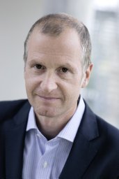 Oliver Stefani合作论文数COAT-Basel28
 Manuel Spitschan合作论文数Departments of Psychology and26Héritier Harris合作论文数Swiss Tropical and Public Health Institute24Werner Strobel合作论文数24
 Reto Pieren合作论文数Swiss Fed Labs Mat Sci & Technol, Empa24Emmanuel Schaffner合作论文数22
 Silvia Frey合作论文数21M. Maire合作论文数20Philippe Peigneux合作论文数20Franziska Rudzik合作论文数19Janine Weibel合作论文数19
 Fabienne Collette合作论文数Cyclotron Research Centre, University of Liège19
 Laurie Thiesse合作论文数18Vera Knoblauch合作论文数18
 Derk-Jan Dijk合作论文数Surrey Sleep Research Centre, University of Surrey;Faculty of Health and Medical Sciences, University of Surrey;School of Biosciences, University of Surrey15
 Stefan Borgwardt合作论文数Department of Psychiatry and Psychotherapy, University of Lübeck15
 Christian Joachim Schmidt合作论文数Detector Laboratory, GSI Helmholtzzentrum für Schwerionenforschung GmbH14
 Vivien Bromundt合作论文数14
 Christian Berthomier合作论文数Département TSI, ENST14
 Klaus Scheffler合作论文数Department High-field Magnetic Resonance, Max-Planck-Institute for Biological Cybernetics;Department of Biomedical Magnetic Resonance, University of Tubingen13
 Lasauskaite Ruta合作论文数Department of Psychology, University of Geneva13Claudia Renz合作论文数13
 Schroder Carmen M合作论文数12Blatter K合作论文数12Kurt Krauchi合作论文数Sleep Laboratory, Psychiatric University Clinic12
 Martin Meyer合作论文数Cognitive Psychology Unit, Alpen-Adria University of Klagenfurt11
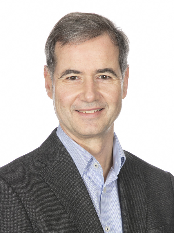 Arno Schmidt-Trucksäss合作论文数Departement für Sport, Bewegung und Gesundheit, Universität Basel;Departement Klinische Forschung, Universität Basel11
 Vincenzo Muto合作论文数GIGA Cyclotron Res Ctr In Vivo Imaging, Univ Liege11Krebs Julia合作论文数11




