Bacterial cell growth involves expansion of the peptidoglycan cell wall. Three mutually exclusive peptidoglycan synthesis mechanisms exist in bacteria: MreB-dependent dispersed growth in many rod-shaped bacteria, polar growth involving specific proteins in actinobacteria and rhizobiales, and septal growth involving FtsZ in many cocci. Here we used imaging, mass spectrometry analysis of peptidoglycan composition, bacterial genetics and colocalization analyses to show that the actinobacterium Streptomyces venezuelae uses both canonical polar peptidoglycan synthesis and MreB-dependent dispersed peptidoglycan synthesis during rapid, exploratory growth. Transmission electron microscopy and peptidoglycan analyses showed changes in cell wall structure and composition with exploratory growth. MreB1 was essential for cell wall integrity and culture viability during exploratory growth and also localized to side walls in regions of new growth. Our results show that MreB1 is required for dynamic cell wall changes over the course of a growth cycle, contributing to a wall that is structurally distinct from that of conventionally growing streptomycetes.
Escherichia coli is arguably the most thoroughly characterized organism, and yet ~20% of its essential genes serve completely unknown functions,1,2 and many core quantitative physiological principles remain unexplained, including the famous nutrient 'growth law' where cell volume seems to depend exponentially on growth rate. Here we develop a platform for massive, multi-generational Optical Pooled Screening (OPS)3-6, and apply it to link image-based phenotypes to genotypes for 133,000 CRISPRi knockdowns of essential genes, tracking tens of millions of lineages and analyzing 1.6 billion cells. Our multi-dimensional dynamic phenotypes correlate exceptionally well with known gene functions, allowing us to identify many unknown roles of essential genes. Quantifying the relation between growth and cell size in turn identifies three distinct variants of the bacterial growth laws, which we explain mechanistically by discovering a new role for (p)ppGpp and SpoT as a sensor of translation elongation. Finally, we propose and systematically test an exceedingly simple, passive mechanism for the nutrient growth law, based on triggering cell division through the accumulation of a protein that, unlike ribosomes, is not controlled by (p)ppGpp. The resulting hyperbolic growth law fits the data from our three variants even better than the previously proposed exponential relationship, and with fewer parameters.
For rod-shaped bacterial model organisms, the division plane is defined by the geometry of the cell. However, for Neisseria gonorrhoeae, a coccoid organism that most commonly exists as a diplococcus and that possesses genes coding for rod-based cell division systems, the relationship between cell geometry and division is unclear. Here, we characterized the organization of N. gonorrhoeae division using a combination of fluorescent probes, genetics, and time-lapse microscopy. We found that the planes of successive cell divisions are orthogonal and temporally overlapping, thereby maintaining diplococcal morphology. Division takes place perpendicular to a long axis in each coccus. In keeping with the ParABS and the MinCDE systems reading the more pronounced long axis of rod-shaped bacteria, in the coccoid N. gonorrhoeae, ParB segregates along this long axis and cells lacking minCDE suffer severe morphological consequences, including an inability to perform orthogonal division and aberrant assembly of the division plane at the cell poles. Taken together, this stresses the central role of even slight dimensional asymmetry as a general organizational principle in coccoid bacterial cell division.
The cell walls of rod-shaped Gram-positive bacteria are thick, multilayered networks that chirally twist as cells elongate. The underlying basis of twisting is not known, but probing the processes underlying this phenomenon may give insights into how cell wall material is inserted, how it evolves during cleavage, and the mechanics within the sacculus. In Bacillus subtilis, we see cell chains lacking hydrolases twist far slower than chains of wild-type cells, indicating that cell wall cleavage modulates the twisting rate. We see that when cells within chains separate, the two nascent ends rotate as they separate. Together, this suggests there is torsional stress within the cell wall that, when unreleased, perturbs overall chain morphology. Unlike Escherichia coli, we see that twisting does not arise from MreB's angle of motion, as its angle is identical in both fast-twisting wild-type cells and slow-twisting hydrolase-deficient cells. Rather, the circumferential insertion of glycans appears to establish this torsional stress, as increasing Rod complex activity by deleting ponA causes cells to twist faster than wild-type cells. Together, these experiments suggest the twisting of B. subtilis cells arises from radial glycan insertion, which somehow causes torsional stress in the wall that is later released by hydrolase activity.
Abstract The first step of cell division is deforming the planar cell membrane inward towards the cytoplasm. As deforming membranes is energetically costly, biology has developed various protein systems to accomplish this task. The mechanisms providing the force to deform bacterial membranes to initiate division remain unknown. In vivo studies have shown the condensation of FtsZ filaments into a sharp ring is required to initiate cell division, an observation mirrored in vitro with FtsZ filaments encapsulated inside liposomes. Similarly, the force for membrane deformation in many eukaryotic deforming systems arises from the local crowding of proteins on the membrane surface. As any membrane deforming system works against the membrane tension, here we modulated the amount of lipid synthesis and thus membrane tension in Bacillus subtilis to examine: 1) if the condensation of FtsZ filaments by FtsZ bundling proteins serves to overcome the cellular membrane tension to deform the membrane inward and 2) how changes to the membrane tension affect the subsequent invagination of the septum. First, we developed methods to simultaneously measure and modulate membrane tension in live cells. Next, we determined how altering the membrane tension affected the cell’s ability to initiate division with reduced levels of FtsZ bundling proteins. While cells depleted of 2 FtsZ bundling proteins were unable to divide, reducing membrane tension to a given threshold restored their ability to initiate division. Likewise, cells with intermediate levels of FtsZ bundling proteins required a lesser decrease in membrane tension to initiate division. We also found that reductions in membrane tension increase the rate of Z ring constriction, with the constriction rate scaling linearly with the membrane tension. Interestingly, while the constriction rate in wild-type B. subtilis is limited by FtsZ treadmilling, the rate of constriction becomes independent of FtsZ’s treadmilling rate when membrane tension is reduced. These experiments give two major insights: First, the filament condensation caused by FtsZ bundling proteins works to overcome membrane tension and deform the membrane inward to initiate division. Second, the rate of septal constriction is limited by membrane tension, suggesting that membrane fluctuations at the tip of the growing septa limit the rate of cell wall synthesis. Finally, our measurements allow the estimation of several physical values of cell division, such as the force required to bend the membrane, but also that the cell membrane provides only 0.1%, a small amount of surface tension relative to the entire cell envelope, indicating 99.9% of the pressure drop occurs across the cell wall. These calculations also indicate that cell division occurs via comparatively very small membrane tension fluctuations relative to the high turgor pressure that exists across the entire cell envelope.
To form a dormant spore, Bacillus subtilis and related endospore-forming bacteria divide asymmetrically to generate daughter cells of unequal size, the smaller of which becomes the spore. The transmembrane protein SpoIIE repositions the cell division machinery and controls cytokinesis during sporulation, but the molecular basis for the precise placement of the asymmetrical division site to the quarter cell point is unknown. Here, we applied live-cell fluorescence microscopy techniques to reveal that SpoIIE localizes with the treadmilling components of the cell division machinery. We found that SpoIIE opposes the inhibitory activity of the MinCD complex, which prevents assembly of Z-rings near the cell poles. Cells expressing a variant of SpoIIE with its transmembrane region replaced by an unrelated transmembrane anchor assembled condensed Z-rings that were unable to initiate constriction. This reveals a new function of SpoIIE and a possible checkpoint licensing cytokinesis downstream of Z-ring condensation. Potentially explaining the role of SpoIIE in cytokinesis, we demonstrated that SpoIIE's transmembrane region interacts with DivIB, an enigmatic structural component of the cell wall synthesis complex required for cytokinesis during sporulation. Finally, we found that FtsZ filaments are unusually short during sporulation, which requires the transmembrane domain of SpoIIE. Together, these results demonstrate that SpoIIE sequentially influences polar divisome assembly at distinct steps to drive asymmetric cell division.
The genus Mycobacterium includes species such as Mycobacterium tuberculosis, which can cause deadly human diseases. These bacteria have a protective cell envelope that can be remodeled to facilitate their survival in challenging conditions. Understanding how such conditions affect membrane remodeling can facilitate antibiotic discovery and treatment. To this end, we describe an optimized fluorogenic probe, N-QTF, that reports on mycolyltransferase activity, which is vital for cell division and remodeling. N-QTF is a glycolipid probe that can reveal dynamic changes in the mycobacterial cell envelope in both fast- and slow-growing mycobacterial species. Using this probe to monitor the consequences of antibiotic treatment uncovered distinct cellular phenotypes. Even antibiotics that do not directly inhibit cell envelope biosynthesis cause conspicuous phenotypes. For instance, mycobacteria exposed to the RNA polymerase inhibitor rifampicin release fluorescent extracellular vesicles (EVs). While all mycobacteria release EVs, fluorescent EVs were detected only in the presence of RIF, indicating that exposure to the drug alters EV content. Macrophages exposed to the EVs derived from RIF-treated cells released lower levels of cytokines, suggesting the EVs moderate immune responses. These data suggest that antibiotics can alter EV content to impact immunity. Our ability to see such changes in EV constituents directly results from exploiting these chemical probes.
Eukaryotic cells have been evolving for billions of years, giving rise to wildly diverse cell forms and functions. Despite their variability, all eukaryotic cells share key hallmarks, including membrane-bound organelles, heavily regulated cytoskeletal networks and complex signaling cascades. Because the actin cytoskeleton interfaces with each of these features, understanding how it evolved and diversified across eukaryotic phyla is essential to understanding the evolution and diversification of eukaryotic cells themselves. Here, we discuss what we know about the origin and diversity of actin networks in terms of their compositions, structures and regulation, and how actin evolution contributes to the diversity of eukaryotic form and function.
Bacterial cell division requires recruitment of peptidoglycan (PG) synthases to the division site by the tubulin homologue, FtsZ. Septal PG synthases promote septum growth. FtsZ treadmilling is proposed to drive the processive movement of septal PG synthases and septal constriction in some bacteria; however, the precise mechanisms spatio-temporally regulating PG synthase movement and activity and FtsZ treadmilling are poorly understood. Here using single-molecule imaging of division proteins in the Gram-positive pathogen Staphylococcus aureus , we showed that the septal PG synthase complex FtsW/PBP1 and its putative activator protein, DivIB, move with similar velocity around the division site. Impairing FtsZ treadmilling did not affect FtsW or DivIB velocities or septum constriction rates. Contrarily, PG synthesis inhibition decelerated or stopped directional movement of FtsW and DivIB, and septum constriction. Our findings suggest that a single population of processively moving FtsW/PBP1 associated with DivIB drives cell constriction independently of FtsZ treadmilling in S. aureus .
The cell wall is a complex structure. For most bacteria, peptidoglycan is an essential component of their cell wall, with different bacteria having evolved distinct biosynthetic strategies. The mechanisms driving bacterial growth can be divided into three, mutually-exclusive categories: (i) dispersed growth, mediated by MreB and employed by many rod-shaped bacteria; (ii) polar growth, driven by distinct proteins in the actinobacteria and rhizobiales; and (iii) septal growth, fueled by FtsZ in many coccoid bacteria. Here, we show that under conditions of rapid growth, the actinobacterial representative Streptomyces venezuelae transcends these categories, simultaneously employing both canonical polar growth, and MreB-mediated dispersed growth. Our results indicate that MreB is essential for cell wall integrity and culture viability under these growth conditions, promotes dynamic cell wall changes over the course of a growth cycle, and contributes to a wall that is structurally distinct from that of conventionally growing streptomycetes. ### Competing Interest Statement The authors have declared no competing interest.
ABSTRACT Across the domains of life, actin homologs are integral components of many essential processes, such as DNA segregation, cell division, and cell shape determination. Archaeal genomes, like those of bacteria and eukaryotes, also encode actin homologs, but much less is known about these proteins’ in vivo dynamics and cellular functions. We identified and characterized the function and dynamics of Salactin, an actin homolog in the hypersaline archaeon Halobacterium salinarum . Live-cell time-lapse imaging revealed that Salactin forms dynamically unstable filaments that grow and shrink out of the cell poles. Like other dynamically unstable polymers, Salactin monomers are added at the growing filament end, and its ATP-bound critical concentration is substantially lower than the ADP-bound form. When H. salinarum’s chromosomal copy number becomes limiting under low-phosphate growth conditions, cells lacking Salactin show perturbed DNA distributions. Taken together, we propose that Salactin is part of a previously unknown chromosomal segregation apparatus required during low-ploidy conditions. IMPORTANCE Protein filaments play important roles in many biological processes. We discovered an actin homolog in halophilic archaea, which we call Salactin. Just like the filaments that segregate DNA in eukaryotes, Salactin grows out of the cell poles towards the middle, and then quickly depolymerizes, a behavior known as dynamic instability. Furthermore, we see that Salactin affects the distribution of DNA in daughter cells when cells are grown in low-phosphate media, suggesting Salactin filaments might be involved in segregating DNA when the cell has only a few copies of the chromosome.
Gram-negative bacteria are surrounded by two membranes. A special feature of the outer membrane is its asymmetry. It contains lipopolysaccharide (LPS) in the outer leaflet and phospholipids in the inner leaflet 1 – 3 . The proper assembly of LPS in the outer membrane is required for cell viability and provides Gram-negative bacteria intrinsic resistance to many classes of antibiotics. LPS biosynthesis is completed in the inner membrane, so the LPS must be extracted, moved across the aqueous periplasm that separates the two membranes and translocated through the outer membrane where it assembles on the cell surface 4 . LPS transport and assembly requires seven conserved and essential LPS transport components 5 (LptA–G). This system has been proposed to form a continuous protein bridge that provides a path for LPS to reach the cell surface 6 , 7 , but this model has not been validated in living cells. Here, using single-molecule tracking, we show that Lpt protein dynamics are consistent with the bridge model. Half of the inner membrane Lpt proteins exist in a bridge state, and bridges persist for 5–10 s, showing that their organization is highly dynamic. LPS facilitates Lpt bridge formation, suggesting a mechanism by which the production of LPS can be directly coupled to its transport. Finally, the bridge decay kinetics suggest that there may be two different types of bridges, whose stability differs according to the presence (long-lived) or absence (short-lived) of LPS. Together, our data support a model in which LPS is both a substrate and a structural component of dynamic Lpt bridges that promote outer membrane assembly.
ABSTRACT Most bacteria are surrounded by their cell wall, containing a highly cross-linked protective envelope of peptidoglycan. To grow, bacteria must continuously remodel their wall, inserting new material and breaking old bonds. Bond cleavage is performed by cell wall hydrolases, allowing the wall to expand. Understanding the functions of individual hydrolases has been impeded by their redundancy: single knockouts usually present no phenotype. We used an exhaustive multiple-knockout approach to determine the minimal set of hydrolases required for growth in Bacillus subtilis. We identified 42 candidate hydrolases. Strikingly, we were able to remove all but two of these genes in a single strain; this “∆40” strain shows only a mild reduction in growth rate, indicating that none of the 40 hydrolases are necessary for growth. The ∆40 strain does not detectably shed old wall, suggesting that turnover is not essential for growth. The remaining hydrolases in the ∆40 strain are LytE and CwlO, previously shown to be synthetically lethal. Either can be removed in ∆40, indicating that either hydrolase alone is sufficient for cell growth. Screening of environmental conditions and biochemistry revealed that LytE activity is inhibited by Mg2+ and that RlpA-like proteins may stimulate LytE activity. Together, these results suggest that the only essential function of cell wall hydrolases in B. subtilis is to enable cell growth by expanding the wall and that LytE or CwlO alone are sufficient for this function. These experiments introduce the ∆40 strain as a tool to study hydrolase activity and regulation in B. subtilis. IMPORTANCE In order to grow, bacterial cells must both create and break down their cell wall. The enzymes that are responsible for these processes are the target of some of our best antibiotics. Our understanding of the proteins that break down the wall— cell wall hydrolases—has been limited by redundancy among the large number of hydrolases many bacteria contain. To solve this problem, we identified 42 cell wall hydrolases in Bacillus subtilis and created a strain lacking 40 of them. We show that cells can survive using only a single cell wall hydrolase; this means that to understand the growth of B. subtilis in standard laboratory conditions, it is only necessary to study a very limited number of proteins, simplifying the problem substantially. We additionally show that the ∆40 strain is a research tool to characterize hydrolases, using it to identify three “helper” hydrolases that act in certain stress conditions.
Macroautophagy/autophagy proteins have been linked with the development of immune-mediated diseases including lupus, but the mechanisms for this are unclear due to the complex roles of these proteins in multiple immune cell types. We have previously shown that a form of noncanonical autophagy induced by ITGAV/alpha(v) integrins regulates B cell activation by viral and self-antigens, in mice. Here, we investigate the involvement of this pathway in B cells from human tissues. Our data reveal that autophagy is specifically induced in the germinal center and memory B cell subpopulations of human tonsils and spleens. Transcriptomic analysis show that the induction of autophagy is related to unique aspects of activated B cells such as mitochondrial metabolism. To understand the function of ITGAV/alpha(v) integrin-dependent autophagy in human B cells, we used CRISPR-mediated knockdown of autophagy genes. Integrating data from primary B cells and knockout cells, we found that ITGAV/alpha(v)-dependent autophagy limits activation of specific pathways related to B cell responses, while promoting others. These data provide new mechanistic links for autophagy and B-cell-mediated immune dysregulation in diseases such as lupus.
Mycobacteriophages are a diverse group of viruses infecting Mycobacterium with substantial therapeutic potential. However, as this potential becomes realized, the molecular details of phage infection and mechanisms of resistance remain ill-defined. Here we use live-cell fluorescence microscopy to visualize the spatiotemporal dynamics of mycobacteriophage infection in single cells and populations, showing that infection is dependent on the host nucleoid-associated Lsr2 protein. Mycobacteriophages preferentially adsorb at Mycobacterium smegmatis sites of new cell wall synthesis and following DNA injection, Lsr2 reorganizes away from host replication foci to establish zones of phage DNA replication (ZOPR). Cells lacking Lsr2 proceed through to cell lysis when infected but fail to generate consecutive phage bursts that trigger epidemic spread of phage particles to neighbouring cells. Many mycobacteriophages code for their own Lsr2-related proteins, and although their roles are unknown, they do not rescue the loss of host Lsr2.
The discovery of new genes regulating essential biological processes has become increasingly important, and CRISPRi has emerged as a powerful tool for achieving this goal. This method has been used in many model organisms to decrease the expression of specific genes and assess their impact on phenotype. Pooled CRISPRi libraries in bacteria have been particularly useful in discovering new regulators of growth, division, and other biological processes. However, these libraries rely on the induction of dCas9 via an inducible promoter, which can be problematic due to promoter leakiness. This is a widespread phenomenon of any inducible promoter that can result in the unwanted downregulation of genes and the emergence of genetic suppressors when essential genes are knocked down. To overcome this issue, we have developed a novel strategy that eliminates dCas9 leakiness and enables reversible knockdown control using the rapamycin-dependent degron system in Bacillus subtilis . This degron system causes rapid degradation of dCas9, resulting in an almost instant reset of the system. Our results demonstrate that it is possible to achieve zero CRISPRi activity in the uninduced state and full activity in the induced state. This improved CRISPRi system will enable researchers to investigate phenotypic changes more effectively while reducing the undesirable effects of leaky expression and noise in their phenotypic data. Moreover, a rapid degradation system could serve as a tool for dynamic perturbation before compensation mechanisms or stress responses kick in. Finally, this approach can be adapted to other organisms and other promoter-inducible systems, potentially opening up strategies for tighter control of gene expression.
Abstract Bacterial cell division is mediated by the tubulin-homolog FtsZ, which recruits peptidoglycan (PG) synthesis enzymes to the division site. Septal PG synthases promote inward growth of the division septum, but the mechanisms governing the spatiotemporal regulation of these enzymes are poorly understood. Recent studies on various organisms have proposed different models for the relationship between the movement and activity of septum-specific PG synthases and FtsZ treadmilling. Here, we studied the movement dynamics of conserved cell division proteins relative to the rates of septum constriction and FtsZ treadmilling in the Gram-positive pathogenStaphylococcus aureus. The septal PG synthesis enzyme complex FtsW/PBP1 and its putative activator protein, DivIB, moved processively, around the division site, with the same velocity. Impairing FtsZ treadmilling did not affect FtsW and DivIB velocities or septum constriction rates. Contrarily, inhibition of PG synthesis slowed down or completely stopped both septum constriction and the directional movement of FtsW/PBP1 and DivIB. Our findings support a model forS. aureusin which a single population of processively moving FtsW/PBP1 remains associated with DivIB to drive cell constriction independently of treadmilling FtsZ filaments.
How bacteria link their growth rate to external nutrient conditions is unknown. To investigate how Bacillus subtilis cells alter the rate at which they expand their cell walls as they grow, we compared single-cell growth rates of cells grown under agar pads with the density of moving MreB filaments under a variety of growth conditions. MreB filament density increases proportionally with growth rate. We show that both MreB filament density and growth rate depend on the abundance of Lipid II and murAA , the first gene in the biosynthetic pathway creating the cell wall precursor Lipid II. Lipid II is sensed by the serine/threonine kinase PrkC, which phosphorylates RodZ and other proteins. We show that phosphorylated RodZ increases MreB filament density, which in turn increases cell growth rate. We also show that increasing the activity of this pathway in nutrient-poor media results in cells that elongate faster than wild-type cells, which means that B. subtilis contains spare ‘growth capacity’. We conclude that PrkC functions as a cellular rheostat, enabling fine-tuning of cell growth rates in response to Lipid II in different nutrient conditions.
All cells must increase their volumes in response to biomass growth to maintain intracellular mass density within physiologically permissive bounds. Here, we investigate the regulation of volume growth in the Gram-positive bacterium Bacillus subtilis. To increase volume, bacteria enzymatically expand their cell envelopes and insert new envelope material. First, we demonstrate that cell-volume growth is determined indirectly, by expanding their envelopes in proportion to mass growth, similarly to the Gram-negative Escherichia coli, despite their fundamentally different envelope structures. Next, we studied, which pathways might be responsible for robust surface-to-mass coupling: We found that both peptidoglycan synthesis and membrane synthesis are required for proper surface-to-mass coupling. However, surprisingly, neither pathway is solely rate-limiting, contrary to wide-spread belief, since envelope growth continues at a reduced rate upon complete inhibition of either process. To arrest cell-envelope growth completely, the simultaneous inhibition of both envelope-synthesis processes is required. Thus, we suggest that multiple envelope-synthesis pathways collectively confer an important aspect of volume regulation, the coordination between surface growth, and biomass growth.
The current understanding of mycobacterial cell envelope remodeling in response to antibiotics is limited. Chemical tools that report on phenotypic changes with minimal cell wall perturbation are critical to understanding such time-dependent processes. We employed a fluorogenic chemical probe to image how antibiotics perturb mycobacterial cell envelope assembly in real-time. Time-lapse microscopy revealed that differential antibiotic treatment elicited unique cellular phenotypes, providing a platform for simultaneously monitoring cell envelope construction and remodeling responses. Our data show that rifampicin, which does not directly inhibit cell wall biosynthesis, affords a readily detected mycomembrane phenotype. The fluorogenic probe revealed the production of extracellular vesicles in response to antibiotics, and analyses of these vesicles indicate that antibiotic treatment elicits the release of agents that attenuate macrophage activation.
 A. Keith Dunker合作论文数School of Medicine, Indiana University16
 Georgia R Squyres合作论文数Harvard Univ, Dept Mol & Cellular Biol, Cambridge, MA 02138 USA14
 Sun Yingjie合作论文数13
 Zoran Obradovic合作论文数Center for Data Analytics and Biomedical Informatics, College of Science and Technology, Temple University;Statistics, Operations and Data Science Department, Fox School of Business, Temple University11
 Alexandre W. Bisson-Filho合作论文数IQ, Universidade de São Paulo;IQ, Universidade de São Paulo10
 Celeste J. Brown合作论文数Department of Biological Sciences, University of Idaho10Wivagg Carl N合作论文数Department of Microbiology and Immunobiology, Harvard Medical School9P Romero合作论文数9
 Sean M Wilson合作论文数Department of Biological Sciences and Center for Reproductive Biology, University of Idaho8
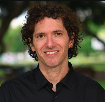 Ariel Amir合作论文数Department of Physics of Complex Systems, The Weizmann Institute of Science8
 Norman M. Ratcliffe合作论文数Faculty of Applied Sciences, University of the West of England7Matthew J Holmes合作论文数6
 Jenny Zheng合作论文数Dept Mol & Cellular Biol, Harvard Univ6
 Suzanne Walker合作论文数Department of Chemistry and Chemical Biology, Faculty of Arts and Sciences, Harvard University;Department of Microbiology, Harvard Medical School, Harvard University6
 Saman Hussain合作论文数From the Molecular and Cellular Biology Department and Faculty of Arts and Sciences (FAS) Center for Systems Biology, Harvard University6
 Benjamin Paul De Lacy Costello合作论文数Institute of Bio-Sensing Technology, University of the West of England6
 Hsu Yen-Pang合作论文数6
 Chris Probert合作论文数Institute of Systems, Molecular & Integrative Biology, Faculty of Health and Life Sciences, University of Liverpool;Institute of Translational Medicine, University of Liverpool6R. Dyche Mullins合作论文数6
 Amy K. Schmid合作论文数Department of Biology, Trinity College of Arts & Sciences, Duke University5
 Charles Lefco Dulberger合作论文数Department of Biochemistry and Molecular Biology,5
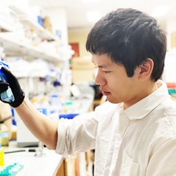 Wong Felix合作论文数Harvard College5
 Kuru Erkin合作论文数Dept Chem, Indiana Univ5Stoddard Patrick R合作论文数5
 Andrew Murray合作论文数Department of Molecular and Cellular Biology, Harvard University;Rowland Institute, Harvard University;Howard Hughes Medical Institute5
 Thomas G. Bernhardt合作论文数Department of Microbiology and Molecular Genetics, Harvard Medical School5
 Eric Rubin合作论文数Department of Immunology and Infectious Diseases, T.H. Chan School of Public Health, Harvard University;Brigham and Women's Hospital;Harvard Medical School5
 Thierry Izore合作论文数Monash Biomed Discovery Inst, Monash Univ4
 Szwedziak Piotr合作论文数Structural Studies Division, MRC Laboratory of Molecular Biology, Cambridge, United Kingdom.4Kapoor Mrinal合作论文数From the Molecular and Cellular Biology Department and Faculty of Arts and Sciences (FAS) Center for Systems Biology, Harvard University4
 Tom Williams合作论文数University of Bristol4
 Yves Brun合作论文数Department of Biology, The College of Arts & Sciences, Indiana University Bloomington;Brun Lab, Département de Microbiologie, Infectiologie et Immunologie, Université de Montréal4Ye-Jin Eun合作论文数Department of Biochemistry, University of Wisconsin-Madison4
 Lars Renner合作论文数Division Polymer Biomaterials Science, Leibniz Institute of Polymer Research Dresden4Albina Orlova合作论文数4Vitold E. Galkin合作论文数4S Smith合作论文数3Edward Hall合作论文数3RW Bailey合作论文数3Charles R. Kissinger合作论文数3
 Sarah R. Barger合作论文数3
 Quincey Justman合作论文数3Rm Williams合作论文数3Kaitlin Schaefer合作论文数3
 Brennan A. Murphy合作论文数Dept Chem, Indiana Univ3Sean Wilson合作论文数Dept Mol & Cellular Biol, Harvard Univ3Junhao Zhu合作论文数3Betheney R Pennycook合作论文数Marine Biol Lab, Physiol Course3X Li合作论文数3




