Upper aerodigestive tract squamous cell carcinoma (UATSCC) is often easy to recognize in its conventional form. However, a subset of tumors loses histologic evidence of squamous differentiation and, instead, become sarcomatoid (spindled). These cases may be difficult or impossible to appropriately classify on biopsy material, even with immunohistochemistry. DNA methylation has emerged as a tool to help in classifying undifferentiated malignancies. We sought to explore whether conventional and sarcomatoid UATSCC harbor the same epigenetic signature. Cases of sarcomatoid UATSCC were collected and reviewed. The conventional and sarcomatoid components were documented. Only specimens with concomitant epithelial dysplasia and/or invasive SCC were included. A panel of keratins (CK AE1/3, CAM5.2 and CK5/6) was performed on the sarcomatoid component of each tumor and scored as follows: 0, < 1
Background Biphenotypic sinonasal sarcoma (BSS) is a low-grade, locally aggressive sarcoma unique to the sinonasal region. BSS is most common in middle aged patients and affects women more frequently than men. It is characterized by a bland spindled cell proliferation with neural and myogenic differentiation. BSS are usually associated with rearrangement t(2;4)(q35;q31.1) resulting in a PAX3::MAML3 fusion. Less commonly, other genes are found in combination with PAX3 and some cases reported in the literature have an unknown fusion partner. Methods A 54-year-old man presented with nasal mass. Endoscopic resection showed a low-grade spindle cell neoplasm with morphologic features of BSS and immunohistochemical and next generation sequencing were performed to confirm the diagnosis. Results The tumor was positive for S100 and smooth muscle actin but negative for SOX10. Next generation sequencing demonstrated a novel PAX3::FOXO6 gene fusion. Conclusions Although a PAX3::FOXO6 gene fusion has never been reported, this finding combined with the morphologic and immunophenotypic features supports the diagnosis of supports the diagnosis of BSS.
Bland thyroid follicles are occasionally identified within cervical lymph nodes, raising the differential diagnosis of benign thyroid inclusions versus metastatic papillary thyroid carcinoma. Proposed criteria for benign thyroid inclusions include lack of cytologic atypia, subcapsular or central nodal location, and involvement of only one or two lymph nodes. We observed several cases of fibrosing variant Hashimoto thyroiditis with multifocal thyroid tissue in cervical lymph nodes despite the absence of identifiable primary carcinoma. The pathology archives from 2014 to 2024 were searched for cases of fibrosing variant Hashimoto thyroiditis. Cases were reviewed for occult papillary thyroid carcinoma and thyroid tissue within sampled lymph nodes. IgG4 staining results were recorded. HBME-1, BRAF V600E, and NRAS Q61R immunostains were performed on nodal thyroid tissue. Thirty-six patients were identified (28 females, 8 males; median age 54 years, range 26–83). Increased IgG4-positive plasma cells were present in 29 cases. Lymph nodes were sampled in 23 patients, and thyroid tissue was identified in 8. No primary papillary thyroid carcinoma was identified in 6 of these 8 patients, including 4 in whom the entire thyroid gland was submitted for microscopic examination. HBME-1, BRAF V600E, and NRAS Q61R were negative in all nodal thyroid tissue. Our findings suggest that multifocal thyroid tissue within cervical lymph nodes is relatively common in fibrosing variant Hashimoto thyroiditis. The absence of primary carcinoma in most cases, together with bland cytology and negative immunohistochemical findings, supports the possibility that these represent benign thyroid inclusions that fall outside traditionally accepted diagnostic criteria.
The current review article deals with the evaluation of the oncocytic/oncocytoid lesions in the salivary gland. The authors will focus on the diagnosis of Warthin tumor (WT) as a launching point to detail important morphologic findings that should prompt designation of an aspirate as oncocytic salivary gland neoplasm of uncertain malignant potential or other Milan categories. Oncocytic cells are defined as cells with a moderate to abundant amount of eosinophilic finely granular cytoplasm, round-to-oval nuclei, and large-distinct nucleoli. In contrast, the term oncocytoid is also frequently used in this discussion and indicates tumor cells with similarly abundant and sometimes granular cytoplasm but lacking all the definitive features of a true oncocyte. Several helpful tips are provided in hopes of improving an accurate diagnosis of WT on an aspirate sample. Using these types allows for consideration of important differential diagnoses, including both benign and malignant entities, when faced with an oncocytic salivary gland neoplasm. The morphological criteria as well as the possible application of ancillary techniques are also discussed.
BACKGROUND:Differentiated thyroid cancer is typically caused by a single oncogenic driver alteration. In PTEN hamartoma tumor syndrome, a pathogenic germline PTEN alteration results in a predisposition to thyroid cancer and adenomatous thyroid nodules. This provides a unique model to study thyroid carcinogenesis in the setting of a baseline "hit" to the PI3K/AKT/mTOR pathway, implicated in RAS-like thyroid tumors. METHODS:RNA sequencing and differential expression analysis were performed on thyroid cancers, adenomatous nodules, and background thyroid tissue in patients with PTEN hamartoma tumor syndrome. BRAFV600E-RAS scores were calculated and compared across histologic subtypes. RNA sequencing data were then integrated with previously published paired exome sequencing data. RESULTS:RNA sequencing was performed on 26 cancers, 27 adenomatous nodules, and 15 background thyroid samples from 20 patients with PTEN hamartoma tumor syndrome. This demonstrated 3 expression clusters: papillary architecture tumors, follicular architecture tumors, and background thyroid tissue. The majority (17, 65.4%) of cancers were RAS-like. Follicular architecture cancers were primarily RAS-like (BRAFV600E-RAS score +0.67) and frequently associated with biallelic PTEN alterations. Papillary architecture cancers were BRAF-like (BRAFV600E-RAS score -0.77), often in absence of biallelic PTEN alterations. Adenomatous nodules had indistinguishable expression profiles from follicular architecture cancers. CONCLUSIONS:PTEN hamartoma tumor syndrome-associated thyroid tumors most frequently have RAS-like expression profiles. This appears to be caused by baseline dysregulation of the PI3K/AKT/mTOR pathway, with predisposition to biallelic PTEN inactivation promoting follicular adenomatous growth and subsequent malignant transformation to follicular architecture cancers. Better understanding malignant potential and tumor progression in PTEN hamartoma tumor syndrome thyroid tissue is essential for optimizing diagnosis, enhanced surveillance, and treatment in this population.
Diagnosis and classification of oral epithelial dysplasia (OED) is critical to identifying and prognosticating patients at risk of squamous cell carcinoma (SCC). However, conventional 3-tiered and 2-tiered grading systems suffer from poor inter-pathologist agreement, and SCC may arise from all grades of OED. This study evaluated pathologist agreement in OED classification as p53 wildtype, p53 abnormal, and HPV-associated based on recent evidence demonstrating the utility of p53/p16 immunohistochemistry (IHC) in this setting and increased risk of p53 abnormal OED progression to SCC, regardless of histologic grade. Fifty digital biopsy specimens were evaluated for diagnosis by 18 subspecialty-trained pathologists, with OED graded utilizing 3-tiered, 2-tiered, and p53 wildtype/p53 abnormal/HPV-associated schemata. Cases were reviewed first without and subsequently with p53/p16 IHC. The cohort consisted of 8 cases of p53 wildtype, 24 cases of p53 abnormal, and 18 cases of HPV-associated OED. Inter-pathologist agreement in OED grading according to 3-tiered (κ=0.32) and 2-tiered (κ=0.39) systems by H&E was poor, but fair-to-good (κ=0.59) in classification as p53 wildtype/p53 abnormal/HPV-associated by H&E and IHC. Classification of OED as p53 wildtype, p53 abnormal, or HPV-associated using p53/p16 IHC outperformed conventional grading in this cohort enriched for p53 abnormal OED, which required correct interpretation of p53 IHC, historically deemed challenging. Routine use of IHC also identifies a wider histologic spectrum of HPV-associated OED than is currently appreciated. More work is needed to determine the efficacy of this classification system in predicting patient outcomes and in guiding management decisions in real-world cohorts.
Diagnosis and classification of oral epithelial dysplasia (OED) is critical to identifying and prognosticating patients at risk of squamous cell carcinoma (SCC). However, conventional 3-tiered and 2-tiered grading systems suffer from poor inter-pathologist agreement, and SCC may arise from all grades of OED. This study evaluated pathologist agreement in OED classification as p53 wildtype, p53 abnormal, and HPV-associated based on recent evidence demonstrating the utility of p53/p16 immunohistochemistry (IHC) in this setting and increased risk of p53 abnormal OED progression to SCC, regardless of histologic grade. Fifty digital biopsy specimens were evaluated for diagnosis by 18 subspecialty-trained pathologists, with OED graded utilizing 3-tiered, 2-tiered, and p53 wildtype/p53 abnormal/HPV-associated schemata. Cases were reviewed first without and subsequently with p53/p16 IHC. The cohort consisted of 8 cases of p53 wildtype, 24 cases of p53 abnormal, and 18 cases of HPV-associated OED. Inter-pathologist agreement in OED grading according to 3-tiered (κ=0.32) and 2-tiered (κ=0.39) systems by H&E was poor, but fair-to-good (κ=0.59) in classification as p53 wildtype/p53 abnormal/HPV-associated by H&E and IHC. Classification of OED as p53 wildtype, p53 abnormal, or HPV-associated using p53/p16 IHC outperformed conventional grading in this cohort enriched for p53 abnormal OED, which required correct interpretation of p53 IHC, historically deemed challenging. Routine use of IHC also identifies a wider histologic spectrum of HPV-associated OED than is currently appreciated. More work is needed to determine the efficacy of this classification system in predicting patient outcomes and in guiding management decisions in real-world cohorts.
Objective Near-infrared autofluorescence (NIRAF) characteristics of parathyroid glands in primary hyperparathyroidism (pHPT) vary, with unclarity regarding the underlying mechanism. Similarly, Tc-99m-sestamibi uptake in diseased parathyroid glands is variable. There is a suggestion that oxyphilic cell content may influence both imaging modalities. This study aims to analyze the relationship between NIRAF imaging characteristics, Tc-99m-sestamibi uptake, and cellular composition in pHPT. Study Design Retrospective analysis of an Institutional Review Board-monitored prospective database. Setting Single tertiary referral center. Methods NIRAF characteristics of parathyroid glands of patients with pHPT between 2019 and 2024 were compared with Tc-99m-sestamibi scan findings from a prospective database. Using third-party software, brightness intensity and heterogeneity index (HI) of the glands were calculated. A subgroup of parathyroid glands obtained from consecutive patients with pHPT in 2020 to 2021 underwent histological analysis. Results A total of 428 patients with 638 diseased parathyroid glands were analyzed. Forty-seven percent of the glands showed an uptake on Tc-99m-sestamibi scans. The brightness intensity of the NIRAF signals from parathyroid glands that were seen versus not seen on sestamibi was 2.1 versus 2.3 (P = .002) and HI 0.18 versus 0.17 (P = .35), respectively. On multivariate analysis, low autofluorescence intensity, high gland volume, and single adenoma were associated with detectability on Tc-99m-sestamibi scan (P < .0001). Intraglandular adipose tissue content was lower in diseased glands that were detected on Tc-99m-sestamibi scans (0% vs 5%, P < .0001). Conclusion Our findings indicate an inverse relationship between autofluorescence intensity and detectability on Tc-99m-sestamibi scans and a lack of correlation between different cell types and autofluorescence properties.
BACKGROUND:Evaluate whether extranodal extension (ENE) extent impacts outcomes in patients with oral cavity squamous cell carcinoma (OCSCC). METHODS:From an institutional database, patients with OCSCC and pathologic ENE who received adjuvant treatment were included. Surgical slides were reviewed to confirm ENE extent. Multivariable Cox regression was used to relate patient/treatment characteristics with disease-free survival (DFS) and overall survival (OS). ENE was analyzed as both a dichotomous and continuous variable. RESULTS:A total of 113 patients were identified. Between major (>2 mm) versus minor ENE (≤2 mm), there was no significant difference in DFS (HR 1.18, 95%CI 0.72-1.92, p = 0.51) or OS (HR 1.17, 95%CI 0.70-1.96, p = 0.55). There was no significant association between ENE as a continuous variable and DFS (HR 0.97 per mm, 95%CI 0.87-1.4, p = 0.96) or OS (HR 0.96 per mm, 95%CI 0.83-1.11, p = 0.58). CONCLUSION:No significant relationship was seen between ENE extent and DFS or OS in individuals with OCSCC.
AIMS:The phosphatase and tensin homologue (PTEN) hamartoma tumour syndrome (PHTS) is a genetic disorder with variable clinical presentation and increased lifetime risk of multiorgan malignancies. The thyroid gland is commonly affected with follicular nodular disease (FND) and follicular cell-derived carcinomas. Histopathological and immunohistochemical assessment of thyroid disease in PHTS is essential to identify patients at-risk. METHODS AND RESULTS:In all, 30 PHTS patients with available thyroidectomy specimen material (2000-2023) and 31 control patients with FND and "adenomatous nodules" were retrieved. Histologic criteria, including the frequency of adenomatous-type nodules versus hyperplastic-type nodules, background and nodular lipomatous metaplasia, chronic lymphocytic thyroiditis, cytoplasmic clearing of follicular cells in nodules, nodule-in-nodule appearance, and spectrum of nuclear atypia between nodules were evaluated in both cohorts and a Thyroid Histomorphologic PHTS Score (THiPS) system was established with a cutoff of 4 points or higher being considered concerning for PHTS. In all, 27 PHTS (90%) and five control (16.1%) cases had THiPS ≥4. A PTEN immunohistochemical stain was evaluated in 25 cases of each cohort and showed nuclear and cytoplasmic loss of expression in all or most of the nodules of 24/25 PHTS cases. In 3/25 control cases, two with THiPS ≥4, had loss of expression in one to multiple nodules. Conventional papillary thyroid carcinomas in PHTS patients retained PTEN cytoplasmic expression. CONCLUSIONS:Our study supports that, although not specific, the finding of multiple histologic features is found more frequently in patients with PHTS compared to the non-PHTS control group. The THiPS system has high sensitivity for thyroid specimens from patients with PHTS.
Palisading adenocarcinoma is a morphologically distinct salivary gland neoplasm that has been recently described with predilection to the sublingual gland. We report our experience with this neoplasm to corroborate and enrich the literature and further clarify its phenotype.
Patients with PTEN hamartoma tumor syndrome (PHTS), a molecular diagnosis for those carrying germline PTEN pathogenic variants, have a high prevalence of benign and malignant thyroid disease. Characterizing the genomic landscape in PHTS thyroid tumors could provide insights into malignant potential and tumor progression to help optimize diagnosis, surveillance, and treatment in this population. To reveal the somatic alterations in PHTS-associated thyroid tumors, we conducted exome sequencing on 58 thyroid tumors (28 cancers, 30 benign nodules) from 19 patients with PHTS. A control cohort of 447 sporadic papillary thyroid cancers (PTC) from The Cancer Genome Atlas was used for comparison. PHTS-associated thyroid tumors had a unique genomic landscape in the setting of a pathogenic germline PTEN mutation, when compared with the general population. PHTS-associated thyroid tumors demonstrated a high frequency of second-hit somatic PTEN alterations, including variants and loss-of-heterozygosity events. Second-hit somatic PTEN alterations were more prevalent in PHTS-associated PTC than sporadic PTC (65.2% vs. 0.067%), occurring frequently in PHTS-associated follicular thyroid cancer (100%) and benign follicular nodules (90%). PHTS-associated PTC additionally harbored somatic alterations in BRAF, RAS family members, and genes associated with DNA double-stranded break repair, as well as somatic arm-level copy-number variations. Together, these findings suggest that biallelic PTEN alterations may function as foundational mutations in PHTS thyroid tissue, promoting benign growth and increasing potential for malignant transformation through impaired DNA double-stranded break repair and increased genomic instability. The unique genomic landscape of PHTS-associated thyroid tumors carries implications for molecular-targeted therapies for patients. Significance: Exome sequencing reveals the distinct mutational landscape of PTEN hamartoma tumor syndrome-associated thyroid cancers from sporadic counterparts, providing insights into tumor progression and behavior that could help improve diagnosis, surveillance, and treatment.
AIMS:The Milan system for reporting salivary gland cytopathology was developed by an international group of experts and first published in 2018 with the goal to standardise reporting of salivary gland aspirates. Seven categories with distinct risks of malignancy were proposed. Core needle biopsies (CNB) of salivary glands are also common, but reporting lacks standardisation. Here we explore the feasibility of a Milan-like reporting system on CNB of the parotid gland. METHODS AND RESULTS:Our laboratory information system was searched for parotid gland CNBs from 2010 to 2021. Reports were translated into a Milan-like reporting system. When available, CNB findings were correlated with cytology and resection specimens. In order to compare the performance of CNB with fine-needle aspirations (FNA), we established a second cohort of cases consisting of parotid FNA with surgical follow-up. The risk of neoplasia (RON) and risk of malignancy (ROM) was calculated for FNA and CNB Milan categories using cases with follow-up resection. We analysed 100 cases of parotid gland CNB. Of these cases, 32 underwent subsequent resection, while 52 had concurrent FNA. A total of 20 cases had concurrent FNA and underwent follow-up resection. In 63 (63%) cases, a specific diagnosis was provided on CNB, with 18 cases undergoing follow-up resection having an accuracy rate of 94%. CONCLUSIONS:This study confirms the feasible of using a Milan-like system in the setting of parotid gland CNB with differentiation in RON and ROM. CNB allows assessment of architectural features that may allow more specific diagnoses in some cases.
BACKGROUND:Molecular triage of indeterminate thyroid aspirates offers the opportunity to stratify the risk of malignancy (ROM) more accurately. Here we examine our experience with ThyroSeq v3 testing.METHODS:We analyzed 276 of 658 (42%) fine needle aspiration samples classified as indeterminate thyroid nodules using ThyroSeq v3 (Sept 2017-Dec 2019). The test provides a ROM and detects specific mutations. Surgical diagnoses were reviewed.RESULTS:Of 276 ThyroSeq-tested cases, 42% (n = 116) harbored genetic alterations, whereas 64% (n = 74) had surgical follow-up. Notably, 79% cases within intermediate to higher risk mutations were highly associated with surgical intervention, resulting in a 77.5% ROM when including both cancer and noninvasive follicular thyroid neoplasia with papillary-like features (cancer+NIFTP) and 68% malignant diagnosis when excluding NIFTP. RAS-like alterations were most common (66%), exhibiting a 73.4% ROM and a 59% malignant diagnosis. Interestingly, this group included 24 encapsulated follicular variant papillary thyroid carcinomas (EFVPTCs), 1 infiltrative FVPTC, 9 follicular carcinomas, and 7 NIFTP. Additionally, three high-risk mutations and eight BRAF/V600E mutations had a 100% ROM, all diagnosed as classic-type papillary thyroid carcinoma (cPTC). Combined analysis of thyroid nodules from Bethesda III and IV categories revealed a 78.2% positive predictive value (PPV) and a 75.9% negative predictive value (NPV).CONCLUSION:ThyroSeq v3 effectively stratifies the ROM in indeterminate thyroid nodules based on specific genetic alterations, guiding appropriate surgical management. Notably, the BRAFV600E/high-risk group and RAS-like groups exhibited ROM of 100% and 77.5%, respectively, with promising predictive accuracy (PPV of 78.2% and NPV of 75.9%).
BACKGROUND AND AIMS:Salivary duct carcinoma (SDC) is an aggressive salivary malignancy with multiple morphological subtypes. Primary salivary squamous cell carcinoma (SCC) requires exclusion of high-grade salivary malignancies and metastatic disease and is considered exceptionally rare. We report six cases of SDC with resemblance to SCC on account of variable, but often extensive, squamous differentiation. METHODS AND RESULTS:A retrospective review (2009-2023) at two institutions of SDC with histological and immunophenotypical evidence of squamous differentiation identified six cases. Medical charts and available glass slides were reviewed. There were five males and one female with a median age of 63 years, with tumours involving the parotid (five of six) and submandibular (one of six) glands. All six tumours showed a conventional SDC component comprising < 5-90% of viable tumour. Squamous differentiation comprised 10-95%+ (> 75% in three of six cases) of total viable tumour, and demonstrated CK5/6, p63 and/or p40 immunoexpression in all cases. A sarcomatoid component, comprising 10-60% of viable tumour, was present in three of six (50%) cases. All tumours were androgen receptor (AR)-positive, but only two of six (33.3%) retained AR immunoreactivity in the squamous component. Metastatic SDC to regional lymph nodes exhibited exclusive squamous differentiation in two of six (33.3%) cases. CONCLUSION:Squamous differentiation, histologically and immunophenotypically, can be extensive in SDC. AR expression may be lost in the squamous component and metastases may demonstrate only squamous differentiation. These findings cast further doubt on the existence of primary salivary SCC. SDC should be considered whenever encountering a carcinoma with squamous differentiation in major salivary glands or within cervical lymph nodes in the setting of a salivary mass.
 Mihir Patel合作论文数Department of Otolaryngology Head and Neck Surgery, School of Medicine, Emory University;Emory University Hospital Midtown27
 Magliocca Kelly R合作论文数25
 Raja R. Seethala合作论文数Department of Pathology, Division of Anatomic Pathology, School of Medicine, University of Pittsburgh23
 Nabil Saba合作论文数Department of Hematology and Medical Oncology, School of Medicine, Emory University;Winship Cancer Institute, Emory University21
 Georgia Chen合作论文数Department of Hematology and Medical Oncology, School of Medicine, Emory University20Mark El-Deiry合作论文数18
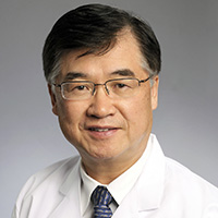 Dong Moon Shin合作论文数Winship Cancer Institute, Emory University;Department of Hematology and Medical Oncology, School of Medicine, Emory University;Department of Otolaryngology, School of Medicine, Emory University17William C. Faquin合作论文数17Ilan Weinreb合作论文数Department of Laboratory Medicine and Pathobiology, Temerty Faculty of Medicine, University of Toronto16
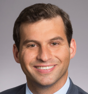 Conor E. Steuer合作论文数Winship Cancer Institute, Emory University;Department of Hematology and Medical Oncology, School of Medicine, Emory University15
 Karen Fritchie合作论文数13
 Amy Chen合作论文数Department of Epidemiology, Rollins School of Public Health, Emory University;Department of Otolaryngology Head and Neck Surgery, Emory University School of Medicine;Department of Health Policy and Management, Rollins School of Public Health, Emory University;Winship Cancer Institute, Emory University School of Medicine13
 Shlomo Koyfman合作论文数Department of Radiation Oncology, Cleveland Clinic;Department of Medicine, School of Medicine, Case Western Reserve University;Case Comprehensive Cancer Center, Case Western Reserve University13Cme Subcommittee合作论文数12Xu Wang合作论文数12
 Sarah Dry合作论文数Department of Pathology and Laboratory Medicine, University of California, Los Angeles;Jonsson Comprehensive Cancer Center, University of California, Los Angeles;Ronald Reagan UCLA Medical Center, UCLA Health;UCLA Santa Monica Medical Center, UCLA Health12
 Adebowale Adeniran合作论文数Yale School of Medicine12Chiosea Simion I合作论文数Dept Pathol, Univ Pittsburgh12
 Vickie Y Jo合作论文数12
 Nicole Anne Cipriani合作论文数12
 Yuri Fedoriw合作论文数Department of Pathology and Laboratory Medicine, School of Medicine, The University of North Carolina at Chapel Hil12Nora Katabi合作论文数12Rhonda Yantiss Chair合作论文数11Stefano La合作论文数11J Lerwill合作论文数11Billie Fyfe-Kirschner合作论文数11Chan Kung-Chao合作论文数11Gregory Bean Govind合作论文数11Matt Quick合作论文数11Gillian Leigh合作论文数11Pantanowitz Carlos合作论文数11Parra-Herran Rajiv合作论文数11Pathologist-In-Training Melinda合作论文数11Kristin Jensen Chair合作论文数11Carla Chair合作论文数11Brock Aaron合作论文数11Pathologist-In-Training Liron合作论文数11M Laganà合作论文数11M Beatriz合作论文数11Hale Suntrea合作论文数11Zabala Blanco合作论文数11
 Lesley Lomo合作论文数11Veronica Klepeis合作论文数11Julie C. Fanburg-Smith合作论文数11Roberto Barrios合作论文数11Anthony J. Gill合作论文数11Tatjana Antic合作论文数11Vasiliki Leventaki合作论文数11Meghan E Kapp合作论文数11




