Aristolochic acids I and II (AA-I/II) are carcinogenic principles of Aristolochia plants, which have been employed in traditional medicinal practices and discovered as food contaminants. While the deleterious effects of AAs are broadly acknowledged, there is a dearth of information to define the mechanisms underlying their carcinogenicity. Following bioactivation in the liver, N-hydroxyaristolactam and N-sulfonyloxyaristolactam metabolites are transported via circulation and elicit carcinogenic effects by reacting with cellular DNA. In this study, we apply DNA adduct analysis, X-ray crystallography, isothermal titration calorimetry, and fluorescence quenching to investigate the role of human serum albumin (HSA) in modulating AA carcinogenicity. We find that HSA extends the half-life and reactivity of N-sulfonyloxyaristolactam-I with DNA, thereby protecting activated AAs from heterolysis. Applying novel pooled plasma HSA crystallization methods, we report high-resolution structures of myristic acid-enriched HSA (HSAMYR) and its AA complexes (HSAMYR/AA-I and HSAMYR/AA-II) at 1.9 Å resolution. While AA-I is located within HSA subdomain IB, AA-II occupies subdomains IIA and IB. ITC binding profiles reveal two distinct AA sites in both complexes with association constants of 1.5 and 0.5 · 106 M-1 for HSA/AA-I versus 8.4 and 9.0 · 105 M-1 for HSA/AA-II. Fluorescence quenching of the HSA Trp214 suggests variable impacts of fatty acids on ligand binding affinities. Collectively, our structural and thermodynamic characterizations yield significant insights into AA binding, transport, toxicity, and potential allostery, critical determinants for elucidating the mechanistic roles of HSA in modulating AA carcinogenicity.
Aristolochic acid (AA) is a potent human carcinogen and nephrotoxin found in preparations of Aristolochia plants used in Chinese Traditional Medicine. Following biotransformation to form N-sulfonyloxyaristolactam (AL-I-NOSO3), this intermediate undergoes heterolytic cleavage of the sulfate group to generate a reactive cyclic nitrenium ion, the ultimate DNA binding species. Recently, we showed that primary human hepatocytes significantly increase renal toxicity of AA in the integrated human liver-kidney “organs on-chips” model. Therefore, we propose that AA is activated in the liver by forming AL-I-NOSO3, which is transported to the kidney protected from decomposition by binding to serum albumin. We employed mass spectrometric, fluorimetric and X-crystallography based approaches to dissect mechanisms of interactions between human serum albumin (HSA), AA, N-hydroxyaristolactam and AL-I-NOSO3. First, we demonstrate that HSA stabilizes otherwise labile N-sulfonyloxyaristolactam. Quenching of the native fluorescence of HSA due to the presence of a sole molecule of tryptophane-214, allowed us to conclude that all three compounds have similar affinities to IIA drug binding pocket of HSA. Subsequently, we obtained a high-resolution X-ray structure of AA bound to HSA in domain IB (1.9°A, pdb: 6HSC). Since prior to crystallization HSA was enriched with sodium myristate and site IB in circulation is occupied by fatty acids, our results imply that the IB pocket is the primary high affinity binding site for AA and its active forms. To assess whether AL-I-NOSO3 covalently binds protein, we incubated human plasma and purified HSA with this active AA. Immunoblotting of reacted HSA using antibodies that recognize aristolactam(AL)-adducted DNA suggests irreversible covalent adduction of AL to HSA. A combined approach using mass spectrometry instruments and enzymatic digestion revealed that AL is adducted to HSA at the following sites: Trp-214, Tyr-138 and Tyr-141. The former amino acid is located in the IIA drug binding site of HSA, while the latter two can be found in our HSA/AA structure in the site IB in the vicinity to AA molecule, corroborating our X-crystallography and fluoremetric data. Based on these studies we propose that AL-I-NOSO3 has a dual mode of interactions with HSA. If AL-I-NOSO3 decomposes prior to HSA binding, aristolactam will become irreversibly trapped with HSA. This binding to HSA would serve as mechanism of detoxication of AA species. However, if AL-I-NOSO3 binds to HSA prior to decomposition, it should be protected by HSA and transported to target tissues in its intact form. Citation Format: Sergei Pomyalov, Radha Bonala, Robert Rieger, Irina Zaitseva, Charles Iden, John Haley, Robert Turesky, Francis Johnson, Thomas Rosenquist, Arthur P. Grollman, Gil Shoham, Viktoriya S. Sidorenko. Molecular mechanisms by which a bioactivated human carcinogen is transported to target tissues [abstract]. In: Proceedings of the American Association for Cancer Research Annual Meeting 2019; 2019 Mar 29-Apr 3; Atlanta, GA. Philadelphia (PA): AACR; Cancer Res 2019;79(13 Suppl):Abstract nr 4660.
Abstract Aristolochic acid, AA, is a natural compound found in Aristolochia plants used in Chinese Traditional Medicine and is a component of many herbal remedies used worldwide. In humans, exposure to AA is associated with kidney toxicity and otherwise rare upper urinary tract cancer (UTUC). AA undergoes bioactivation creating reactive species of AA that bind covalently to proteins and DNA. Formation of mutagenic AA-DNA adducts leads to AA-induced cancers. We identified the mutational signature associated with AA exposure in patients with UTUC in Taiwan, as well as in AA-initiated cancers in liver, bladder and kidneys. Meanwhile, the mechanism of AA nephrotoxicity remained a mystery, with covalent and non-covalent binding of AA or its metabolites to proteins hypothesized to be involved in its nephrotoxic effects. As millions of people in Asia have ingested AA and no effective therapies are known, establishing the molecular mechanism(s) of AA nephrotoxicity is a topic of major importance. A series of AA analogs were used to design affinity probes for target identification. Structure activity relationship studies in human cells aided in identifying a position in AA suitable for introducing biotinylated, aminopropyl and alkyne linkers. In addition, we have developed and validated monoclonal antibodies against AA-bovine serum albumin and/or AA-DNA covalent adducts. Combined with proteomics techniques, AA-affinity probes and monoclonal antibodies provide powerful tools for defining AA targets in human kidneys. We attached the aminopropyloxy derivative of AA to magnetic beads coated with N-hydroxysuccinimido acyl linkers. These probes, in parallel with unmodified, “control” beads were incubated with renal cortex lysates obtained from C3H mice selected for their sensitivity to the effects of AA. Bound proteins were digested "on bead", then identified by mass spectroscopic peptide analysis, with a total of 316 bound proteins detected. Spectral counts for 128 proteins bound to AA-beads were higher than those for control beads. After correction for nonspecific binding, 18 of these proteins had spectral counts of 10 or higher. These proteins are localized to various organelles, indicating high coverage of the major subcellular compartments. Among these proteins, Cryzl2, quinone oxidoreductase-like 2, was the leading candidate. Based on homology, Cryzl2 belongs to the family of the quinone oxidoreductase-like protein and bears a mitochondrial localization sequence. This protein retains conserved domains for both quinone oxidoreductase activity and NAD(P) binding sites, which is of particular interest since other oxidoreductases, such as NQO1, are known to catalyze nitroreduction, the initial step in the bioactivation of AA. In summary, our studies provide guidelines for design of AA-affinity probes and reveal an enzyme that may be responsible for AA activation in mitochondria, by generating active intermediates and inducing mitochondrial damage in renal tissue. Citation Format: Viktoriya S. Sidorenko, Kathleen G. Dickman, Thomas Rosenquist, Radha Bonala, Sivaprasad Attaluri, Irina Zaitseva, Charles Iden, Francis Johnson, Arthur P. Grollman. Using affinity probes to explore the nephrotoxicity of aristolochic acid [abstract]. In: Proceedings of the American Association for Cancer Research Annual Meeting 2017; 2017 Apr 1-5; Washington, DC. Philadelphia (PA): AACR; Cancer Res 2017;77(13 Suppl):Abstract nr 5241. doi:10.1158/1538-7445.AM2017-5241
Aristolochic acids (AA) are implicated in the development of chronic renal disease and upper urinary tract carcinoma in humans. Using in vitro approaches, we demonstrated that N-hydroxyaristolactams, metabolites derived from partial nitroreduction of AA, require sulfotransferase (SULT)-catalyzed conjugation with a sulfonyl group to form aristolactam-DNA adducts. Following up on this observation, bioactivation of AA-I and N-hydroxyaristolactam I (AL-I-NOH) was studied in human kidney (HK-2) and skin fibroblast (GM00637) cell lines. Pentachlorophenol, a known SULT inhibitor, significantly reduced cell death and aristolactam-DNA adduct levels in HK-2 cells following exposure to AA-I and AL-I-NOH, suggesting a role for Phase II metabolism in AA activation. A gene knockdown, siRNA approach was employed to establish the involvement of selected SULTs and nitroreductases in AA-I bioactivation. Silencing of SULT1A1 and PAPSS2 led to a significant decrease in aristolactam-DNA levels in both cell lines following exposure to AA-I, indicating the critical role for sulfonation in the activation of AA-I in vivo Since HK-2 cells proved relatively resistant to knockdown with siRNAs, gene silencing of xanthine oxidoreductase, cytochrome P450 oxidoreductase and NADPH:quinone oxidoreductase was conducted in GM00637 cells, showing a significant increase, decrease and no effect on aristolactam-DNA levels, respectively. In GM00637 cells exposed to AL-I-NOH, suppressing the SULT pathway led to a significant decrease in aristolactam-DNA formation, mirroring data obtained for AA-I. We conclude from these studies that SULT1A1 is involved in the bioactivation of AA-I through the sulfonation of AL-I-NOH, contributing significantly to the toxicities of AA observed in vivo.
The magnitude and nature of lesion-induced energetic perturbations empirically correlate with mutagenicity/cytotoxicity profiles and can be predictive of lesion outcomes during polymerase-mediated replication in vitro. In this study, we assess the sequence and counterbase-dependent energetic impact of the Thymine glycol (Tg) lesion on a family of deoxyoligonucleotide duplexes. Tg damage arises from thymine and methyl-cytosine exposure to oxidizing agents or radiation-generated free-radicals. The Tg lesion blocks polymerase-mediated DNA replication in vitro and the unrepaired site elicits cytotoxic lethal consequences in vivo. Our combined calorimetric and spectroscopic characterization correlates Tg -induced energetic perturbations with biological and structural properties. Specifically, we incorporate a 5R-Tg isomer centered within the tridecanucleotide sequence 5'-GCGTACXCATGCG-3' (X = Tg or T) which is hybridized with the corresponding complementary sequence 5'-CGCATGNGTACGC-3' (N = A, G, T, C) to generate families of Tg -damaged (Tg ·N) and lesion-free (T·N) duplexes. We demonstrate that the magnitude and nature of the Tg destabilizing impact is dependent on counterbase identity (i.e., A ∼ G < T < C). The observation that a Tg lesion is less destabilizing when positioned opposite purines suggests that favorable counterbase stacking interactions may partially compensate lesion-induced perturbations. Moreover, the destabilizing energies of Tg ·N duplexes parallel their respective lesion-free T·N mismatch counterparts (i.e., G < T < C). Elucidation of Tg-induced destabilization relative to the corresponding undamaged mismatch energetics allows resolution of lesion-specific and sequence-dependent impacts. The Tg-induced energetic perturbations are consistent with its replication blocking properties and may serve as differential recognition elements for discrimination by the cellular repair machinery.
Plants from the Aristolochia genus have been recommended for the treatment of a variety of human ailments since the time of Hippocrates. However, many species produce the highly toxic aristolochic acids (AAs), which are both nephrotoxic and carcinogenic. For the purposes of extensive biological studies, a versatile approach to the synthesis of the AAs and their major metabolites was devised based primarily on a Suzuki–Miyaura coupling reaction. The key to success lies in the preparation of a common ring-A precursor, namely, the tetrahydropyranyl ether of 2-nitromethyl-3-iodo-4,5-methylendioxybenzyl alcohol (27), which was generated in excellent yield by oxidation of the aldoxime precursor 26. Suzuki–Miyaura coupling of 27 with a variety of benzaldehyde 2-boronates was accompanied by an aldol condensation/elimination reaction to give the desired phenanthrene intermediate directly. Deprotection of the benzyl alcohol followed by two sequential oxidation steps gave the desired phenanthrene nitrocarboxylic acids. This approach was used to synthesize AAs I–IV and several other related compounds, including AA I and AA II bearing an aminopropyloxy group at position-6, which were required for further conversion to fluorescent biological probes. Further successful application of the Suzuki–Miyaura coupling reaction to the synthesis of the N-hydroxyaristolactams of AA I and AA II then allowed the synthesis of the putative, but until now elusive, N-acetoxy- and N-sulfonyloxy-aristolactam metabolites.
Aristolochic acids are natural nitro-compounds found globally in the plant genus Aristolochia that have been implicated in the severe illness in humans termed aristolochic acid nephropathy (AAN). Aristolochic acids undergo nitroreduction, among other metabolic reactions, and active intermediates arise that are carcinogenic. Previous experiments with rats showed that aristolochic acid I (AA-I), after oral administration or injection, is subjected to detoxication reactions to give aristolochic acid Ia, aristolactam Ia, aristolactam I, and their glucuronide and sulfate conjugates that can be found in urine and feces. Results obtained with whole rats do not clearly define the role of liver and kidney in such metabolic transformation. In this study, in order to determine the specific role of the kidney on the renal disposition of AA-I and to study the biotransformations suffered by AA-I in this organ, isolated kidneys of rats were perfused with AA-I. AA-I and metabolite concentrations were determined in perfusates and urine using HPLC procedures. The isolated perfused rat kidney model showed that AA-I distributes rapidly and extensively in kidney tissues by uptake from the peritubular capillaries and the tubules. It was also established that the kidney is able to metabolize AA-I into aristolochic acid Ia, aristolochic acid Ia O-sulfate, aristolactam Ia, aristolactam I, and aristolactam Ia O-glucuronide. Rapid demethylation and sulfation of AA-I in the kidney generate aristolochic acid Ia and its sulfate conjugate that are voided to the urine. Reduction reactions to give the aristolactam metabolites occur to a slower rate. Renal clearances showed that filtered AA-I is reabsorbed at the tubules, whereas the metabolites are secreted. The unconjugated metabolites produced in the renal tissues are transported to both urine and perfusate, whereas the conjugated metabolites are almost exclusively secreted to the urine.
Aristolochic acids (AAs) are a structurally related family of nephrotoxic and carcinogenic nitrophenanthrene compounds found in Aristolochia herbaceous plants, many of which have been used worldwide for medicinal purposes. AAs have been implicated in the etiology of so-called Chinese herbs nephropathy and of Balkan endemic nephropathy. Both of these disease syndromes are associated with carcinomas of the upper urinary tract (UUC). 8-Methoxy-6-nitrophenanthro-[3,4-d]-1,3-dioxolo-5-carboxylic acid (AA-I) is a principal component of Aristolochia herbs. Following metabolic activation, AA-I reacts with DNA to form aristolactam (AL-I)-DNA adducts. We have developed a sensitive analytical method, using ultraperformance liquid chromatography-electrospray ionization/multistage mass spectrometry (UPLC-ESI/MS(n)) with a linear quadrupole ion-trap mass spectrometer, to measure 7-(deoxyadenosin-N(6)-yl) aristolactam I (dA-AL-I) and 7-(deoxyguanosin-N(2)-yl) aristolactam I (dG-AL-I) adducts. Using 10 μg of DNA for measurements, the lower limits of quantitation of dA-AL-I and dG-AL-I are, respectively, 0.3 and 1.0 adducts per 10(8) DNA bases. We have used UPLC-ESI/MS(n) to quantify AL-DNA adducts in tissues of rodents exposed to AA and in the renal cortex of patients with UUC who reside in Taiwan, where the incidence of this uncommon cancer is the highest reported for any country in the world. In human tissues, dA-AL-I was detected at levels ranging from 9 to 338 adducts per 10(8) DNA bases, whereas dG-AL-I was not found. We conclude that UPLC-ESI/MS(n) is a highly sensitive, specific and robust analytical method, positioned to supplant (32)P-postlabeling techniques currently used for biomonitoring of DNA adducts in human tissues. Importantly, UPLC-ESI/MS(n) could be used to document exposure to AA, the toxicant responsible for AA nephropathy and its associated UUC.
Acrolein is an alpha,beta-unsaturated aldehyde that is a major environmental pollutant, as well as a product of cellular metabolism. DNA bases react with acrolein to form two regioisomeric exocyclic guanine adducts, namely gamma-hydroxy-propanodeoxyguanosine (gamma-OH-PdG) and its positional isomer alpha-hydroxy-propanodeoxyguanosine (alpha-OH-PdG). The gamma-OH-PdG isomer adopts a ring-opened conformation with minimal structural perturbation of the DNA host duplex. Conversely, the alpha-OH-PdG isomer assumes a ring-closed conformation that significantly disrupts Watson-Crick base-pair alignments within the immediate vicinity of the damaged site. We have employed a combination of calorimetric and spectroscopic techniques to characterize the thermodynamic origins of these lesion-induced structural alterations. Specifically, we have assessed the energetic impact of alpha-OH-PdG centered within an 11-mer duplex by hybridizing the adduct-containing oligonucleotide with its complementary strand harboring a central base N [where N = C or A], yielding a pair of duplexes containing the nascent lesion (alpha-OH-PdG.C) or mismatched adduct (alpha-OH-PdG.A), respectively. Our data reveal that the nascent lesion is highly destabilizing, whereas its mismatched counterpart partially ameliorates alpha-OH-PdG-induced destabilization. Collectively, our data provide energetic characterizations of the driving forces that modulate error-free versus error-prone DNA translesion synthesis. The biological implications of our findings are discussed in terms of energetically probing acrolein-mediated mutagenicity versus adduct-induced genotoxicity.
Aristolochic acids I and II (AA-I, AA-II) are found in all Aristolochia species. Ingestion of these acids either in the form of herbal remedies or as contaminated wheat flour causes a dose-dependent chronic kidney failure characterized by renal tubulointerstitial fibrosis. In approximately 50% of these cases, the condition is accompanied by an upper urinary tract malignancy. The disease is now termed aristolochic acid nephropathy (AAN). AA-I is largely responsible for the nephrotoxicity while both AA-I and AA-II are genotoxic. DNA adducts derived from AA-I and AA-II have been isolated from renal tissues of patients suffering from AAN. We describe the total synthesis, de novo, of the dA and dG adducts derived from AA-II, their incorporation site-specifically into DNA oligomers and the splicing of these modified oligomers into a plasmid construct followed by transfection into mouse embryonic fibroblasts. Analysis of the plasmid progeny revealed that both adducts blocked replication but were still partly processed by DNA polymerase(s). Although the majority of coding events involved insertion of correct nucleotides, substantial misincorporation of bases also was noted. The dA adduct is significantly more mutagenic than the dG adduct; both adducts give rise, almost exclusively, to misincorporation of dA, which leads to AL-II-dA-->T and AL-II-dG-->T transversions.
The 2'-deoxyguanosine adduct of the dietary mutagen 2-amino-1-methyl-6-phenylimidazo[4,5-b]pyridine (PhIP) has been synthesized and incorporated into DNA using solid state synthesis technology. The key step to obtaining the C8-dG adduct is a palladium (Xantphos-chelated)-catalyzed N-arylation (Buchwald-Hartwig reaction) of PhIP by a suitably protected 8-bromo-2'-deoxyguanosine derivative. The reaction proceeded in good yield without complicating side products, and the adduct was converted to the required 5'-O-DMT-3'-O-phosphoramidite by standard methods. This modified deoxynucleoside was used to synthesize three oligodeoxynucleotides in which the C8-PhIP-dG adduct was incorporated at a single site. The oligomers were purified by reverse phase HPLC and characterized by mass spectrometry.
Thymine glycols (Tg) are major pyrimidine oxidation products produced by chemical agents and ionizing radiation. Recent improvements in purification procedures gave us the opportunity to examine the incision of DNA duplexes containing a single (5S,6R)- or (5R,6S)-Tg lesion by mouse NTH1 DNA glycosylase and mammalian cell nuclear extracts. Time course experiments and steady state enzyme kinetics indicated that mNTH1 discriminates between the cis-Tg isomers. In addition, a variety of mammalian cell nuclear extracts showed a similar discrimination between the cis-Tg isomers. Trapping of Schiff base intermediates with sodium borohydride demonstrated that a single protein-DNA complex was formed in the presence of the nuclear extracts. The electrophoretic mobility of trapped complexes formed with both Tg isomers was identical to one another and similar to that of the complex formed with recombinant mNTH1. These results suggest that among all Tg-active DNA glycosylases, NTH1 is the major enzyme in mammalian cell nuclear extracts responsible for incision of duplexes containing cis-Tg isomers.
ADVERTISEMENT RETURN TO ISSUEPREVBook ReviewNEXTMass Spectrometry in Drug Discovery Edited by David T. Rossi and Michael W. Sinz. Marcel Dekker, Inc., New York. 2002. viii + 420 pp. 16 × 23.5 cm. ISBN 0-8247-0607-2. $165.00.Charles R. IdenView Author Information Department of Pharmacological Sciences School of Medicine SUNY-Stony Brook Stony Brook, New York 11794-3400Cite this: J. Med. Chem. 2002, 45, 19, 4375–4376Publication Date (Web):September 5, 2002Publication History Published online5 September 2002Published inissue 1 September 2002https://pubs.acs.org/doi/10.1021/jm020305jhttps://doi.org/10.1021/jm020305jbook-reviewACS PublicationsCopyright © 2002 American Chemical SocietyRequest reuse permissionsArticle Views74Altmetric-Citations-LEARN ABOUT THESE METRICSArticle Views are the COUNTER-compliant sum of full text article downloads since November 2008 (both PDF and HTML) across all institutions and individuals. These metrics are regularly updated to reflect usage leading up to the last few days.Citations are the number of other articles citing this article, calculated by Crossref and updated daily. Find more information about Crossref citation counts.The Altmetric Attention Score is a quantitative measure of the attention that a research article has received online. Clicking on the donut icon will load a page at altmetric.com with additional details about the score and the social media presence for the given article. Find more information on the Altmetric Attention Score and how the score is calculated. Share Add toView InAdd Full Text with ReferenceAdd Description ExportRISCitationCitation and abstractCitation and referencesMore Options Share onFacebookTwitterWechatLinked InRedditEmail Other access optionsGet e-Alertsclose SUBJECTS:Drug discovery,Mass spectrometers,Mass spectrometry,Pharmaceuticals,Quantitative analysis Get e-Alerts
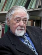 Francis Johnson合作论文数Department of Pharmacological Sciences, Renaissance School of Medicine, Stony Brook University17
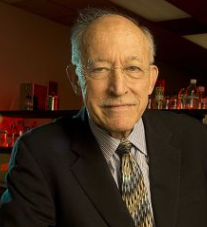 Arthur P. Grollman合作论文数Department of Pharmacological Sciences, Renaissance School of Medicine, Stony Brook University13
 Radha Bonala合作论文数11Sivaprasad Attaluri合作论文数9
 Viktoriya S Sidorenko合作论文数8Rosenquist Thomas A合作论文数6Zaitseva Irina合作论文数5Robert A. Rieger合作论文数5Kathleen G. Dickman合作论文数4
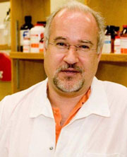 Robert J. Turesky合作论文数Masonic Cancer Center’s Analytical Biochemistry shared resource, University of Minnesota;Turesky Lab, Department of Medicinal Chemistry, College of Pharmacy, University of Minnesota4
 Conceicao Minetti合作论文数Rutgers—The State University of New Jersey, Department of Chemistry and Chemical Biology, Piscataway, NJ 08854, USA3
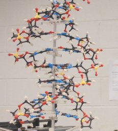 David P. Remeta合作论文数Department of Biology and Biocalorimetry Center, The Johns Hopkins University3Masaaki Moriya合作论文数3
 Kenneth J. Breslauer (Kenneth Breslauer)合作论文数Robert Wood Johnson Medical School, Rutgers University;Department of Chemistry and Chemical Biology, School of Arts and Sciences, Rutgers University3
 B. H. Yun合作论文数College of Sciences, Yeungnam University3Sergei Pomyalov合作论文数2Gil Shoham合作论文数Department of Inorganic Chemistry and the Laboratory for Structural Chemistry and Biology, The Hebrew University of Jerusalem2M. Cecilia Torres合作论文数2Chung-Hsin Chen合作论文数2Rahda Bonala合作论文数1Yoshimitsu Oda合作论文数1Ra Rieger合作论文数1In-Young Yang合作论文数1MM McTigue合作论文数1Orlando Schärer合作论文数1Mark Lukin合作论文数Department of Pharmacological Sciences, Stony Brook University1S Brodsky合作论文数1
 Dan Gu合作论文数1
 P. Lee Ferguson合作论文数Department of Civil & Environmental Engineering, Pratt School of Engineering, Duke University1Haley J D合作论文数1Goligorsky Michael S合作论文数1
 Shinya Shibutani合作论文数Department of Pharmacological Sciences, Renaissance School of Medicine, Stony Brook University1
 Yeong-Shiau Pu (蒲永孝)合作论文数Department of Urology, National Taiwan University Hospital;Graduate Institute of Medical Medicine, College of Medicine, National Taiwan University1
 Keiji Hashimoto合作论文数Faculty of Science and Engineering, Kinki University1
 Yoshiharu Okuno合作论文数1Avalyn Lewis合作论文数1
 Irina Zaitseva合作论文数1Ivan Habus合作论文数1Monica M. Mctigue合作论文数1
 David R Taft合作论文数1Robert Gentles合作论文数1Sue Ellen Gerchman合作论文数1Urszula Golebiewska合作论文数Department of Physiology and Biophysics, Stony Brook University1J.H. Kycia合作论文数1M. A. Barbieri合作论文数1Yujing Wen合作论文数1Jung Eun Yeo合作论文数1
 Bruce Brownawell合作论文数Marine Sciences Research Center, State University of New York1Horacio A. Priestap合作论文数1




