The impaired regenerative capacity of osteoporotic individuals poses a significant challenge to the repair of bone defects. In the osteoporotic microenvironment, low pH, excessive reactive oxygen species (ROS), and chronic inflammation create a self-perpetuating vicious cycle that impedes healing. However, conventional therapies fail to sustainably improve the damaged microenvironment. Here, a pH/ROS dual responsive nanocomposite hydrogel (Z-QCDs@M2-Migs@OHA-PP) was developed based on oxidized hyaluronic acid (OHA), phenylboronic acid-grafted ε-polylysine (PP), quercetin-derived carbon dots (QCDs) loaded ZIF-8 (Z-QCDs) and M2 macrophage-derived migrasomes (M2-Migs), which possesses intrinsic antioxidant and osteogenic differentiation-promoting capabilities. Due to the presence of dynamic. Schiff base bonds and boronate bonds, the hydrogel exhibited injectability and pH/ROS dual responsiveness. OHA-PP releases Z-QCDs and M2-Migs on demand in response to changes in pH and ROS levels. Z-QCDs exhibit strong antioxidant and nanozyme activity, capable of scavenging ROS, suppressing inflammatory responses, and promoting M2 macrophage polarization. Furthermore, the introduction of M2-Migs as an osteogenic activator further enhances the capacity for osteogenic differentiation. Transcriptomic and Western blot analyses revealed that the hydrogel promotes osteogenic differentiation by activating the PI3K-AKT signaling pathway. In a mouse osteoporotic bone defect model, the nanocomposite hydrogel effectively inhibited ferroptosis, modulated inflammation, and promoted new bone formation. Therefore, this hydrogel system, which combines therapeutic rationale with microenvironmental regulation, offers a promising strategy for the regeneration of osteoporotic bone defects.
N6 methyladenosine (m6A) is one of the most prevalent epitranscriptomic modifications of mRNAs, and plays a critical role in various bioprocesses. Bone-derived mesenchymal stem cells (BMSCs) can attenuate apoptosis of nucleus pulposus cells (NPCs) under compression; however, the underlying mechanisms are poorly understood. This study showed that the level of m6A mRNA modifications was decreased, and the autophagic flux was increased in NPCs under compression when they were cocultured with BMSCs. We report that under coculture conditions, RNA demethylase ALKBH5-mediated FIP200 mRNA demethylation enhanced autophagic flux and attenuated the apoptosis of NPCs under compression. Specific silencing of ALKBH5 results in impaired autophagic flux and a higher proportion of apoptotic NPCs under compression, even when cocultured with BMSCs. Mechanistically, we further identify that the m6A "reader" YTHDF2 is likely to be involved in the regulation of autophagy, and lower m6A levels in the coding region of FIP200 lead to a reduction in YTHDF2-mediated mRNA degradation of FIP200, a core molecular component of the ULK1 complex that participates in the initiating process of autophagy. Taken together, our study reveals the roles of ALKBH5-mediated FIP200 mRNA demethylation in enhancing autophagy and reducing apoptosis in NPCs when cocultured with BMSCs.
Tissue infections such as osteomyelitis and infected skin wounds require therapies that can both eradicate drugresistant bacteria and promote tissue regeneration. Antimicrobial peptide therapy is an emerging anti-infective strategy, but it still faces two major challenges: achieving more rapid and potent antibacterial activity, and endowing peptides with dual functions of killing bacteria and promoting tissue repair. Here, we design a piezoelectric hexapeptide of Phe-Phe-Citrulline-Glu-Ser-Val (FFCitESV), which has ultrasound-activatable antimicrobial and promoting tissue healing activities. FFCitESV exhibits robust piezoelectricity and generates abundant singlet oxygen under low-intensity ultrasound, enabling rapid killing of MRSA in vitro. The peptide significantly enhanced hBMSCs osteogenic differentiation, ultrasound further amplified this effect via piezoelectric stimulation. In a rat tibial osteomyelitis model and a mouse infected skin-wound model, FFCitESV combined with ultrasound effectively controlled MRSA infection, reduced local and systemic inflammation, and markedly accelerated bone and skin defect repair compared with standard treatments. Multi-omics and histological analyses further clarified mechanisms. Transcriptomic analysis showed that FFCitESV plus ultrasound activated PI3K-Akt, Wnt, and calcium-related pathways associated with osteogenic differentiation and bone regeneration. In infected skin wounds, RNA sequencing revealed enhanced keratinocyte differentiation and activation of PI3K-Akt and JAK-STAT pathways, which together facilitated re-epithelialization and dermal repair. Overall, FFCitESV represents a biodegradable piezoelectric hexapeptide that enables on-demand disinfection and tissue regeneration, thereby serving as a versatile therapeutic platform for refractory osteomyelitis, infected skin wounds, and other infection-associated tissue defects.
Extracellular vesicles (EVs) have emerged as key mediators of intercellular communication. However, the mechanisms governing their degradation remain poorly understood. In this study, we demonstrated that EVs are predominantly degraded via the lysosomal pathway. Mechanistically, MAP1LC3B recognizes SNX18 on the surface of endosome-escaped EVs to facilitate their sorting into the autolysosomal pathway for degradation. Leveraging this mechanism, we optimized the lysosomal sorting efficiency of EVs by surface display of LIR motifs and constructed an EV-based targeted protein degradation nanoplatform. The EV-based nanoplatform is highly modular and can be combined with monoclonal antibodies in a plug-and-play manner. It demonstrated remarkable efficiency and selectivity in degrading EGFR, PD-L1, and VEGF. Moreover, the nanoplatform demonstrated multi-targeting capability by simultaneously degrading EGFR and VEGF. Our findings uncover a previously unrecognized mechanism of EVs degradation and provide a novel strategy to harness the EVs degradation machinery as a nature-inspired nanoplatform for the degradation of multiple targeted proteins.
Macrophages eliminate apoptotic cells produced daily in the body through efferocytosis. Restricted clearance can cause inflammation-related diseases. In intervertebral discs (IVDs), apoptotic nucleus pulposus cells (NPCs) are difficult to effectively remove, and their accumulation can cause changes in the inflammatory microenvironment, disrupt IVD homeostasis, and lead to IVD degeneration (IDD). Here, we present chimeric antigen receptor-M-like engineered macrophages (CAR-eMs) with enhanced efferocytosis capacity for IDD treatment. Macrophages undergo phenotypic transformation and a reduction in phagocytic ability after phagocyting apoptotic NPCs, but their efferocytosis capacity recovers with upregulated brain-specific angiogenesis inhibitor 1 (BAI1) expression. We develop a CAR-eM system with enhanced BAI1 expression and an IVD circular microneedle (MN) delivery system that utilizes arrays of MNs to deliver CAR-eMs into the deep IVD layers, thereby clearing apoptotic NPCs, ameliorating the inflammatory microenvironment, and repairing damaged IVDs. Our study explores the therapeutic potential of CAR-eM efferocytosis for IDD treatment.
The maintenance and regulation of cancer stem cell (CSC) stemness are crucial for tumor progression; however, the mechanisms underlying tumor stemness regulation remain poorly understood. Herein, we discovered that the enhanced hypoxia-induced transforming growth factor beta induced protein (TGFBI) in bladder cancer (BLCA) promotes the establishment of a stemness loop in the tumor microenvironment, facilitating the maintenance of CSC stemness and malignant proliferation. Clinically, the upregulation of hypoxic TGFBI in BLCA correlates with malignant BLCA features and poor prognosis. Mechanically, TGFBI can stabilize the structural integrity of disulfide bonds in Cys48 and Cys77 of growth differentiation factor 15 (GDF15), leading to aberrant function activity of GDF15 and secretion. Interestingly, secreted GDF15 consequently not only further upregulates CSC-related gene expression but also induces the activation of cancer-associated fibroblasts through the transforming growth factor beta receptor type 2 (TGFBR2)–transforming growth factor β (TGFβ)–TGFBI self-regulatory feedback loop to promote stemness in BLCA. TGFBI knockdown or GDF15 inhibition results in a decrease in functional proteins associated with stemness maintenance, which suppresses bladder CSCs’ self-renewal and effectively improves the efficacy of chemotherapy. Together, these findings demonstrate the pivotal role of TGFBI in BLCA’s stemness maintenance and BLCA progression, highlighting that the inhibition of the TGFBI/GDF15 axis is a potential therapeutic strategy for the amelioration of cancer chemotherapy.
Complex craniocervical malformations pose significant challenges to surgical fixation. The biomechanical advantage of occipital plate fixation versus short-lever modified C1 lateral mass screw fixation remains controversial, and finite element analysis (FEA) is a reliable tool for implant performance evaluation. To compare biomechanical characteristics of occipital plate fixation and modified C1 lateral mass screw fixation in AOZ-BI and AOZ-AAD models via FEA, and guide surgical decision-making. A validated healthy occipito-atlantoaxial (C0-C2) FEA model was established using CT data. Two pathological models were constructed: AOZ-BI (Group A, atlantoaxial distance [ADI] < 5 mm) and AOZ-AAD (Group B, ADI ≥ 5 mm with transverse ligament dysfunction), each divided into occipital plate and modified C1 lateral mass screw subgroups. Static loads (40 N preload + 1.5 N·m torque) simulated flexion (Fe), extension (Ex), lateral bending (LB), and axial rotation (AR). C1-C2 range of motion (ROM) and screw-rod peak Von Mises stress (PVMS) were measured. Modified C1 lateral mass screw fixation reduced C1-C2 ROM by 19.67
Intervertebral disc degeneration (IDD), a primary cause of low back pain, currently lacks therapeutic strategies capable of halting its progression or promoting disc regeneration. Vascular ingrowth is the hallmark pathological feature of IDD, which arises from annulus fibrosus (AF) injury and accelerates degenerative processes through intricate interactions with inflammation and extracellular matrix degradation. This review summarizes the characteristics of vascular ingrowth in IDD, including differential vascular distribution between normal and degenerative discs, spatiotemporal dynamics of progressive invasion, and its association with pain via neurovascular co-invasion. The key underlying mechanisms involve the activation of pro-angiogenic factors (e.g., vascular endothelial growth factor [VEGF]), regulation by miRNA networks (e.g., miR-140-5p), macrophage-mediated coupling of inflammation and angiogenesis, and the dual roles of stress pathways and hypoxia-inducible factor signaling in driving pathological vascularization. Current research employs in vivo models (e.g., puncture- or fixation-induced degeneration), in vitro vascular co-culture systems, as well as advanced imaging techniques to dissect the process of vascular ingrowth in IDD. Aimed at counteracting pathological angiogenesis and halting the progression of IDD, emerging therapeutic strategies have been developed, including VEGF interventions, miRNA-targeted therapies, modulation of the inflammatory microenvironment, and multi-target combinatorial regimens. Despite substantial advances in understanding vascular ingrowth, several critical unresolved issues remain, including the unclear causal relationship between vascular ingrowth and IDD progression, species-specific disparities in preclinical models, and challenges in optimizing therapeutic timing and target selection. Future research will focus on addressing these gaps, with key priorities including single-cell analysis of vascular heterogeneity, mechanobiological coupling with vascularization, biomaterial-based precision regulation, and the establishment of standardized clinical translation pathways. In summary, vascular ingrowth is a critical driver of IDD, and mechanistic insights gained herein support its potential as a therapeutic target. Addressing current challenges will accelerate the translation of novel strategies into clinical practice for effective IDD management.
Adolescent idiopathic scoliosis (AIS) involves significant sagittal plane abnormalities, most consistently characterized by reduced thoracic kyphosis (TK), with compensatory changes in lumbar lordosis (LL) and cervical lordosis (CL). Brace treatment effectively controls coronal curve progression but commonly reduces TK and LL, potentially leading to a flatback deformity, while global sagittal balance is often maintained through spinal and pelvic compensation. Surgical intervention, particularly posterior spinal fusion, reliably improves sagittal alignment, with the most pronounced restoration of TK and CL observed in patients with preexisting sagittal malalignment. Despite regional alterations, global sagittal balance typically remains stable posttreatment, underscoring the body’s inherent compensatory mechanisms to maintain an energy-efficient upright posture. Pelvic parameters exhibit considerable individual variability but adapt to help sustain overall spinal equilibrium, with a tendency toward retroversion when compensating for spinal imbalances. Clinical management of AIS should integrate personalized, three-dimensional assessment and correction strategies, balancing coronal correction with the imperative of achieving optimal sagittal alignment to ensure favorable long-term outcomes.
Sulfation is a ubiquitous modification in glycobiology, yet its enzymology and biological significance in glycosylated natural products remain poorly understood. Saccharomicin A, a potent oligosaccharide antibiotic, carries 17 sugars including a unique sulfated fucose. Here, we report the identification of Sam10 as an unprecedented fucose-specific sulfotransferase through genetic studies, in vitro reconstitution, and structural analysis, establishing its role in saccharomicin sulfation. Comparative bioassays revealed that sulfation contributes to antibacterial potency, including activity against diverse multidrug-resistant pathogens. We also characterized Sam35 as an efficient adenylyl-sulfate kinase that boosts cellular 3 '-phosphoadenosine-5 '-phosphate sulfate (PAPS) supply. Our findings define the biochemical and structural basis of saccharomicin sulfation and provide enzymatic tools for engineering novel sulfated oligosaccharide antibiotics.
Infected wounds present a high clinical incidence, and current treatments are inadequate in both effectively eliminating bacterial biofilms and continuously promoting tissue regeneration. To address these challenges, we develop a microwave-responsive thermoelectric nanocomposite of zinc oxide-bismuth telluride (ZnO-Bi2Te3) via hydrothermal synthesis for the treatment of biofilm infected wounds. The heterointerface within the nanocomposite enhances phonon scattering, while the ZnO component provides additional charge carrier transport pathways, collectively significantly improving the thermoelectric conversion performance. Under microwave irradiation, the ZnO-Bi2Te3 rapidly generates substantial amounts of reactive oxygen species (ROS), enabling efficient penetration and eradication of biofilms with an antibacterial rate of 99.2
Venous thromboembolism (VTE) remains a critical clinical challenge due to limitations in conventional therapies, including poor thrombus targeting, inadequate imaging, and bleeding risks. This study develops nanoparticles composed of polylactic-co-glycolic acid, perfluoropentane, ferric oxide, and a macrophage membrane (designated as PLGA-PFP-Fe3O4-MO NPs) for targeted thrombolysis and dual-modal imaging. The NPs integrate macrophage membrane coating to enhance immune evasion and thrombus targeting, a perfluoropentane core enabling phase-change capabilities under low-intensity focused ultrasound (LIFU), and Fe3O4 for magnetic targeting and photoacoustic imaging. Evaluations in vitro and in a rat deep vein thrombosis model demonstrate superior thrombolytic efficacy, imaging performance, and long-term safety. The NPs reduce residual thrombus area to 15.57%, outperforming urokinase at 30.25% and low-molecular-weight heparin, while restoring venous blood flow and eliminating pulmonary embolism incidence. Long-term safety assessments over 28 days confirm no systemic toxicity or organ damage. LIFU-triggered phase transitions enable contrast-enhanced ultrasound imaging with prolonged durability exceeding 8 min surpassing SonoVue, and high-resolution photoacoustic localization. These findings establish PLGA-PFP-Fe3O4-MO as a multifunctional platform for precise VTE management, offering non-invasive thrombolysis, real-time imaging guidance, and robust biosafety for clinical translation.
Bladder perfusion chemotherapy remains the standard treatment for bladder cancer, yet its effectiveness is frequently limited by rapid drug clearance through urinary excretion and inadequate infiltration of immune cells into bladder tissue. To address these challenges, we developed an active-targeting nano-drug delivery system specifically designed for bladder tumors. This system utilizes a sialic acid-targeted poly (lactic-co-glycolic acid) (PLGA) platform to co-deliver doxorubicin (DOX) and the purified protein derivative (PPD) of bacillus Calmette-Guérin (BCG-PPD). By leveraging the selective binding of phenylboronic acid to sialic acid, the system enhances tumor-specific drug uptake, significantly amplifying DOX’s therapeutic efficacy and inducing immunogenic cell death. Furthermore, BCG-PPD exerts potent immunostimulatory effects, promoting dendritic cell (DC)-mediated tumor antigen processing and presentation, which in turn drives robust cytotoxic T lymphocyte (CTL) infiltration into the tumor microenvironment. The superior anti-tumor performance of this system was validated in an orthotopic bladder cancer mouse model. In conclusion, by synergistically combining targeted drug delivery with chemo-immunotherapy, our nanoparticle system presents a highly effective and promising new paradigm for bladder cancer therapy.
Spinal aging is a progressive degenerative process involving structural and functional decline of the spine as a whole and of its individual components, ultimately contributing to the development of degenerative spinal diseases (DSDs). Previous studies have largely focused on local spinal tissues or the intervertebral disc (IVD). However, recent advances in geroscience indicate that spinal degeneration is not an isolated local event, but is influenced by systemic aging through a multi-organ aging network. Accordingly, this review proposes a “from local to systemic” framework to better understand the mechanisms underlying spinal aging. At the local level, spinal aging follows heterogeneous trajectories, with intervertebral disc degeneration (IVDD) often serving as a central process. Different DSDs share common pathological mechanisms, and their anatomical proximity allows them to interact biomechanically. Therefore, the spine should be regarded as an integrated organ system within systemic aging. Based on this concept, we propose that vertebral osteoporosis and paraspinal muscle degeneration should also be incorporated into the framework of spinal aging and DSDs. At the systemic level, immunosenescence, skeletal muscle aging, neural and vascular aging, digestive system aging, endocrine system aging, and nutrient-sensing dysregulation may collectively affect the spine through circulating inflammatory factors, senescence-associated secretory phenotypes, metabolic mediators, endocrine signals, and mechanical loading, thereby influencing the susceptibility, progression, clinical phenotypes, and treatment responses of DSDs. We further discuss clinical translation in spinal aging, including anatomical barriers to drug delivery and emerging strategies to overcome them, and summarize therapeutic approaches based on multi-organ aging. Ultimately, this review emphasizes that future DSDs treatment should achieve more precise stratification, move beyond lesion-centered local interventions, and incorporate systemic anti-aging strategies to optimize long-term clinical outcomes in patients with DSDs.
Intervertebral disc degeneration (IDD), a major contributor to low back pain, is closely associated with oxidative stress-induced nucleus pulposus cell (NPC) senescence. Current clinical treatments primarily alleviate symptoms without targeting the underlying pathological mechanisms. Herein, we present a dual-polyphenol extracellular matrix (ECM)-mimetic hydrogel platform (Zn@MOF-gel) that integrates a gallic acid-derived metal-organic framework (Zn@MOF) with a tannic acid-crosslinked hydrogel (TA-gel). This ECM-mimetic injectable hydrogel platform is designed to enable localized and sustained release of both tannic acid (TA) and Zn@MOF within disc. In vitro, the Zn@MOF-gel effectively scavenged ROS, stabilized mitochondrial membrane potential, and reduced the secretion of pro-inflammatory cytokines in NPCs. Importantly, the Zn@MOF-gel further suppressed NPC senescence and restored ECM metabolic homeostasis, thereby disrupting the vicious cycle of NPC senescence and ECM dysregulation. Transcriptomic analysis revealed that the therapeutic effects are mediated through inhibition of the NF-kappa B-mediated TNF alpha and IL-6/JAK/STAT3 signaling pathways. In a rat needle-puncture IDD model, the Zn@MOF-gel effectively attenuated IDD, as confirmed by radiographic and histopathological analyses. Moreover, to better evaluate clinical translational potential beyond rodent models, a goat discectomy-induced IDD model was established. In this large animal model, the Zn@MOF-gel not only successfully filled disc defects but also facilitated functional tissue repair. Overall, this dual-polyphenol ECM-mimetic hydrogel platform represents a promising and translationally relevant strategy for the treatment of IDD.
Photodynamic therapy (PDT) is a promising anticancer strategy but remains limited by inefficient reactive oxygen species (ROS) generation and insufficient subcellular targeting. Here, we report a cationic engineering strategy to enhance mitochondrial targeting and ROS-amplified photodynamic tumor therapy. A neutral aggregation-induced emission (AIE) photosensitizer (PS) was stepwise converted into monocationic (PS-Bu) and dicationic (PS-PPh) derivatives, enabling precise regulation of molecular charge density. Cationic modification modulates excited-state energetics, promotes intersystem crossing, and simultaneously enhances Type I and Type II ROS generation under visible-light irradiation. The increased positive charge drives preferential mitochondrial accumulation, leading to pronounced mitochondrial membrane depolarization, intracellular ROS amplification, cell-cycle arrest, and apoptosis. Consequently, PS-PPh exhibits markedly enhanced phototoxicity toward HepG2 cells while maintaining negligible dark toxicity. In vivo, PS-PPh achieves superior tumor growth suppression in HepG2 xenograft models, accompanied by reduced proliferation, enhanced apoptosis, and alleviation of tumor hypoxia, without detectable systemic toxicity. This work establishes cationic modulation as an effective molecular design paradigm for mitochondria-targeted and ROS-enhanced photodynamic cancer therapy.
Background: This study aimed to evaluate the efficacy of unilateral biportal endoscopy-unilateral laminectomy bilateral decompression (UBE-ULBD) relative to microscopic anterior cervical discectomy and fusion (ACDF) for cervical spondylotic myelopathy (CSM). Methods: In this matched cohort study, 32 patients undergoing UBE-ULBD were compared with 96 patients undergoing ACDF (1:3 ratio). Radiologic parameters, clinical outcomes, surgical data, and laboratory markers were analyzed. Results: The mean follow-up duration was 24.09 months in the UBE-ULBD group and 24.47 months in the ACDF group. At the final follow-up, the ACDF group exhibited significant decreases in C2-C7 Cobb angle, cervical curvature index, range of motion (ROM), and adjacent segment disc height, and remarkable increase in the segmental Cobb angle of adjacent levels, segmental degeneration index, T1 slope, and sagittal vertical axis. These radiologic changes were not observed in the UBE-ULBD group. Baseline characteristics and improvements in Japanese Orthopaedic Association scores, visual analog scale (VAS) scores, short form-12 scores, and neck disability index, as well as reoperation rate, were comparable between groups. However, UBE-ULBD was associated with significantly shorter operative time, reduced blood loss, decreased length of hospital stay, lower serum creatine kinase and C-reactive protein levels, and lower complication rate. Conclusions: Both UBE-ULBD and ACDF yielded favorable clinical outcomes for CSM. Nonetheless, UBE-ULBD demonstrated potential advantages in preserving cervical alignment and ROM, mitigating adjacent segment degeneration, reducing postoperative complications, and minimizing surgical trauma.
Intervertebral disc degeneration is the leading cause of low back pain, which is prevalent in aging individuals. Numerous pathogenic factors, including loading burden, injury, and aging, contribute to intervertebral disc degeneration via various mechanisms, which are tightly regulated at different levels, including by post-transcriptional regulation. Emerging evidence suggests that RNA processing is crucial for the regulation of these factors and that multiple post-transcriptional regulatory pathways work independently of one another to enable precise control of transcripts throughout intervertebral disc homeostasis maintenance. Moreover, aberrant post-transcriptional regulation plays a critical role in the pathogenesis of intervertebral disc degeneration, broadening insights into how post-transcriptional alterations affect the homeostasis and function of intervertebral discs. In this review, the mechanisms and characteristics of post-transcriptional regulatory mechanisms are introduced, and their emerging functions in intervertebral disc degeneration are discussed. The summarized mechanisms and interconnections of post-transcriptional regulation provide a strong basis for the development of new therapeutics for repairing degenerative discs.
The sterility and controllable biological functionalities of implantable medical devices determine their lifespan, safety, and therapeutic efficacy. Inspired by the thermal effect of working electronic chips, which consist of various heterostructures, we have developed an electronic chip-mimetic sonothermal platform through constructing metal-semiconductor heterogeneous interface. Through magnetron sputtering on the surface of the sandblasted/acid-etched pretreated titanium (pTi), we find that semiconductor coatings (TiO2, Si, ZnO, and Te) endow pTi with different in situ sonothermal effects (ΔT > 18°C, 15 min) under ultrasound (US) irradiation, whereas conductor coatings do not. The sonothermal mechanism of pTi-semiconductor is associated with US-activated electron and phonon transport within the heterogeneous interface of implants, which is determined by the electrical and phonon characteristics of pTi-semiconductor, including their matching degree, thermal conductivity, defected structure, and type of semiconductor. Clinical titanium screws were introduced with defected structure in oxygen layer (TiO2-x) and bone-derived whitlockite, which shows great sonothermal/sonodynamic effects for efficient elimination of biofilm infection and improved osseointegration. In addition, the prepared NiTi-TiO2-x guidewire enables rapid thrombolysis (30 min) in the deep vein thrombosis of beagles after 10 min of sonothermal/urokinase treatment. The electronic chip-mimetic sonothermal platform provides a promising and widespread clinical application prospect.
Background: This study investigates the feasibility and effectiveness of a fusion cage with ipsilateral-impaction bone grafting (IIBG) for treating lumbar spinal stenosis (LSS) combined with segmental instability. Methods: From January 2018 to December 2021, 282 LSS patients undergoing minimally invasive transforaminal lumbar interbody fusion (MIS-TLIF) at Union Hospital were enrolled (164 IIBG vs. 118 controls). The cohort comprised 97 males and 185 females (mean age 62.75 ± 6.88 years; body mass index [BMI] 24.02 ± 2.58 kg/m 2 ). Surgical levels included 218 single-level (L3–L4: 5, L4–L5: 113, L5–S1: 100), 42 double-level (L3–L5), and 22 multi-level (L3–S1) fusions. Visual analogue scale (VAS) and Oswestry disability index (ODI) were used to assess the severity of pain and spinal nerve function before surgery, as well as at one month, six months, and 12 months after surgery. Besides, the MacNab criteria were used to evaluate clinical efficacy at the last follow-up. Imaging indicators refer to the preoperative and postoperative ratio of intervertebral height and bone grafting area to vertebral body area. Fusion status was evaluated via the Bridwell Grade on 1-year computed tomography (CT). Results: The mean operative time was 94.11 ± 38.11 min (range 68–238 min), with an intraoperative blood loss of 90.82 ± 40.35 mL (range 20–150 mL). Follow-up visits were conducted among all 282 patients, with an average follow-up time of 22.44 ± 6.84 months (range 12–34 months). Preoperative VAS of lower back pain and lower limb pain was 6.99 ± 1.42 points and 7.80 ± 1.33 points, respectively, which showed statistical differences ( F = 2290.00, p < 0.001; F = 3307.17, p < 0.001) compared with postoperative results. The scores decreased to 2.20 ± 0.85 and 2.03 ± 0.84 at one month after surgery, 1.56 ± 0.60 and 1.51 ± 0.50 at six months after surgery, 0.89 ± 0.69 and 0.89 ± 0.69 at one year after surgery, and 0.85 ± 0.70 and 0.87 ± 0.69 at the last follow-up. ODI was 54.47 ± 2.89% (range 50%–59%) before surgery, 25.50 ± 2.88% (range 21%–30%) at one month after surgery, 15.02 ± 3.14% (range 10%–20%) at six months after surgery, 12.50 ± 1.71% (range 10%–15%) at one year after surgery, and 12.51 ± 1.71% (range 50%–59%) at the last follow-up; the difference was significant ( F = 734.432, p < 0.001). MacNab criteria indicated excellent outcomes in 209 cases, good outcomes in 58 cases, and fair outcomes in 15 cases, with excellent and good results of 92.74% among all 282 patients. Mean intervertebral height was 5.72 ± 1.39 mm (range 3.14–8.34 mm) before surgery, 13.21 ± 0.43 mm (range 12.46–13.98 mm) after surgery, and 11.62 ± 0.22 mm (range 11.10–11.99 mm) at the last follow-up; the difference was significant ( F = 6082.37, p < 0.001). Postoperative CT revealed that the bone grafting area (including cage) at the ipsilateral level accounted for 33.63 ± 3.42% (range 32.56%–39.02%) of the vertebral endplate area in the IIBG group, which was significantly different ( T = 31.35, p < 0.001) from the control group (22.83 ± 1.79%, range 18.57%–26.30%). CT at one year after surgery pointed out that significantly more patients in the IIBG group (91.5%, range 87.3%–95.7%) achieved successful fusion (Grade I or II in Bridwell) than the control group (76.53%, range 69.1%–83.9%), with a K value of 12.50 and a p value less than 0.001. Furthermore, a 3-year postoperative CT follow-up in a subset of patients consistently demonstrated more robust and extensive bridging bone formation across the entire disc space in the IIBG group, in contrast to the control group, where bridging bone was primarily confined within the cage windows. Besides, eight cases (4.8%) and 11 cases (9.3%) in the two groups appeared to have cage subsidence, but the difference was insignificant ( K = 2.03, p = 0.154). Three patients (1.0%) developed bone graft particles breaking through the anterior longitudinal ligament, but this did not cause vascular injury. Complications included CSF leakage (0.7%), pedicle screw misplacement (0.7%), and screw revision (0.3%). Conclusions: A fusion cage with ipsilateral impaction bone grafting ensures not only a large bone graft area outside the cage but also a shorter bone bridging distance between endplates and denser impaction of bone graft particles. Thus, this technique appears to be a safer impaction bone grafting method that provides a better environment for intervertebral fusion.
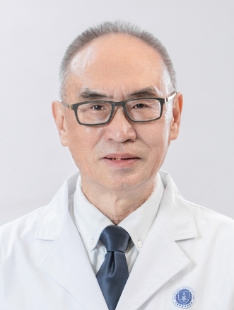 Shuhua Yang (杨述华)合作论文数Union Hospital, Tongji Medical College, Huazhong University of Science and Technology188
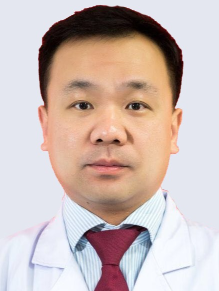 Yukun Zhang (张宇坤)合作论文数Union Hospital, Tongji Medical College, Huazhong University of Science and Technology121
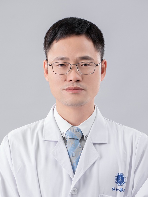 Xinghuo Wu (吴星火)合作论文数Union Hospital, Tongji Medical College, Huazhong University of Science and Technology115
 Yu Song合作论文数108Kun Wang合作论文数108Shuai Li合作论文数99
 Wenbin Hua合作论文数93Xiaobo Feng合作论文数85
 Weihua Xu (许伟华)合作论文数Union Hospital, Tongji Medical College, Huazhong University of Science and Technology70
 Jin Li (李进)合作论文数Union Hospital, Tongji Medical College, Huazhong University of Science and Technology68Zhiwei Liao合作论文数67
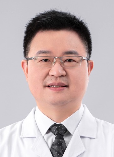 Yong Gao (郜勇)合作论文数Union Hospital, Tongji Medical College, Huazhong University of Science and Technology66
 Bingjin Wang合作论文数64Gaocai Li合作论文数61
 Zengwu Shao (邵增务)合作论文数Union Hospital, Tongji Medical College, Huazhong University of Science and Technology53
 Rongjin Luo合作论文数49Wencan Ke合作论文数49
 Haitao Ran (冉海涛)合作论文数The Second Affiliated Hospital of Chongqing Medical University45Kangcheng Zhao合作论文数44Huaizhen Liang合作论文数39Liang Ma合作论文数38
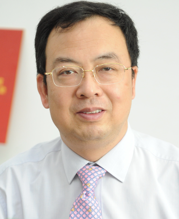 Yingbin Liu (刘颖斌)合作论文数Renji Hospital Affiliated to Shanghai Jiaotong University School of Medicine;Shanghai Cancer Institute38
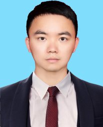 Lei Tan (谭磊)合作论文数Department of Orthopaedics, Union Hospital, Tongji Medical College, Huazhong University of Science & Technology33
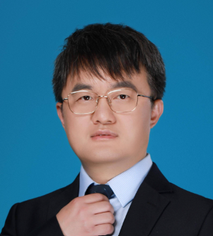 Maolan Li (李茂岚)合作论文数Renji Hospital, Shanghai Jiao Tong University School of Medicine33YiJun Shu (束翌俊)合作论文数32Zhewei Ye合作论文数31Run-Fa Bao合作论文数30
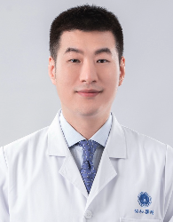 Xianzhe Liu (刘先哲)合作论文数Union Hospital, Tongji Medical College, Huazhong University of Science and Technology30
 Ji Tu (涂计)合作论文数29
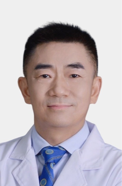 Guohui Liu (刘国辉)合作论文数Union Hospital, Tongji Medical College, Huazhong University of Science and Technology28
 Zhenfeng Duan合作论文数Department of Orthopaedics, Miller School of Medicine, University of Miami28Xiangsong Wu (吴向嵩)合作论文数27
 Zhigang Wang (王志刚)合作论文数The Second Affiliated Hospital of Chongqing Medical University27Xianlin Zeng合作论文数25Weifeng Zhang合作论文数25
 Henry J. Mankin合作论文数Harvard Medical School;Department of Orthopaedic Surgery, Massachusetts General Hospital25
 Di Wu (吴棣)合作论文数24Wen-Guang Wu合作论文数23
 Jie Lei合作论文数Department of Orthopaedics, Union Hospital, Tongji Medical College, Huazhong University of Science and Technology, Wuhan 430022, China.23Hao Weng (翁昊)合作论文数23
 Wei Liu (刘卫)合作论文数College of Life Science and Technology, Huazhong University of Science and Technology;National Engineering Research Center for Nanomedicine23
 Kang Liang合作论文数22Hu Yunping合作论文数22
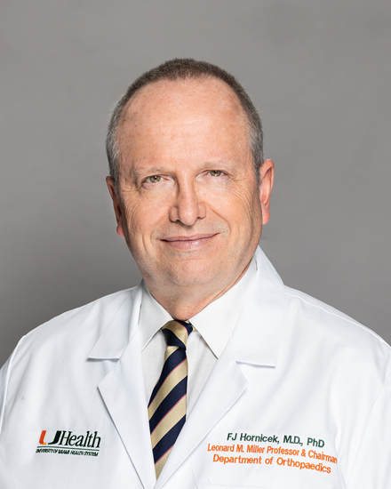 Francis John Hornicek合作论文数Sarcoma Research Laboratory, University of Miami22
 Edwin Choy合作论文数Departments of Medicine and Cell Biology, New York University School of Medicine22
 Baojun Xiao (肖宝钧)合作论文数Union Hospital, Tongji Medical College, Huazhong University of Science and Technology21Jiasheng Mu (穆嘉盛)合作论文数21Shunan Ye (叶树楠)合作论文数21Huipeng Yin合作论文数20




