Abstract Glioblastoma (GBM) is the most common primary brain tumor in adults with a median survival of less than 15 months with maximal safe surgical resection, radiation, and the chemotherapy temozolomide. Addition of Tumor Treating Fields (TTFields), or alternating electromagnet fields therapy, to temozolomide was shown to extend the survival of GBM patients by approximately 4.9 months. TTFields disrupt mitosis to inhibit cell growth, but we also determined that TTFields alter the cellular kinome. Using a PamStation, we identified kinases that are predicted to be activated and repressed by TTFields treatment in newly diagnosed and recurrent GBM models that are sensitive or resistant to temozolomide or irradiation, respectively. While the growth of all GBM cells tested was significantly decreased by TTFields, there was a relatively limited set of kinases that were commonly altered in newly diagnosed and temozolomide-resistant GBM cells with little similarly across irradiation resistant GBMs. These kinase data are reminiscent of published data demonstrating kinome variability in radioresistant GBM xenografts. We did find that TTFields were predicted to activate PDGFRα in both newly diagnosed and temozolomide-resistant GBM cells: when combined with TTFields, a blood brain barrier penetrant PDGFR inhibitor, crenolanib, significantly decreased GBM cell growth. Subsequent studies have identified additional kinases to be evaluated in combination with TTFields in radioresistant GBM cells. Using the Novocure inovivo system, we plan to test these novel kinase inhibitor combinations with TTFields in mouse models bearing intracranial GBMs. We hope to identify a kinase inhibitor based treatment strategy that can be translated to the clinic to further improve TTFields mediated increases in patient survival. Citation Format: Taylor Lynn Schanel, Amber Jones, Rhea Pandit, Johsua C. Anderson, Patricia H. Hicks, Corinne Griguer, Braden C. Mcfarland, Christopher D. Willey, Anita B. Hjelmeland. Tumor Treating Fields remain effective in therapy-resistant glioblastoma with kinome shifts revealing novel therapeutic opportunities [abstract]. In: Proceedings of the American Association for Cancer Research Annual Meeting 2026; Part 1 (Regular Abstracts); 2026 Apr 17-22; San Diego, CA. Philadelphia (PA): AACR; Cancer Res 2026;86(7 Suppl):Abstract nr 5878.
Abstract Introduction: Glioblastoma (GBM) is the most common malignant brain tumor in adults, and resistance to radiation therapy remains a major therapeutic obstacle. Although genetic alterations contribute to GBM progression, they do not fully explain the emergence of radiation resistance, suggesting contributions from non-genetic mechanisms. Notably, radiation-resistant GBM exhibits elevated collagen deposition. Discoidin domain receptor tyrosine kinase 1 (DDR1), a collagen-binding receptor regulated by ectodomain shedding, is highly expressed in GBM. Here, we demonstrate that radiation-resistant GBM alters DDR1 activity and shedding in response to collagen to promote tumor cell proliferation. Methods and Results: To investigate DDR1 regulation, we utilized a GBM patient-derived xenoline pair, JX39P (radiation-sensitive) and JX39P-RT (radiation-resistant). JX39P-RT displayed significantly higher DDR1 mRNA and protein expression. When cells were embedded in collagen I to mimic the extracellular environment, DDR1 phosphorylation at Y792 increased more rapidly and to a greater extent in JX39P-RT than in JX39P. Collagen embedment also led to the accumulation of a short DDR1 fragment in JX39P-RT, indicating enhanced ectodomain shedding. Western blotting of conditioned media confirmed increased release of the DDR1 N-terminal fragment and confirmed that ectodomain shedding occurred only in JX39P-RT in response to collagen. Treatment of JX39P-RT cells with marimastat, a broad-spectrum MMP inhibitor, diminished both the N-terminal fragment shed in the media and the short C-terminal fragment in cell lysates. Consistent with elevated DDR1 expression and activation, JX39P-RT exhibited increased phospho-ERK1/2 levels when embedded in collagen I, indicating enhanced downstream signaling pathways. Furthermore, cell proliferation assays and Ki67 staining showed that JX39P cells proliferate more rapidly than JX39P-RT in liquid culture; however, when embedded in collagen I, JX39P showed no significant change in Ki67 staining, whereas JX39P-RT demonstrated a time-dependent increase, indicating that radiation-resistant GBM cells proliferate in response to collagen- and DDR1-mediated signaling. Conclusions: Together, our results identify DDR1 upregulation and ectodomain shedding as key adaptive mechanisms that promote proliferation and survival of radiation-resistant GBM cells. These regulatory processes enable resistant cells to sustain growth under therapeutic stress, providing a potential mechanism of therapeutic evasion. Future studies will investigate how DDR1 shedding and downstream signaling integrate to maintain radiation resistance and explore strategies to sensitize GBM to radiation therapy. Citation Format: Sophia Dunlap, Caitlin A. Harvey, Christopher D. Willey, Ahn Erin. The role of DDR1 ectodomain shedding in glioblastoma radiation resistance [abstract]. In: Proceedings of the American Association for Cancer Research Annual Meeting 2026; Part 1 (Regular Abstracts); 2026 Apr 17-22; San Diego, CA. Philadelphia (PA): AACR; Cancer Res 2026;86(7 Suppl):Abstract nr 3306.
During glioblastoma (GBM) progression, therapeutic resistance is influenced by a heterogeneous network of tumor- and tumor-promoting subpopulations within the tumor microenvironment. Therapeutic interventions against tumor cells, such as chemoradiotherapy, can further induce adaptive defense mechanisms mediated by sophisticated intercellular communication and aberrant signaling pathways. Tunneling nanotubes (TNTs) have emerged as key mediators of this process, promoting tumor cell survival through metabolic rescue while enabling the recruitment and reprogramming of surrounding normal cells into tumor-supportive phenotypes. This dynamic is exemplified in GBM, where TNT-mediated interactions between brain tumor-initiating cells (BTICs) and normal human astrocytes (NHAs) highlight the need to elucidate the molecular mediators underlying this crosstalk. Myristoylated Alanine Rich C-Kinase Substrate (MARCKS) has never been investigated as a potential regulator of TNTs despite several overlapping signaling pathways and its influence on GBM therapeutic resistance. In the present study, we demonstrate a role for the MARCKS effector domain (ED) and PKC activation in the formation and functionality of TNTs between PTEN-null GBM BTICs and NHAs. We employ a MARCKS phosphorylation site (MPS) peptide derived from MARCKS effector domain (MED2), PKC-targeting drugs, and an inducible MARCKS ED U87 model to elucidate a potential role for MARCKS phosphorylation and PKC in TNT regulation between GBM cells (i.e., BTICs or U87s) and NHAs.
PURPOSE:Radiosurgical treatment of multiple intracranial targets using a single isocenter is more efficient than sequential isocenter treatment. The primary concern with single-isocenter, multitarget (SIMT) stereotactic radiosurgery (SRS) is that rotational error may cause underdosing of peripheral lesions and/or increased normal tissue toxicity, particularly for targets distant from the isocenter. Many centers mitigate this risk by adding a planning target volume (PTV) margin, which increases dose to normal brain tissue. This study examined whether distance from the isocenter affects local tumor control or toxicity in patients treated with SIMT SRS using a 0-mm PTV margin. METHODS AND MATERIALS:We retrospectively evaluated 429 patients with 3039 intracranial tumors treated with SIMT SRS or fractionated SRS using single-isocenter volumetric modulated arc therapy plans with a 0-mm clinical target volume/PTV expansion from gross tumor volume. All treatments used the Varian HD-120 multileaf collimators on a 6-degree-of-freedom couch. Local failure was defined as ≥25% increase in maximum tumor diameter (minimum 3 mm) or viable tumor cells at salvage surgery. Toxicity was defined as National Cancer Institute Common Terminology Criteria for Adverse Events version 5.0 central nervous system grade ≥3. RESULTS:Of the 3039 tumors evaluated, 1510 and 1034 tumors had clinical and radiographic follow-up at 6 months and 1 year, respectively. Median distance from the isocenter was 4.9 cm (range, 0.04-13.4 cm) and 70% of tumors in this study were ≥4 cm from the isocenter. One-year local control was 95.1% (95% CI, 94.0-96.1) and freedom from grade ≥3 toxicity was 98.4% (95% CI, 97.8-99.0). Larger tumor volume correlated with both worse local control (hazard ratio, 1.017; 95% CI, 1.000-1.034; P = .047) and higher toxicity (hazard ratio, 1.088; 95% CI, 1.056-1.121; P < .001). Increased distance from the isocenter was not associated with worse local failure or toxicity on multivariable analysis. CONCLUSIONS:In this retrospective analysis of SIMT volumetric modulated arc therapy radiosurgery using HD-120 multileaf collimators, 6-degree-of-freedom couch, intrafraction surface monitoring, and 0-mm clinical target volume/PTV margin, increased distance from the isocenter did not adversely impact short-term local tumor control or increase high-grade toxicity. These findings are currently being tested in a prospective randomized trial.
PURPOSE:We previously reported short-term (2 months) efficacy results of an advanced pneumatic compression device (APCD) versus usual care (UC) for the treatment of head and neck cancer survivors with symptomatic treatment-naive lymphedema (HNLEF). Herein we report the long-term (4 and 6 months) outcomes of that trial. METHODS AND MATERIALS:This multisite, prospective randomized clinical trial was conducted at academic and community-based sites. Eligibility criteria included: head and neck cancer survivors without evidence of cancer, previously untreated HNLEF evaluable on exam or imaging, and ≥1 associated symptom with severity of ≥4 out of 10. Participants were randomly assigned 1:1 to either daily use of an APCD for 6 months or UC per institutional standards. Measurement tools included: patient-reported outcome measures, Clinician-reported outcome (CRO) measures, digital photographs, and computed tomography (CT). Measures were at baseline, 2, 4 (no CT), and 6 months. RESULTS:A total of 236 participants were enrolled (119 APCD, 117 UC). Tumor distribution by group was as follows: APCD = larynx 29.4%, salivary glands 1.7%, oral cavity 37.8%, paranasal sinuses 1.7%, pharynx 25.2%, unknown primary 4.2%; UC = larynx 16.2%, salivary glands 5.1%, oral cavity 48.7%, paranasal sinuses 1.7%, pharynx 21.4%, unknown primary 6.8%. Of the UC group, 17.1% (n = 20) underwent bilateral neck dissection, as did 12.6% (n = 15) of the APCD group. Symptom improvement garnered during initial treatment was maintained over time in both groups with no significant difference between groups. CRO measures demonstrated improvement in internal HNLEF (Modified Patterson Scale, P < .01 both groups) and external HNLEF (grading criteria, APCD P < .01; UC P = .06) in the APCD group. Statistically significant differences at 2 of 19 anatomic subsites favored the APCD group. At 6 months, the digital photography showed improvement with no between-group difference. CT findings at 6 months verified significant improvement in soft tissue swelling in both groups (P < .01 for both groups) that was not present at 2 months, with no between-group difference. CONCLUSIONS:At 6 months, the analysis indicated that APCD and UC resulted in improved symptom control of similar magnitude. CROs, imaging, and digital photography demonstrated improvement in anatomic lymphedema in both groups over time. Select CRO outcome measures demonstrated marginal differences between groups that favored the APCD. Both interventions provided long-term benefit to patients with treatment-naive lymphedema.
PET imaging targeting immune cells can be used to dynamically monitor intratumoral immune modulation in tissues. Radiation therapy is known to alter the tumor immune microenvironment; therefore, this study demonstrates how CD8 immunoPET imaging can optimize combination immunotherapy and radiation therapy by stratifying tumors who could derive the greatest benefit from immunotherapy following radiation therapy. A radiation resistant triple negative breast cancer cell line was derived through repeat irradiation of the radiosensitive parental 4T1 cell line prior to in vivo studies, until a radiation resistant subclone (RR-4T1) was isolated. CD8 immunoPET imaging was used to image immune cell infiltration in response to fractionated radiotherapy in radiation sensitive and radiation resistant 4T1 breast cancer models. In this genetically matched radiation sensitive and resistant model, we explore how radiation resistance alters radiation-induced immune modulation and CD8 T cell trafficking with flow cytometry, while response to combination radiation and immunotherapy was assessed in the radiosensitive parental 4T1 model. CD8 immunoPET was utilized to stratify for long-term therapeutic response to immunotherapy based on post-radiation therapy changes in CD8 tissue infiltration. Radiosensitive parental 4T1 tumors show increased CD8 immunoPET signal (SUV) when treated with radiation therapy, relative to control tumors (p < 0.01) whereas radiation resistant 4T1 tumors showed no change following radiation therapy (p = 0.99), which was validated with flow cytometry. When tumors were stratified for high or low CD8 minibody uptake, CD8-high radiosensitive parental 4T1 tumors treated with radiation and immunotherapy had significantly increased sensitivity to immunotherapy compared to CD8-low radiosensitive parental 4T1 tumors (p < 0.05). Radiation therapy enhanced CD8 + expression in tumors and CD8 immunoPET effectively stratifies tumors that are more likely to respond to subsequent immunotherapy. CD8 immunoPET provides an approach to optimize combination immunotherapy following radiation treatment in triple-negative breast cancer.
Glioblastoma (GBM) is a highly lethal brain cancer with limited therapeutic durability, where the majority of patients develop recurrent or persistent disease after standard chemoradiotherapy. Meanwhile, tRNA-derived fragments (tRFs) have become increasingly relevant to cancer biology; however, their clinical relevance in GBM remains undefined. Here, we report that a specific family of tRFs, 5'-tRNA halves (tiR5s) dominates the small RNA landscape of GBM patient tumors and associates with worse overall survival, post-therapeutic disease persistence, and pro-invasive proteogenomic pathways across two independent GBM patient cohorts. This association between elevated tiR5 levels and therapeutic resistance re-emerges in radiation-resistant GBM xenograft models. Our findings reveal that tiR5s are an underappreciated molecular feature of highly aggressive GBM tumors, supporting further investigation into their biological roles and prognostic utility in GBM. Highlights:tiR5s are the predominant tRF family in primary GBM patient tumorsElevated tiR5 expression distinguishes primary GBM tumors that develop persistent disease after first-line therapyRadiation-resistant GBM PDX models show elevated tiR5 expressionElevated tiR5 expression associates with poor overall patient survival and pro-invasive molecular programs in GBM patient tumors.
BACKGROUND:Two-month outcomes of advanced pneumatic compression device (APCD) and usual care (UC) in Head and Neck Cancer survivors with previously untreated lymphedema were compared. METHODS:Participants in this multisite, randomized clinical trial were randomized to APCD or UC. The primary endpoint was severity of lymphedema symptoms. Secondary endpoints were anatomical lymphedema changes, biopsychosocial outcomes, and barriers to care. RESULTS:Two hundred thirty-six participants were enrolled (119 APCD, 117 UC). Analysis was intention-to-treat. Lymphedema-associated symptom burden measured using the VHNSS and LSIDS was improved to a similar degree in both groups. APCD demonstrated a statistically significant improvement in external soft tissue swelling assessed by digital photography. No difference in CT imaging measures of lymphedema was noted. UC participants experienced barriers to care. CONCLUSIONS:APCD is an effective treatment for lymphedema in HNCS. The APCD addresses clinically significant barriers to therapist guided treatment. A hybrid approach may be complementary and optimize patient outcomes. TRIAL REGISTRATION:NCT04797390.
Anaplastic thyroid cancer is an aggressive malignancy, for which prompt treatment is necessary. Surgical resection is the recommended initial treatment for resectable cases and most patients require postoperative radiation therapy (PORT). NCCN Guidelines state that adjuvant radiation should ideally begin 2–3 weeks postoperatively, however, limited evidence exists to support this guideline. Herein, we examine the National Cancer Database (NCDB) to assess practice patterns with regard to PORT timing, and to assess whether the time interval between surgery and the start of RT impacts overall survival (OS). An NCDB data file was obtained and condensed to only include stage IVa and IVb anaplastic thyroid cancer cases during the years 2004 to 2018. Patients were included if they were treated with surgery followed by radiation (6000 cGy-7020 cGy), and the radiation commenced no later than 90 days after surgery. Patients were grouped according to PORT: Early PORT (≤ 21 days after surgery) and Late PORT (> 21 days after surgery). Several variables were tested using univariate and multivariate binary logistic regression to determine their effect on whether patients received Early PORT vs. Late PORT. These variables were also used to conduct univariate and multivariate Cox regression to determine effects on survival for the cohort. The covariate variables included: presence or absence of a chemotherapy regimen in the treatment plan, total thyroidectomy or alternate surgery, type of treatment facility (Academic vs. Other), resection margin status (R0/R1 vs. R2), Charlson-Deyo comorbidity score, tumor stage, age, race/ethnicity and sex. The analysis included 277 patients after exclusions. The Early PORT group included 57 patients, and the Late PORT group included 220 patients. Multivariate binary logistic regression analysis showed that patients with R2 resection margins were less likely to receive Late PORT compared to patients with R0/R1 resection margins (OR: 0.306 (0.123–0.764)), and patients with stage IVb disease were less likely to receive Late PORT compared to patients with stage IVa disease (OR: 0.496 (0.257–0.957)). Multivariate cox regression analysis displayed survival advantages for patients who received Late PORT (HR: 0.563 (0.401–0.790)) and patients with R0/R1 resection margins (HR: 1.901 (1.161–3.114)), while treatment at an academic center trended toward significance (p = 0.053). The majority of patients in this cohort received Late PORT, later than the proposed guidance by the NCCN. Resection margin status and disease stage appears to influence how physicians select the timing of PORT. Within the limitations of our retrospective study, delayed PORT beyond NCCN recommendations is not associated with a detriment to OS. Further prospective analyses may be necessary to identify the ideal timing of adjuvant radiation therapy.
Abstract Glioblastoma (GBM) is the most aggressive primary brain tumor. Despite advances in immunotherapy for other solid tumors, T-cell checkpoint blockades have failed to improve overall survival in GBM patients, largely due to low T-cell infiltration. In contrast, tumor-associated macrophages (TAMs) comprise 82-97% of the immune cell population in newly diagnosed GBM. TAMs, however, often fail to engulf tumor cells, partly due to the phagocytosis inhibitory “do not eat me" signals such as CD47, which interacts with macrophage SIRPα to suppress phagocytosis. Although, CD47 blockade enhances phagocytosis in preclinical models, clinical trials of anti-CD47 therapies such as magrolimab were halted due to increased mortality and limited efficacy in acute myeloid leukemia patients, highlighting the need for alternative phagocytosis-inducing therapies. To identify novel tumor-expressed genes that inhibit macrophage phagocytosis, we performed a genome-wide CRISPR screen using two rounds of coculture of radioresistant GBM tumor cells (MGG18-RR) and tumor-conditioned human peripheral blood (hPBMCs)-derived macrophages. Tumor-conditioned hPBMCs macrophages were considered more reliable, as our cytometry imaging results revealed distinct polarization states compared to unpolarized (M0) macrophages. In the first round, MGG18-RR GBM cells were co-cultured with tumor-conditioned hPBMCs-derived macrophages at a 1:5 ratio for 7 days, with media refreshed every 2 days. Parallel tumor-only cultures served as controls. Tumor cells that survived this first co-culture were pooled and subjected to a second 7-day co-culture with freshly differentiated and polarized macrophages. After the second round, the remaining tumor cells were collected, and genomic DNA was extracted for library prep and sequencing. Among the top 100 hits, several known regulators of phagocytosis, including CD47 and KRAS were enriched, validating the reliability of the screen. We identified Epithelial Membrane Protein 2 (EMP2) as a novel tumor-expressed gene that suppresses macrophage phagocytosis. EMP2 showed high expression in glioma cells in publicly available single-cell sequencing data for GBM patients. EMP2 is highly expressed in GBM and associated with tumor progression and poor survival. Functional validation using the pHrodo in-vitro phagocytosis assay demonstrated a significant increase in the engulfment of EMP2-knockout GBM cells (JX14P-RT) by both hPBMCs-derived macrophages and RAW246.7 macrophages (p < 0.05), compared to wild-type tumor cells, suggesting that EMP2 acts as an anti-phagocytic regulator in GBM. Ongoing studies aim to validate that EMP2 knockout enhances macrophage phagocytosis in syngeneic murine GBM model. Collectively, our findings highlight EMP2 as a novel regulator of macrophage-mediated tumor clearance and a promising therapeutic target for macrophage-based immunotherapy in GBM. Citation Format: Amr Elkholy, Eshika Kudaravalli, Mostafa Mohamed, Hasan Alrefai, Saeed Zakakhosravi, Satoru Osuka, Christopher D. Willey, Ahn Erin. EMP2 is a potential immunotherapeutic target to modulate macrophage-induced phagocytosis in glioblastoma [abstract]. In: Proceedings of the American Association for Cancer Research Annual Meeting 2026; Part 1 (Regular Abstracts); 2026 Apr 17-22; San Diego, CA. Philadelphia (PA): AACR; Cancer Res 2026;86(7 Suppl):Abstract nr 7017.
This study evaluated the safety and effectiveness of an intraoral light-emitting diode (LED)-based photobiomodulation (PBM) device to reduce the incidence and severity of oral mucositis (OM) from intensity modulated radiation therapy (IMRT) for head and neck cancer (HNC). This randomized, double-blind, sham-controlled trial enrolled patients with HNC undergoing high-dose IMRT over 6–8 weeks, with or without concurrent chemotherapy. Participants received daily 10-min PBM or sham treatments immediately before IMRT sessions. Assessments were conducted at baseline, daily and weekly during IMRT, and two weeks post-IMRT. Eighty-five participants (42 PBM; 43 sham) were enrolled across 12 US sites. No device-related adverse events were observed, and 99.5
Gene Ontologies (GOs) are standardized descriptions of gene functions in terms of biological processes, molecular functions, and cellular components, capturing their Parent-Child relationships in a structured framework and advancing cancer biological modeling to provide consistent and meaningful insights into functional genomics analysis. The conventional GO hierarchical structure is defined by human curation experts, with levels determined by the shortest path to the root term. However, grouping GOs poses challenges due to the uneven distribution of gene members within GO terms and inconsistencies in the level of detail across terms at the same GO level. In this work, we introduce Gene Ontology Analysis using Layered Shells (GOALS), a novel tool that discretizes GOAs into optimal layers. GOALS creates scalable GO layers while maintaining a balanced number of genes across GOs in each layer. Unlike existing tools, the GOALS framework organizes GO terms using a bottom-up approach based on their co-membership network, discretizing GOs to achieve an exponential fit with GO’s gene member size. Meanwhile, GOALS reveals clusters or supersets reflecting biological relevance by unsupervised clustering of GO’s latent projections. In a case study on mouse natural killer (NK) cell development, GOALS identified distinct GO functional clusters with multi-GO layers to reveal multiple levels of detail from specific to abstract contexts to maximize signal discovery and uncover those signals’ associations with trajectory divergence. More importantly, GOALS enhances enrichment analysis by introducing additional GO stratification and latent GO map that enables more accurate classification of functional differences. GOALS offers a robust and innovative framework for exploring disordered GO clusters, mining GO activities, and analyzing potential GO-GO interplays. By addressing critical challenges in functional genomics, GOALS provides a powerful tool for advancing our understanding of cell heterogeneity and potentially uncovering actionable insights for therapeutic development. ### Competing Interest Statement The authors have declared no competing interest.
Purpose Adjuvant radiation therapy for atypical meningiomas (AMs) aids in local control following surgery and salvage after recurrence. The role of fractionated stereotactic radiosurgery (FSRT) in this population remains an area of active study with many unanswered clinical questions. This single-institution retrospective study evaluates the local control, marginal control, and toxicity of FSRT in treating AM. Methods Between 2009 and 2022, 39 patients with WHO grade 2 AM underwent FSRT via marginless, frameless volumetric-modulated arc therapy (VMAT) at doses of 27.5-30 Gy in five fractions. Local recurrence was defined as an increase of 20% in the greatest cross-sectional diameter on MRI or CT, following RECIST criteria. Cavity and marginal recurrences were defined as any new lesion outside the prescription volume but within the resection cavity or within 2 cm of the resection cavity, respectively. High-grade toxicity was defined per Common Terminology Criteria for Adverse Events (CTCAE) v5. Resection for radionecrosis with viable residual tumor was considered a local failure. Results Twenty-six AMs were treated post-subtotal resection (STR), 16 post-gross total resection (GTR) with recurrence, and five treated definitively. Patient characteristics included a mean age of 54 years, 20 (51%) male patients, and 31 (79%) patients with ECOG 0-1. The three-year local control rate was 84.0%. Larger tumors were more likely to fail locally (p > 0.001). Two (5%) patients experienced high-grade toxicity necessitating resection. The three-year marginal control rate was 92.3%, and recurrent tumors post-GTR failed marginally more often compared to those treated after STR (p = 0.009). One (4%) tumor treated after STR failed marginally, while four (33%) tumors treated after GTR recurrence failed marginally. The three-year control rate of the unirradiated cavity was 88%. Conclusion The rate of high-grade toxicity in AM patients receiving FSRT was low. Local control appeared comparable to historical rates, which may suggest the potential need for dose escalation with longer-term follow-up. Recurrent tumors were more prone to marginal failures. Further investigation is needed to determine which patients may benefit from whole-cavity treatment, additional CTV margin, or prolonged fractionated dose schedules. Newer imaging studies, including DOTATATE PET, should be explored to assess whether improvements in targeting accuracy can enhance outcomes.
Background:Brain metastases (BMs) patients often receive multiple courses of radiotherapy (RT). Comparing follow-up imaging with RT plans is time-intensive and currently not possible in picture archiving and communication system (PACS). In this study, we aim to develop an automated tool to overlay pre-RT tumor contours onto follow-up MRI in the PACS to support response assessment and identification of untreated metastases, reducing review complexity and time. Methods:We built an AI-driven workflow that registers planning CT to follow-up MRI and propagates treated-lesion contours; outputs are exported as PACS-compliant DICOM. Performance was evaluated in 40 patients: 20 underwent quantitative comparison between manual and automated registrations using Dice similarity coefficient (DSC) and mean surface distance (MSD), following AAPM TG-132 guidelines; 20 additional patients (5-35 lesions per patient) underwent clinical evaluation of follow-up and pre-RT lesions contours visualization in PACS. Three physicians assessed these cases to measure review time and inter-observer agreement in treatment response classification (improved/stable vs indeterminate). Results:The workflow successfully registered all CT-MRI pairs. Mean DSC/MSD was: brain 0.97 ± 0.01/0.0 ± 0.0 mm, brainstem 0.89 ± 0.03/0.1 ± 0.1 mm, and gross tumor volumes 0.65 ± 0.18/0.6 ± 0.4 mm. Average physician review time per case decreased from 7.97 to 3.95 min with the automated workflow, and full inter-physician agreement increased from 72.4% to 93.5%. Conclusion:We developed and validated an AI-based tool that accurately fuses pre-RT contours with follow-up MRI for BMs, addressing a key gap in current PACS systems. The workflow significantly reduced review time per lesion and enhanced inter-physician agreement, and has the potential to enable faster, more consistent multidisciplinary follow-up assessment.
Development of acquired therapeutic resistance limits the efficacy of cancer treatments and accounts for therapeutic failure in most patients. How resistance arises, varies across cancer types and differs depending on therapeutic modalities is incompletely understood. Novel strategies that address and overcome the various and complex resistance mechanisms necessitate a deep understanding of the underlying dynamics. We are at a crucial time when innovative technologies applied to patient-relevant tumour models have the potential to bridge the gap between fundamental research into mechanisms and timing of acquired resistance and clinical applications that translate these findings into actionable strategies to extend therapy efficacy. Unprecedented spatial and time-resolved high-throughput platforms generate vast amounts of data, from which increasingly complex information can be extracted and analysed through artificial intelligence and machine learning-based approaches. This Roadmap outlines key mechanisms that underlie the acquisition of therapeutic resistance in cancer and explores diverse modelling strategies. Clinically relevant, tractable models of disease and biomarker-driven precision approaches are poised to transform the landscape of acquired therapy resistance in cancer and its clinical management. Here, we propose an integrated strategy that leverages next-generation technologies to dissect the complexities of therapy resistance, shifting the paradigm from reactive management to predictive and proactive prevention. Acquired therapeutic resistance is a key contributor to cancer treatment failure, requiring new approaches to address its complex mechanisms. In this Roadmap, Soragni, Knudsen and colleagues discuss the mechanisms of acquired resistance and the models to better study it. Finally, they promote integration of biomarker-driven strategies and cutting-edge technologies to advance predictive and proactive prevention in cancer therapy.
Purpose:Preoperative stereotactic radiosurgery (SRS) has been proposed as a strategy to reduce nodular leptomeningeal disease (nLMD) after resection of brain metastases by devitalizing tumor cells before surgical manipulation. This study aimed to determine the safety of preoperative SRS via a phase 1 dose escalation trial and compare outcomes-including nLMD, classical LMD, local control, and overall survival (OS)-between preoperative and postoperative treatment cohorts. Methods and Materials:A phase 1 trial evaluated the safety of single-fraction preoperative SRS at escalating doses (12 and 15 Gy) in patients with tumors 2 to 6 cm. Adverse events were assessed per National Cancer Institute Common Terminology Criteria for Adverse Events v4.0, with dose-limiting toxicity (DLT) defined as high-grade neurologic or wound complications. Retrospective analysis included an expanded preoperative cohort (n = 95) and a historical postoperative cohort (n = 107). Outcomes were analyzed with Kaplan-Meier and Cox proportional hazards models. Results:Fifteen Gy was well tolerated in the phase 1 cohort, with no DLTs in larger tumors (4-6 cm) and 3 DLTs in smaller tumors, not meeting thresholds for dose de-escalation. In the expanded analysis, preoperative SRS significantly reduced rates of nLMD (7.4% vs 27.1%, P = .002), while rates of classical LMD (4.2% vs 4.5%) and local failure (14.6% vs 18.7%) were similar between groups. OS was also similar (median 12.8 vs 12.3 months). Multivariable analysis confirmed preoperative SRS as protective against nLMD (hazard ratio = 0.18, 95% CI, 0.07-0.43, P < .001). Conclusions:Preoperative SRS to 15 Gy is safe for tumors 2 to 6 cm and significantly reduces nLMD without compromising local control or OS. These findings support preoperative SRS as a viable treatment strategy and justify further investigation into optimal dosing and patient selection.
Ultra-high dose rate (FLASH-RT) and conventional proton beam radiotherapy to intracranial glioblastoma PDX were compared for tumor control and normal tissue toxicity via DNA and RNA damage response markers. Both conventional and FLASH radiotherapy yielded similar survival benefits; however, conventional radiotherapy resulted in greater normal tissue DNA and RNA damage.
Kinases, at the signaling level, dynamically mediate uncontrolled cellular growth, survival and other cancer supporting processes. This, paired with the inherent druggability of kinases, points to the importance of measuring kinase activity, and that of inhibitors against them, directly, and to analyze this accurately. High-throughput kinome profiling technologies, such as the PamStation®12, allow researchers to kinetically capture kinase activity, against a multitude of peptide targets simultaneously. Yet, the complex datasets produced often require advanced computational tools and bioinformatics expertise to properly analyze that are not intuitive or readily available. To address this gap, we developed KinoViz, a web-based application to simplify analysis and visualization of kinome array data. KinoViz offers a suite of interactive tools that enables users to upload raw peptide phosphorylation datasets and conduct in-depth analyses without the need for coding knowledge. Key features include modules for visualizing kinetic phosphorylation curves, identifying statistically significant peptide changes, exploring individual peptide profiles, and generating insightful visualizations such as heatmaps, network diagrams, and dimensionality reduction plots (PCA, UMAP). By making complex kinomic data more accessible and interpretable, KinoViz allows researchers to rapidly generate interactive visualizations and comparative analyses. We aim to expand KinoViz's analytical capabilities for more advanced use, including use in direct translational drug discovery.
Purpose:Single isocenter stereotactic radiosurgery (SRS) efficiently delivers radiation to patients with multiple brain metastases. Although several fractionated SRS (fSRS) regimens show acceptable local control and toxicity, few studies directly compare them. This retrospective study evaluates 2 common regimens-6 Gy × 5 fractions and 9 Gy × 3 fractions-for their effects on local control and toxicity. Methods and Materials:A retrospective review was conducted of 1215 brain tumors from 251 patients receiving either 9 Gy × 3 fx or 6 Gy × 5 fx fSRS. All tumors were treated with single isocenter volumetric modulated arc therapy. Recurrent tumors and postoperative cavities were excluded from the analysis. Local tumor failure was defined as 25% increase in maximum tumor diameter (minimum 3 mm) or more than scant tumor cells at time of salvage surgery. Toxicity included CTCAE V5.0 central nervous system (CNS) grade 3 or greater events. Local tumor control and freedom from toxicity were calculated using Kaplan-Meier method and Cox regression models. Results:Overall local control was 93% at 1 year and 88% at 2 years. The 3-fraction regimen had superior 1-year local control compared with the 5-fraction regimen (97% vs. 91%, P = .001). Tumors <2 cm had significantly better control with 3 fractions (99% vs. 95%, P = .004), whereas tumors 2-4 cm showed no significant difference. One-year freedom from grade 3+ toxicity was similar between regimens (99% for 3-fx vs. 96% for 5-fx, P = .097). Conclusions:In this study, 9 Gy × 3 fx for brain metastases had improved tumor control and comparable toxicity to 6 Gy × 5, particularly among tumors <2 cm. 9 Gy × 3 fx may be the preferred regimen when treating multiple tumors with one prescription using single isocenter radiosurgery as it improves efficiency and local control while having similar toxicity.
 Joshua Anderson合作论文数88
 Markus Bredel合作论文数37
 Eddy S Yang合作论文数Department of Radiation Medicine, College of Medicine, University of Kentucky33
 John Fiveash合作论文数Department of Radiation Oncology, Heersink School of Medicine, The University of Alabama at Birmingham;O'Neal Comprehensive Cancer Center, Heersink School of Medicine, The University of Alabama at Birmingham29
 James Bonner合作论文数Department of Radiation Oncology, Heersink School of Medicine, The University of Alabama at Birmingham25
 G. Yancey Gillespie合作论文数Heersink School of Medicine, The University of Alabama at Birmingham21
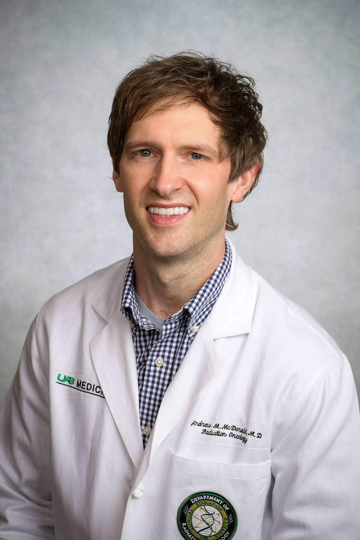 Andrew McDonald合作论文数Department of Radiation Oncology, Heersink School of Medicine, The University of Alabama at Birmingham19
 Anita Hjelmeland合作论文数Department of Cell, Developmental and Integrative Biology, University of Alabama at Birmingham;O'Neal Comprehensive Cancer Center, School of Medicine, University of Alabama at Birmingham;Center for Glial Biology in Medicine, University of Alabama at Birmingham19
 Richard A Popple合作论文数19
 Hicks Patricia H合作论文数18
 John S Jarboe合作论文数18
 James Macdowell Markert合作论文数Department of Neurosurgery, School of Medicine, University of Alabama, Birmingham17
 Christian Stackhouse合作论文数Dept Radiat Oncol, Univ Alabama Birmingham17Dobelbower Michael C合作论文数17Hasan Alrefai合作论文数16Dennis Hallahan合作论文数Washington University in St. Louis15Nicholas Eustace合作论文数Sch Med, Univ Alabama Birmingham15
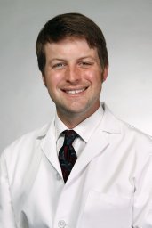 Boggs Drexell Hunter合作论文数Department of Radiation Oncology, University of Maryland Medical Center15Andee M Beierle合作论文数14Rohrbach Timothy D合作论文数14
 Kristen E. Riley合作论文数13
 Guru P. Sonpavde合作论文数Department of Medical Oncology, Dana-Farber/Harvard Cancer Center, Dana-Farber Cancer Institute12Christine W. Duarte合作论文数12Kristen O. Riley合作论文数11
 Gurudatta Naik合作论文数Comprehensive Cancer Center, University of Alabama at Birmingham;National Cancer Institute, National Institutes of Health11Sharon Spencer合作论文数11
 Samantah Anne Spencer合作论文数11Hoa Q. Trummell合作论文数11Bruce A. Julian合作论文数10
 Jake Chen合作论文数College of Arts and Sciences, The University of Alabama at Birmingham;Department of Biomedical Engineering, The University of Alabama at Birmingham;MedeoLinx, LLC;Institute of Electrical and Electronics Engineers;BIOKDD;American Computing Machinery10Catherine P. Langford合作论文数10Taylor L. Schanel合作论文数9Zhi-Qiang Huang合作论文数9Samuel R Marcrom合作论文数9Patricia J Hicks合作论文数9
 Dussaq Alex M合作论文数The Department of Pathology, The University of Alabama at Birmingham9Gilbert Ashley N合作论文数The Department of Radiation Oncology, The University of Alabama at Birmingham9Stewart Jerry E合作论文数9
 Dinesh Thotala合作论文数Radiation Oncology, Vanderbilt University9Elizabeth A. Beierle合作论文数9
 Dongquan Chen合作论文数Institute of Applied Physics and Computational Mathematics 100088 Beijing China9
 Zongliang Yue合作论文数8J.C. Anderson合作论文数8
 Colin Quinn合作论文数8
 Bo Lu合作论文数University of Missouri-Columbia;Fischel Cancer Center, University of Missouri Health Care8Welaya Karim合作论文数8
 Evan Marshall Thomas合作论文数Department of Radiation Oncology, University of Alabama at Birmingham;Department of Neurosurgery, University of Alabama at Birmingham;University of Alabama at Birmingham, University of Alabama at Birmingham8
 William E. Grizzle合作论文数Heersink School of Medicine, The University of Alabama at Birmingham8




