CryoSeek is a research strategy that employs cryo-electron microscopy (cryo-EM) to discover bio-entities from accessible sources, supplemented with AI-facilitated data processing and bioinformatic analyses. Here we report CryoSeek-based characterization of more glycofibrils isolated from the Tsinghua Lotus Pond (TLP), named TLP-0/2/3/4b/IPT, with resolutions ranging from 3.0-3.5 Å. These glycofibrils, all covered with dense glycoshells, have various mass percentiles of the central protein components. TLP-0 has no protein at all, TLP-2 and TLP-4b each only have a linear chain of di- or tetra-peptide repeats, respectively; the central stem of TLP-3 is a trimer of linear tripeptide repeats, and IPT (immunoglobulin-like, plexins, and transcription factors) refers to the tandem domain that constitutes the central stem of TLP-IPT. Glycan-mediated interactions determine the structural assembly of the glycofibrils that lack folded protein component. Our previous and current studies reveal the diversity of the structural folds of glycans, advance our understanding of glycan architecture, and further demonstrate CryoSeek as a structure-first paradigm for discovery.
The serotonin transporter (SERT) is responsible for the reuptake of the neurotransmitter serotonin (5-HT), utilizing the Na⁺ gradient across the cell membrane to drive substrate transport. It is proposed that 5-HT is cotransported with one Na⁺ and one Cl⁻, while a potassium ion (K⁺) is transported in the opposite direction. Despite recent structural advances in SERT, the precise intracellular K⁺ binding site still requires experimental structural confirmation. In this study, we capture two previously unreported states of SERT: an inward-open conformation bound with one K⁺ ion and one Cl⁻ ion, and an outward-open conformation free of any ions. These findings provide comprehensive insights into the ion-coupled transport cycle of SERT.
Approximately one-third of clinical drugs mediate their therapeutic effects through G protein-coupled receptors (GPCRs), highlighting their immense therapeutic relevance. Novel approaches to modulate GPCR activity have the potential to yield unique pharmacological profiles. Conventionally, the G protein and β-arrestin signaling pathways downstream of GPCRs have been viewed as mutually exclusive. Using the in-house developed survival pressure selection (SPS) method, a high-throughput platform for GPCR agonist discovery, we identified an allosteric ligand that stabilizes a GPCR-G protein-β-arrestin megacomplex, thereby mediating sustained receptor signaling following internalization. Remarkably, this compound, atazanavir, exhibits pan-receptor activation across multiple family A GPCRs, including GPR119, β1AR, and β2AR, demonstrating the broad applicability of this regulatory mechanism. This discovery uncovers a distinct mechanism of GPCR regulation, opening alternative avenues for the development of therapeutics targeting GPCRs.
The chemical complexity and nontemplated biosynthesis of glycans have posed considerable challenges for establishing sequence-structure relationships. Here we report cryo-electron microscopy structures of tubular mastigonemes from a golden alga species, Ochromonas danica, in which a large number of N- and O-glycans are resolved at 1.8- to 2.2-angstrom resolution. Beyond high-mannose and complex N-glycans, we identify a noncanonical N-glycan on the Ala-Asn-Asp (AND) motif. The surface spikes comprise dense O-glycans coating PSXX tetrapeptide repeats, with two glycans linked on trihydroxylated proline and one on serine per repeat. In addition to various types of sugars and their covalent modifiers, water molecules (>10% of resolved volume) and cations are clearly resolved and mediate the structural assembly. Our study establishes a framework for investigating glycan folding in high-order biological assemblies.
Cryo-electron microscopy (Cryo-EM) single particle analysis (SPA) is currently the primary method for determining structures of macromolecular complexes ranging from tens of kilodaltons (kDa) to several megadaltons (MDa). Traditional cryo-EM three-dimensional (3D) reconstruction and refinement methods are based on maximum likelihood approaches. Recently, deep learning-based methods began to emerge in the field of cryo-EM reconstruction. Here, we propose cryoFIFA, a deep curriculum learning-based approach for cryo-EM fine and fast 3D reconstruction. CryoFIFA enables the ab-initio reconstruction of macromolecules ranging from ~150 kDa to over 2 MDa, and significantly enhances reconstructing speed through supporting multi-GPU processing. It also demonstrates superior performance in the reconstruction of near-atomic resolution map for G-protein coupling receptors (GPCR) complex. CryoFIFA provides a framework for exploring new paradigms in deep learning-driven cryo-EM reconstruction, highlighting the potential for future applications of deep learning in cryo-EM analysis.
Deciphering transducer selectivity in G protein-coupled receptors (GPCRs) is essential for developing next-generation therapeutics with improved safety profiles. Here, we identify (R)-141, a μ-opioid receptor (μOR) agonist with a distinct scaffold that exhibits exceptional G protein bias. To decode the underlying mechanism, we determined the cryo-EM structures of μOR bound to (R)-141 in complex with Gi and with GRK2. Our structural, functional and dynamics data together reveal that (R)-141 achieves this selectivity through a stepwise gating mechanism, in which the conformational dynamics of TM7 serves as a terminal checkpoint. This "conformational veto" by TM7 provides a mechanism to modulate β-arrestin recruitment at the final step. Collectively, our work provides a systemic vision of transducer selectivity and a framework for rational biased drug design.
Recent breakthroughs in protein structure prediction have opened new avenues for genome-wide drug discovery, yet existing virtual screening methods remain computationally prohibitive. We present DrugCLIP, a contrastive learning framework that achieves ultrafast and accurate virtual screening, up to 10 million times faster than docking, while consistently outperforming various baselines on in silico benchmarks. In wet-lab validations, DrugCLIP achieved a 15% hit rate for norepinephrine transporter, and structures of two identified inhibitors were determined in complex with the target protein. For thyroid hormone receptor interactor 12, a target that lacks holo structures and small-molecule binders, DrugCLIP achieved a 17.5% hit rate using only AlphaFold2-predicted structures. Finally, we released GenomeScreenDB, an open-access database providing precomputed results for ~10,000 human proteins screened against 500 million compounds, pioneering a drug discovery paradigm in the post-AlphaFold era.
Collagen, a fundamental constituent of the extracellular matrix, has long remained elusive to high-resolution structural characterization. Using a tailored system and optimized cryo-electron microscopy processing for long-period filaments, we determined the structure of native collagen fibrils from the porcine vitreous body, with local resolutions extending from 2.6 to 7 angstroms. Each 67-nanometer periodic unit contains type II, V/XI, and IX collagen triple helices together with opticin, at a stoichiometry of 8:4:4:4, which reveals their detailed higher-order molecular packing. Abundant galactose-glucose disaccharides modify hydroxylysine residues in conserved -glycine-X-hydroxylysine- motifs, mediating fibril packing and structural stability. Our structure uncovers the glycan-mediated assembly principle of collagen fibrils and clarifies the structure-function basis of collagens in the vitreous body.
Last year, we reported CryoSeek, a research strategy that employs cryo-electron microscopy (cryo-EM) to discover novel bio-entities from any accessible source, supplemented with AI-facilitated data processing and bioinformatic analyses. Here we report CryoSeek characterization of five additional glycofibrils isolated from the Tsinghua Lotus Pond (TLP), named TLP-IPT, TLP-12, TLP-3, TLP-2, and TLP-0, with overall resolutions ranging from 3.0-3.5 Å. These five glycofibrils, all covered with dense glycoshells, have decreasing ratios of the central protein components, with TLP-0 with no protein at all. IPT (immunoglobulin-like, plexins, transcription factors) refers to the tandem domain that constitutes the central filament of this type of glycofibrils. In TLP-12, the central cylindrical stem is made of a trimer of dodecapeptide repeats that weave to β-sheet ribbons. The central stem of TLP-3 is also a trimer, but of linear tripeptide repeats. TLP-2, similar to our previously reported TLP-4, only has a linear chain of dipeptide repeats, and glycosylation occurs to a phosphoserine in each repeat. Glycan-mediated interactions are essential for the assembly of all five glycofibrils. Our previous and present studies demonstrate the diversity in the high-order structure and folding of glycans. ### Competing Interest Statement The authors have declared no competing interest. National Natural Science Foundation of China, 92478205 National Natural Science Foundation of China, 32330052
Approximately 1/3 of all clinical drugs exert their therapeutic effects by modulating the activity of G protein-coupled receptors (GPCRs). Thus, there is a constant interest in finding novel ways to modulate GPCR activity. In this work, through a newly established Survival Pressure Selection (SPS) method for high-throughput screening of GPCR agonists, we discover that atazanavir functions as an agonist for GPR119. Further studies suggest that atazanavir is capable of activating a number of Family A GPCRs, including the β1 adrenergic receptor (β1AR), the β2 adrenergic receptor (β2AR) and the μ opoioid receptor (μOR). Cryo-EM structures reveal that atazanavir binds to an allosteric pocket near TM6/7 in both GPR119 and the β1AR. Pharmacological studies suggest that atazanavir mediates non-canonical signaling of Family A GPCRs. During this process, G protein and β-arrestin are spatially close, but β-arrestin forms a complex with the receptor and G protein instead of desensitizing G protein signaling, thus resulting in sustained G protein signaling. The work expands the signal transduction modes and pharmacological properties of allosteric modulators for GPCRs and provides a general starting point for developing therapeutics targeting this allosteric site. ### Competing Interest Statement The authors have declared no competing interest.
There is growing interest in peptide or small protein based drugs targeting G protein-coupled receptors (GPCRs) for improved subtype selectivity over small molecules. Naturally occurring toxins represent rich sources of such ligands. AdTx1 (ρ-Da1a), a three-finger toxin (3FTx) from the green Mamba Snake, selectively binds and antagonizes α-adrenoceptors. Here, we present the cryo-electron microscopy structure of α1A-adrenoceptor in complex with AdTx1. The structure reveals the molecular mechanism of the subtype selectivity and antagonist activity of AdTx1 for α1A-adrenoceptor, which is different from those revealed by the only 3FTx-GPCR structure reported so far, the Muscarinic toxins 7 (MT7) - Muscarinic acetylcholine receptor 1 (M1AChR) structure. Based on the structural information, we further engineered the AdTx1 and enhanced its antagonist activity by introducing three mutations. The results highlight the potential of developing potent toxin drugs towards GPCRs based on the 3FTx scaffold and structural information.
FKS1 is a β−1,3-glucan synthase critical for fungal cell wall formation and a target for antifungal drugs such as echinocandin and ibrexafungerp. However, the mechanisms regulating FKS1 activity remain largely unknown. Here, we reveal that transfer RNA (tRNA) acts as an endogenous inhibitor, whereas GSR1 functions as a stabilizer of FKS1. The cryo-EM structure of FKS1 adopts a tRNA-mediated homodimer configuration, representing a quiescent state of β−1,3-glucan synthase. Unexpectedly, the copurified endogenous tRNA is identified as a potent inhibitor that suppresses FKS1 activity. Moreover, high-resolution cryo-EM density analysis enable the identification of GSR1 as an additional binding partner of FKS1. Mutagenesis experiments confirm the interaction between FKS1 and GSR1. Evolutionarily conserved GSR1 is found to increase the stability of FKS1 in β−1,3-glucan biosynthesis. Collectively, our findings identify both tRNA and GSR1 as intrinsic modulators of β−1,3-glucan biosynthesis, thereby providing opportunities for the further development of FKS1-targeted antifungal drugs. This study discovers that tRNA directly binds and regulates the membrane protein FKS1, a β-1,3-glucan synthase essential for fungal cell wall formation. Cryo-EM and biochemical analyses show how tRNA and GSR1 control FKS1 activity and fungal growth.
GPCRs signal through both G protein pathways and β–arrestin pathways. Previous work suggests that β–arrestins bind to GPCRs through different modes, including core engagement and tail engagement. Core engagement competes with G proteins and terminates G protein signaling, while tail engagement can coexist with G proteins, mediating sustained intracellular activation of the receptor – a process dependent on the high affinity between β–arrestin and the phosphorylated C-terminus of the receptor. In this study, we determined the structure of a GPCR - G protein - β-arrestin-1 complex stabilized by an allosteric modulator. The compound, atazanavir, acts like molecular glue to anchor β-arrestin-1 to the receptor′s TM6 and TM7 regions. This ′pendulum′binding mode is structurally compatible with simultaneous G protein binding. We further demonstrate that the atazanavir–mediated β–arrestin recruitment does not require the receptor′s C-terminal region. This work illustrates a novel paradigm of GPCR–G protein–β–arrestin1 megacomplex assembly and opens up new avenues for modulating GPCR function. ### Competing Interest Statement The authors have declared no competing interest.
Thyroid hormones (THs), including T4 (3,5,3′,5′-tetraiodo-L-thyronine) and T3 (3,5,3′-triiodo-L-thyronine), play critical roles in regulating tissue development and basal metabolism. Monocarboxylate transporter 8 (MCT8) is a key player in TH transport, known for its high specificity and affinity for THs and its direct association with Allan-Herndon-Dudley syndrome (AHDS) caused by pathogenic mutations. In this study, we present the cryo-EM structures of human MCT8 bound to the substrate T3 or the inhibitor silychristin, both in an outward-open conformation at resolutions of 3.0-3.2 Å. MCT8 forms a homodimer with a lipid molecule positioned at the dimerization interface. The carboxyl group of T3 is recognized by Arg371, while its three iodine atoms interact with distinct hydrophobic cavities. Silychristin is also recognized by Arg371, competing with T3 for binding. Complemented by structure-guided biochemical analyses, our research elucidates the mechanisms of substrate recognition and transport, as well as the mode of action of the inhibitor silychristin. These findings may offer insights for developing targeted therapies for TH-related disorders. Thyroid hormones are crucial for metabolism and development. Here, authors resolve the cryo-EM structure of MCT8, a key transporter for thyroid hormones, revealing how it interacts with substrates and inhibitors. These insights may guide future treatments for thyroid-related diseases.
β-1,3 Glucan synthase (GS) is essential for fungal cell wall biosynthesis. The GS holoenzyme comprises the glycosyltransferase FKS1 and its regulatory factor Rho1, a small GTPase. However, the mechanism by which Rho1 activates FKS1 in a GTP-dependent manner remains unclear. Here, we present two cryo-EM structures of FKS1, apo and in complex with Rho1. FKS1 adopts a cellulose synthase-like conformation. The interaction between Rho1 and FKS1 is enhanced in the presence of GTPγS. Rho1 is positioned within a pocket between the glycosyltransferase domain of FKS1 (GT domain) and the transmembrane helix spanning TM7-15. Comparison of the two structures reveals extensive conformational changes within FKS1. These alterations suggest that Rho1’s GTP/GDP cycling may act as a molecular pump, promoting a dynamic transition between the resting and active states of FKS1. Notably, Rho1 triggers FKS1 conformational changes that may push the growing glucan chain into FKS1’s transmembrane channel, thereby facilitating β-1,3-glucan elongation. Cryo-EM structures of apo-FKS1 and the FKS1-Rho1 complex reveal how Rho1 activates FKS1, driving conformational changes of FKS1 in β−1,3- glucan synthesis.
The signal peptide peptidase (SPP) remains the only intramembrane protease family that is yet to be structurally characterized. Here, we report the cryoelectron microscopy (cryo-EM) structures of human SPPL2a in two functional states: ligand-free and inhibitor-bound, at average resolutions of 3.3 and 3.6 Å, respectively. SPPL2a contains nine transmembrane helices with a conserved fold for the SPP and presenilin families. In the ligand-free state, an antiparallel β-hairpin is already formed near the active site, reminiscent of presenilin 1 (PS1) in its substrate-bound state. Binding by the small molecule inhibitor L685,458 triggers further conformational rearrangement in SPPL2a. Together with the cryo-EM structure of compound E-bound PS1, our findings reveal insights into selective inhibitor recognition and substrate gating by aspartyl intramembrane proteases. Structure-based sequence analysis unveils key differences between the SPP and presenilin families that underlie their function and assembly.
Successive cleavages of amyloid precursor protein C-terminal fragment with 99 residues (APP-C99) by γ-secretase result in amyloid-β (Aβ) peptides of varying lengths. Most cleavages have a step size of three residues. To elucidate the underlying mechanism, we determined the atomic structures of human γ-secretase bound individually to APP-C99, Aβ49, Aβ46, and Aβ43. In all cases, the substrate displays the same structural features: a transmembrane α-helix, a three-residue linker, and a β-strand that forms a hybrid β-sheet with presenilin 1 (PS1). Proteolytic cleavage occurs just ahead of the substrate β-strand. Each cleavage is followed by unwinding and translocation of the substrate α-helix by one turn and the formation of a new β-strand. This mechanism is consistent with existing biochemical data and may explain the cleavages of other substrates by γ-secretase.
Mastigonemes, the hair-like lateral appendages lining cilia or flagella, participate in mechanosensation and cellular motion, but their constituents and structure have remained unclear. Here, we report the cryo-EM structure of native mastigonemes isolated from Chlamydomonas at 3.0 Å resolution. The long stem assembles as a super spiral, with each helical turn comprising four pairs of anti-parallel mastigoneme-like protein 1 (Mst1). A large array of arabinoglycans, which represents a common class of glycosylation in plants and algae, is resolved surrounding the type II poly-hydroxyproline (Hyp) helix in Mst1. The EM map unveils a mastigoneme axial protein (Mstax) that is rich in heavily glycosylated Hyp and contains a PKD2-like transmembrane domain (TMD). Mstax, with nearly 8,000 residues spanning from the intracellular region to the distal end of the mastigoneme, provides the framework for Mst1 assembly. Our study provides insights into the complexity of protein and glycan interactions in native bio-architectures.
High serum urate levels are the major risk factor for gout. URAT1, the primary transporter for urate absorption in the kidneys, is well known as an anti-hyperuricemia drug target. However, the clinical application of URAT1-targeted drugs is limited because of their low specificity and severe side effects. The lack of structural information impedes elucidation of the transport mechanism and the development of new drugs. Here, we present the cryoelectron microscopy (cryo-EM) structures of human URAT1(R477S), its complex with urate, and its closely related homolog OAT4. URAT1(R477S) and OAT4 exhibit major facilitator superfamily (MFS) folds with outward- and inward-open conformations, respectively. Structural comparison reveals a 30° rotation between the N-terminal and C-terminal domains, supporting an alternating access mechanism. A conserved arginine (OAT4-Arg473/URAT1-Arg477) is found to be essential for chloride-mediated inhibition. The URAT1(R477S)-urate complex reveals the specificity of urate recognition. Taken together, our study promotes our understanding of the transport mechanism and substrate selection of URAT1.
 Yigong Shi (施一公)合作论文数School of Life Sciences, Westlake University61
 Nieng Yan (颜宁)合作论文数37
 Jianlin Lei (雷建林)合作论文数School of Life Sciences, Tsinghua University;Beijing Frontier Research Center for Biological Structure31
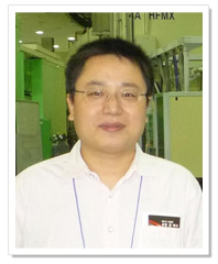 Jiawei Wang (王佳伟)合作论文数School of Life Sciences, Tsinghua University21
 Ping Yin (殷平)合作论文数National Key Laboratory of Crop Genetic Improvement, College of Life Science and Technology, Huazhong Agricultural University19
 Ruixue Wan (万蕊雪)合作论文数School of Life Sciences, Westlake University16Dong Deng (邓东)合作论文数12Fang Kong合作论文数School of Life Science, Westlake University11Rui Zhou合作论文数11
 Rui Bai (白蕊)合作论文数School of Life Sciences, Westlake University11Xiechao Zhan合作论文数10Gaoxingyu Huang合作论文数Institute of Biology, Westlake Institute for Advanced Study, 18 Shilongshan Road, Hangzhou, Zhejiang, China. huanggaoxingyu@westlake.edu.cn.10Qiang Zhou (周强)合作论文数10
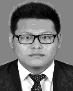 Wenqi Li合作论文数10
 Angqi Zhu合作论文数Newstead Lab, Department of Biochemistry, University of Oxford9Qi Hao合作论文数8Tian Xie (谢田)合作论文数8
 Guanghui Yang (杨光辉)合作论文数College of Biological Sciences, China Agricultural University8
 Xinqi Gong (龚新奇)合作论文数School of Mathematics, Renmin University of China8
 Xiaofeng Zhang (张晓峰)合作论文数School of Life Sciences, Division of Life Sciences and Medicine, University of Science and Technology of China7Jing Hang (杭婧)合作论文数7Xuechen Zhu (朱薛辰)合作论文数6Peilong Lu (卢培龙)合作论文数6Yanyu Zhao合作论文数Institute of Biology, Westlake Institute for Advanced Study, 18 Shilongshan Road, Hangzhou, Zhejiang, China.6Xuefei Guo合作论文数6Shuhao Zhang合作论文数6
 Minhao Liu (刘旻昊)合作论文数Westlake University;Tsinghua University6Jianping Wu (吴建平)合作论文数6Qi Hu (胡奇)合作论文数6
 Qinghua Tao (陶庆华)合作论文数School of Life Sciences, Tsinghua University6Dan Ma (马丹)合作论文数5
 Linfeng Sun (孙林峰)合作论文数School of Life Sciences, University of Science and Technology of China5
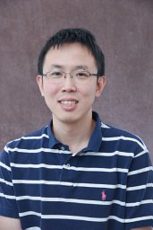 Xiao-Chen Bai合作论文数MRC Laboratory of Molecular Biology, Cambridge CB2 0QH, UK.5
 Yafei Yuan (袁亚飞)合作论文数Tsinghua University5Xin Jiang合作论文数5
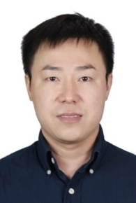 Feng Wang (王丰)合作论文数School of Life Sciences, Beijing Institute of Technology5
 Sjors Scheres合作论文数Scheres Lab, MRC Laboratory of Molecular Biology;Structural Studies Division, MRC Laboratory of Molecular Biology5
 Catherine C L Wong (黄超兰)合作论文数4Xiaoqiu Yuan合作论文数4Nan Wang合作论文数4Guifeng Lu合作论文数4Qifan Wang合作论文数4Jialu Li合作论文数4Di Wu合作论文数4
 Min Huang (黄敏)合作论文数Shanghai Institute of Materia Medica, Chinese Academy of Sciences;University of Chinese Academy of Sciences4Meng Ke合作论文数4Xiaojing Pan (潘孝敬)合作论文数4Yanqing Zhang合作论文数4
 Yuxuan Pang合作论文数4




