Objective To compare the cost-effectiveness of sialendoscopy with gland excision for the management of submandibular gland sialolithiasis. Study Design Cost-effectiveness analysis. Setting Outpatient surgery centers. Methods A Markov decision model compared the cost-effectiveness of sialendoscopy versus gland excision for managing submandibular gland sialolithiasis. Surgical outcome probabilities were found in the primary literature. The quality of life of patients was represented by health utilities, and costs were estimated from a third-party payer’s perspective. The effectiveness of each intervention was measured in quality-adjusted life-years (QALYs). The incremental costs and effectiveness of each intervention were compared, and a willingness-to-pay ratio of $150,000 per QALY was considered cost-effective. One-way, multivariate, and probabilistic sensitivity analyses were performed to challenge model conclusions. Results Over 10 years, sialendoscopy yielded 9.00 QALYs at an average cost of $8306, while gland excision produced 8.94 QALYs at an average cost of $6103. The ICER for sialendoscopy was $36,717 per QALY gained, making sialendoscopy cost-effective by our best estimates. The model was sensitive to the probability of success and the cost of sialendoscopy. Sialendoscopy must meet a probability-of-success threshold of 0.61 (61%) and cost ≤$11,996 to remain cost-effective. A Monte Carlo simulation revealed sialendoscopy to be cost-effective 60% of the time. Conclusion Sialendoscopy appears to be a cost-effective management strategy for sialolithiasis of the submandibular gland when certain thresholds are maintained. Further studies elucidating the clinical factors that determine successful sialendoscopy may be aided by these thresholds as well as future comparisons of novel technology.
Treatment options for unilateral glottic carcinoma include radiation therapy, partial laryngectomy, and endoscopic cordectomy. We use partial laryngectomy with imbrication laryngoplasty (PLIL) as a single modality curative approach in a selected group of patients with unilateral glottic carcinoma. PLIL includes a composite resection of the entire vocal fold with its ligament, muscle, adjacent paraglottic tissues, and the adjacent block of thyroid cartilage. A neocord is reconstructed by imbricating the remaining thyroid cartilage strips and covering them with a false vocal fold flap. PLIL provides a rapid recovery of oral/nasal airway and swallowing, excellent voice quality, and a disease-control rate similar or better than other treatment modalities.
General Principles Otology, Neuro-Otology, and Cradial Base Surgery Paranasal Sinus Surgery Head and Neck Surgery/Plastic and Reconstructive Surgery Laryngology and Bronchoesophagology Inde
Background: Ganglioside GM1 is a membrane glycolipid typical of nerve cell membranes, where it partakes in neu- rotransmitter release and is catabolized by the lysosomal -galactosidase (GM1ase) (EC 3.2.1.23). After demon- strating a novel degenerative disease of the parotid gland in mice deficient in GM1ase, mimicking the human stor- age disease GM1 gangliosidosis, we studied GM1ase and ganglioside GM1 content in the human parotid glands. Study Design: Levels of GM1ase and ganglioside GM1 were determined in samples of parotid tissues and neigh- boring muscle (as a negative control) for 3 subjects. Tis- sues were also processed for histochemical demonstra- tion of GM1ase. Results: The mean specific activity of GM1ase was more than 6-fold higher in the healthy human parotid tissues (1.4±0.5 nmol of 4-methylumbelliferone per minute per milligram of protein) relative to the neighboring muscle tis- sue (0.23±0.07 nmol of 4-methylumbelliferone per minute permilligramofprotein).ActivityofGM1asewashistochemi- cally localized mainly to striated duct and acinar cells of the parotid gland. Ganglioside GM1 content in the parotid gland was on average 30-fold higher relative to muscle. Conclusions: Our results are consistent with previous findings reported in the mouse and the rabbit, and prob- ably reflect a general property of the mammalian pa- rotid glands. The novel mechanism we previously pro- posed for the mouse parotid saliva secretion, mimicking neurotransmitter release in ganglioside GM1-containing nerve cells, is probably applicable also to the human pa- rotid gland. Similarly, the human parotid gland is prob- ably also severely affected in GM1 gangliosidosis. Arch Otolaryngol Head Neck Surg. 2001;127:1381-1384
A new model for the subcellular basis of parotid secretion is presented in this article. GM(1)-ganglioside, typically found in neural tissues, is shown to be abundant in the parotid gland. This ganglioside may play a central role in membrane turnover mechanisms underlying exocytosis/endocytosis in its role as a promoter of membrane fusion or a fusogen. The lysosome and lysosomal hydrolases also play a central role in this model in catabolism of GM(1)-ganglioside. Consequently, high levels of the lysosomal hydrolase acidic beta-galactosidase are demonstrated in the salivary gland. GM(1)-gangliosidosis of the parotid glands, as described in mice, appears to be the first single-gene heritable disease found so far in the salivary glands.
Several transnasal, transnasal-transseptal, and external approaches for dacryocystorhinostomy have been described. The first two and especially the first have the disadvantage of limited visualization. The third leaves an external scar, disrupts the horizontal canilicular system, and requires manipulation of the medial canthal tendon. The method described, using nasal endoscopes, affords excellent visualization and precise control, while avoiding an external scar, the canilicular system, and the medial canthal tendon.
A variety of diagnostic imaging techniques are currently available for the investigation of parotid gland disorders. At this time, for the investigation of mass lesions, computed tomography is the most valuable. Over the past several years, we have been comparing magnetic resonance imaging with computed tomographic scanning of our parotid gland lesions to evaluate the relative merits of both systems. This current study illustrates our current experience and shows the relative strengths and weaknesses of both methods.
Osteosarcomas and chondrosarcomas are rare malignant neoplasms in the head and neck. Eighteen cases seen at the UCLA Center for the Health Sciences are reviewed. Osteosarcomas of the maxilla tended to occur a decade later than those of the mandible or cranial bones. Several followed prior irradiation for retinoblastoma. Complete excision offers the only realistic chance for cure. Adjuvant therapy has little to offer these patients at this time.
The experience of the Intergroup Rhabdomyosarcoma Study in the treatment of this relatively common pediatric malignancy is described, and the group's standardized staging system is outlined.
Verrucous squamous cell carcinoma is a distinctive clinicopathologic entity. The lesion's biologic activity places it between conventional (nonverrucous) carcinomas and nonautochthonous hyperplasias of squamous epithelium. The diagnosis of the lesion requires full communication and cooperation between surgeon and pathologist. Once the diagnosis is confirmed, the selection of surgery over radiotherapy should be based on the recorded effectiveness of each modality and not on the phenomenon of anaplastic transformation reported to follow radiotherapy.
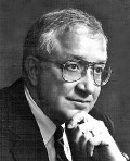 John G. Batsakis合作论文数Division of Pathology, University of Texas, M.D. Anderson Cancer Center7
 Alvin R. Solomon合作论文数3R Hybels合作论文数2Steven D. Schaefer合作论文数2Kokot Niels合作论文数2Gady Har-El合作论文数2Joseph H. Zernik合作论文数2Nakisa Nowroozi合作论文数2Anita N. Newman合作论文数2Hiroyuki Warita合作论文数2Elliot Abemayor合作论文数2Akihisa Segawa合作论文数2paul c denny合作论文数1K D McClatchey合作论文数1Donald R. Howard合作论文数1JH Zernik合作论文数1R O BRIERE合作论文数1Reuben C. Setliff合作论文数1Ashley C. Hsu合作论文数1sung kim合作论文数1
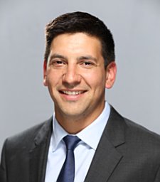 Joseph R Acevedo合作论文数1Randal C. Paniello合作论文数1
 James S Reilly合作论文数1David Kennedy合作论文数1Herbert Silverstein合作论文数1Jeffrey Yu合作论文数1Culton G L合作论文数1Gerwin J M合作论文数1
 Daniel H. Kwon合作论文数Life Sciences Division, E. O. Lawrence Berkeley National Laboratories;Life Sciences Division, E. O. Lawrence Berkeley National Laboratories1Monfared Ashkan合作论文数1Nowroozi N合作论文数1Lanza Donald C合作论文数1
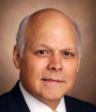 James Netterville合作论文数Vanderbilt Institute for Global Health, Vanderbilt University Medical Center;Department of Otolaryncology, Vanderbilt University Medical Center1Joseph A. Regezi合作论文数1
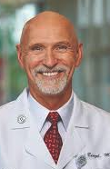 Edward C Benzel合作论文数Department of Neurosurgery, Neurological Institute, Cleveland Clinic;Cleveland Clinic Lerner College of Medicine, Case Western Reserve University1Daniel H. Kim合作论文数1
 John Crissman合作论文数Department of Pathology, Wayne State University1
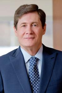 Paul C Mccormick合作论文数Irving Medical Center, Columbia University;NewYork-Presbyterian Allen Hospital;College of Physicians and Surgeons, Columbia University;The Och Spine Hospital, The Neurological Institute of New York1
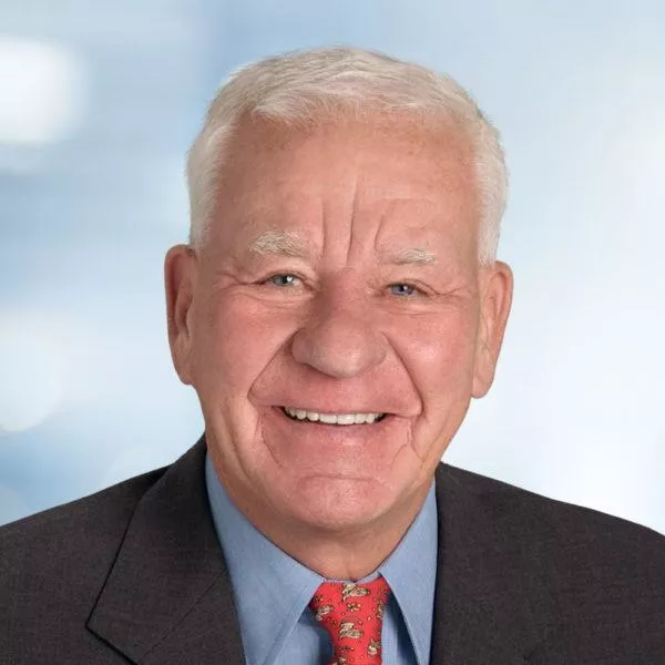 Volker K. H. Sonntag合作论文数Barrow Neurological Institute1Kashif Mazhar合作论文数Keck School of Medicine, University of Southern California1Wayne F. Larrabee合作论文数1Heinz Stammberger合作论文数1Toshitsugu Kawata合作论文数1S. James Zinreich合作论文数1Rodney P. Lusk合作论文数1Christopher H. Rassekh合作论文数1William L. Meyerhoff合作论文数1Goutham Gorti合作论文数1Jerome Vener合作论文数1




