Survival data of U-LMS based on RNase H2 status and RNASEH2B/RB1 HomDel status A) Overall survival of U-LMS patients based on loss of RNase H2 (n=23) or intact RNase H2 (n=85) by IHC B) Overall survival of ULMS patients with or without RNASEH2B HomDels, based on the presence or absence of RB1 HomDels.
Anatomic location of ST-LMS and RNase H2 status. Absolute number and frequency of cases of RNase H2 loss in the ST-LMS cohort based on primary anatomic location.
Supplementary Table S3. Patient demographics of patients enrolled in the GEIS-32 clinical trial.
Supplementary Figure S2. Progression-free survival probability of the patients treated within the GEIS-32 clinical trial, according to the 7 gene-based score. Time is defined in months.
Single-cell landscape of TME and epithelial cells from longitudinally collected tumor samples at baseline and during trial therapy.
PURPOSE:Immune checkpoint inhibitors (ICI) have transformed cancer therapy, but their efficacy in sarcomas remains limited. A seven-gene predictive signature (CD86, CHI3L1, CXCL10, CXCL9, LAG3, NR4A1, and VCAM1) was previously identified as a biomarker for response to combined antiangiogenic and PD-1 inhibitor therapy in soft-tissue sarcomas. This study aimed to externally validate the predictive utility of this signature [SIGNature for immuno-oncology PD-1 inhibitors in sarcoma (SIGNIOS)] in an independent cohort of patients with sarcoma treated with ICIs. EXPERIMENTAL DESIGN:Using RNA sequencing data from pretreatment tumor samples from the SARC028 phase II trial (pembrolizumab for advanced sarcomas) and the GEIS-32 phase II trial (pazopanib in advanced solitary fibrous tumor), we calculated the SIGNIOS score. Patients were stratified into low (score 0-4) and high (score 5-7) risk groups. RESULTS:The SARC028 validation cohort comprised 31 patients, with a median age of 33 years, and 45.2% were female. The most common histologic subtypes included osteosarcoma (25.8%) and Ewing sarcoma (19.4%). The seven-gene signature significantly stratified patients into groups with significantly different progression-free survival (PFS): Those with scores of 0 to 4 had a median PFS of 49 days, whereas those with scores of 5 to 7 had a median PFS of 170 days [HR = 0.71 (95% confidence interval, 0.55-0.91), P = 0.007]. CONCLUSIONS:This external validation confirms the seven-gene signature as a potential biomarker for predicting ICI efficacy in advanced sarcomas. Prospective studies are needed to determine the prognostic versus predictive value of SIGNIOS, refine its use across sarcoma subtypes, and explore its applicability in other tumor types.
Abstract Desmoplastic Small Round Cell Tumor (DSRCT) is an ultra-rare, aggressive sarcoma primarily affecting young adults. Characterized by a pathognomonic EWSR1::WT1 gene fusion, DSRCT typically presents with extensive peritoneal metastasis and has a 5-year overall survival rate of 15-25%. Histologically, these tumors exhibit marked peritumoral desmoplasia and minimal immune infiltrate (“immune-cold”). However, substantial heterogeneity among and within tumors has hindered the identification of secondary molecular mechanisms and the discovery of novel therapeutic targets. We constructed a first-of-its-kind DSRCT tissue microarray (TMA), comprising over 260 specimens from 63 patients spanning 13 years. We applied sequential multiplexed immunofluorescence imaging using a 20-antibody panel (selected based on prior studies) to map tumor, stroma, and immune cell components simultaneously. Our image analysis pipeline utilized tissue and cell segmentation to extract proteomic expression data from nuclear and cytoplasmic compartments of each cell. We applied Harmony batch correction and Leiden clustering to assign putative cell identities based on multi-marker expression. Our analysis of over 800,000 cells revealed significant inter- and intra-tumoral heterogeneity. We observed a variety of tumor nest morphologies, including well-circumscribed and trabecular architectures, as well as a range of stromal densities. We observed distinct marker expression phenotypes, with some cores dominated by AR-positive, epithelial-like (Pan-Keratin+) cells, while others display prominent neural markers (NSE+/SYP+). Estimated TMA core compositions quantitatively confirmed a range of tumor phenotypes and highlighted variable stromal predominance and a sparse immune infiltrate. Spatial analysis revealed distinct cellular neighborhood patterns and spatial arrangements among the tumor, stromal, and immune components. Finally, we correlated spatial signatures with clinical outcomes. Our work provides the first comprehensive spatial proteomic atlas of DSRCT at a single-cell resolution using a TMA. This foundational resource aims to elucidate the complex cellular environment of DSRCT, uncover novel biological insights into tumor-driving mechanisms, and ultimately identify new therapeutic vulnerabilities in DSRCT. Ongoing work will incorporate spatial transcriptomics to augment cell phenotyping and identify signalling pathways implicated in the tumor and microenvironment. Citation Format: Kevin A. Murgas, Jiaqian Fan, Diana Shamsutdinova, Khalida Wani, Davis R. Ingram, Alexander J. Lazar, Danh D. Truong, Joseph A. Ludwig. Distinct cellular phenotypes and spatial neighborhoods constitute the heterogeneous microenvironment of desmoplastic small round cell tumor [abstract]. In: Proceedings of the American Association for Cancer Research Annual Meeting 2026; Part 1 (Regular Abstracts); 2026 Apr 17-22; San Diego, CA. Philadelphia (PA): AACR; Cancer Res 2026;86(7 Suppl):Abstract nr 804.
Biallelic alterations in other DDR genes across all sarcomas in TCGA dataset. Assessment of the number of sarcomas with biallelic alterations in other DDR pathway members.
Leiomyosarcoma is a smooth muscle-derived malignancy marked by significant clinical heterogeneity. The extent and nature of cellular heterogeneity and molecular underpinnings remain poorly understood. To address this at transcriptomic and epigenomic levels, we performed single-nucleus multiome sequencing on untreated primary leiomyosarcoma tissues. Malignant cells segregated almost exclusively into two previously unrecognized and epigenetically distinct states: a dedifferentiated, mesenchymal-like subtype (MES) and a differentiated smooth muscle-enriched subtype (SMC). Chromatin accessibility profiling revealed strong enrichment of nuclear factor I (NFI) transcription factor motifs in MES cells, whereas AP-1 family motifs-most prominently FOSL2-were selectively accessible in SMC cells. Established leiomyosarcoma cell lines faithfully recapitulated these subtypes, and targeted depletion of NFI or AP-1 factors suppressed proliferation, invasion, and in vivo tumor growth, demonstrating functional dependency on these transcriptional programs. Spatial transcriptomics across 328 tissue cores from 128 leiomyosarcomas showed that immunosuppressive macrophages preferentially cluster around MES regions, revealing a subtype-specific tumor-immune niche. Clinically, MES-dominant tumors were associated with significantly worse patient outcomes. Through an epigenetic inhibitor screen, we identify and validate SMARCA4/2 inhibition as a promising therapeutic vulnerability for MES leiomyosarcomas. Together, this work defines two epigenetically driven, transcription factor-regulated, and clinically relevant states of leiomyosarcoma, revealing mechanistic underpinnings of tumor heterogeneity and uncovering actionable therapeutic strategies.
Overall survival from treatment initiation with ixazomib plus gemcitabine plus doxorubicin.
SNiPDx results of U-LMS and ST-LMS samples in the MDACC cohort, with RNase H2 IHC status also reported.
Supplementary Figure S1. Correlation between the 7-gene-based signature score and best response. The test of Mann-Whitney was used for the correlation analyses.
Well-differentiated and dedifferentiated liposarcoma (WDLPS and DDLPS) exhibit markedly different clinical behaviors, with DDLPS showing greater aggressiveness, higher recurrence and metastasis rates, and worse outcomes. Using single-nucleus multiome sequencing, epigenomic profiling, and spatial transcriptomics, we characterized cellular and epigenetic heterogeneity between these subtypes at single-cell and spatial resolution. We found distinct phenotypic states reflecting altered lineage differentiation and plasticity: DDLPS is dominated by early-differentiated progenitor-like cells, sclerotic WDLPS displays broader mesenchymal lineage plasticity, and adipocytic WDLPS contains abundant committed adipocytes. The DDLPS immune microenvironment was dominated by immunosuppressive macrophages, whereas WDLPS harbored more T cells and inflammatory macrophages. Notably, sclerotic WDLPS displayed intermediate cellular and molecular features, suggesting it may represent a distinct WDLPS subtype. Importantly, we identified novel gene regulatory circuits underlying each state, including FABP4/PPARG programs in adipocytic WDLPS, GLI2/TCF7L2/RBPJ/KLF7 programs in sclerotic WDLPS, and KLF7/FOSL2/SP3/GLI2/RBPJ programs in DDLPS. H3K27ac-marked enhancers were enriched near adipocytic marker genes in WDLPS and mesenchymal markers in DDLPS. Together, these findings reveal the cellular heterogeneity of tumor and immune compartments across liposarcoma subtypes and identify regulatory programs driving their differentiation states.
 Alexander J. Lazar合作论文数Department of Anatomical Pathology, Division of Pathology-Lab Medicine, The University of Texas M.D. Anderson Cancer Center;Department of Genomic Medicine, Division of Basic Science Research, The University of Texas MD Anderson Cancer Center;Department of Translational Molecular Pathology, Division of Pathology/Lab Medicine, The University of Texas MD Anderson Cancer Center136
 Wei-Lien Wang合作论文数Department of Anatomical Pathology, Division of Pathology-Lab Medicine Div, The University of Texas M. D. Anderson Cancer Center;Departments of Pathology and Translational Molecular Pathology, The University of Texas M. D. Anderson Cancer Center83
 Meliouh Khalida合作论文数61
 Keila E Torres合作论文数39
 Rossana Lazcano合作论文数Department of Pathology, The University of Texas MD Anderson Cancer Center35
 Christina L. Roland合作论文数Department of Surgical Oncology, The University of Texas MD Anderson Cancer Center;Division of Surgery, The University of Texas MD Anderson Cancer Center;Department of Cancer Network, The University of Texas MD Anderson Cancer Center35
 Neeta Somaiah合作论文数33
 Andy Futreal合作论文数Department of Genomic Medicine, Division of Cancer Medicine, The University of Texas MD Anderson Cancer Center33Hannah Cheung Beird合作论文数Univ Texas MD Anderson Canc Ctr, Dept Genom Med, Houston, TX 77030 USA27
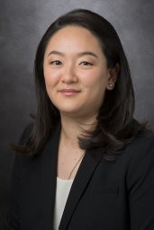 Emily Z. Keung合作论文数25Joseph A. Ludwig合作论文数Department of Sarcoma Medical Oncology, MD Anderson Cancer Center23
 Najat C. Daw合作论文数Division of Pediatrics, Department of Pediatrics, The University of Texas M.D. Anderson Cancer Center;Children's Memorial Hermann Hospital23Chia-Chin Wu合作论文数Univ Texas MD Anderson Canc Ctr, Dept Genom Med, Houston, TX 77030 USA22
 Vinod Ravi合作论文数Department of Sarcoma Medical Oncology, Division of Cancer Medicine, The University of Texas MD Anderson Cancer Center;Department of Genomic Medicine, Division of Cancer Medicine, The University of Texas MD Anderson Cancer Center20
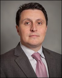 Anthony P. Conley合作论文数Department of Sarcoma Medical Oncology, Division of Cancer Medicine, The University of Texas MD Anderson Cancer Center20
 Shreyaskumar R. Patel合作论文数Department of Sarcoma Medical Oncology, Division of Cancer Medicine, The University of Texas MD Anderson Cancer Center19
 J. Andrew Livingston合作论文数Division of Cancer Medicine (MDACC), Houston, TX 77005, USA.18
 Kelly K. Hunt合作论文数Department of Breast Surgical Oncology, Division of Surgery, The University of Texas MD Anderson Cancer Center18Jianhua Zhang合作论文数17
 Elizabeth Demicco合作论文数Department of Laboratory Medicine and Pathobiology, Temerty Faculty of Medicine, University of Toronto16Sannaa Ghadah Al合作论文数16
 Danh Truong合作论文数Department of Bioengineering, University of Texas at Arlington;Joint Biomedical Engineering Program, University of Texas Southwestern Medical Center;University of Texas at Arlington, University of Texas Southwestern Medical Center16
 Barry W. Feig合作论文数Department of Surgical Oncology, Division of Surgery, The University of Texas MD Anderson Cancer Center16
 Kunal Rai合作论文数Univ Texas MD Anderson Canc Ctr, Dept Genom Med, Houston, TX 77030 USA15
 Jennifer Wargo合作论文数Department of Surgical Oncology, Division of Surgery, The University of Texas MD Anderson Cancer Center;Department of Genomic Medicine, Division of Cancer Medicine, The University of Texas MD Anderson Cancer Center15Linghua Wang (王凌华)合作论文数School of Life Sciences and Biotechnology, Shanghai Jiao Tong University15Grace Mathew合作论文数14Dina C Lev合作论文数14Genovese Giannicola合作论文数Univ Texas MD Anderson Canc Ctr14Heather Lin合作论文数13Jianjun Gao (高建军)合作论文数13Sharon M Landers合作论文数13
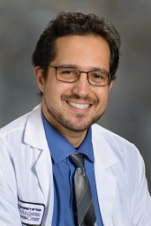 Pavlos Msaouel合作论文数Department of Genitourinary Medical Oncology, The University of Texas MD Anderson Cancer Center13
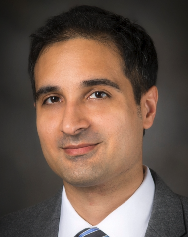 Rahul A. Sheth合作论文数Department of Interventional Radiology, Division of Diagnostic Imaging, The University of Texas MD Anderson Cancer Center13
 Nizar M. Tannir合作论文数Department of Genitourinary Medical Oncology, Division of Cancer Medicine, The University of Texas MD Anderson Cancer Center13
 Jillian Losh合作论文数13
 Cara Haymaker合作论文数Translational Molecular Pathology, University of Texas MD Anderson Cancer Center, Houston, Texas, USA13
 Karki Menuka合作论文数13Curtis Gumbs合作论文数13Ravin Ratan合作论文数12Rao Priya合作论文数12Luigi Perelli合作论文数11Fei Duan合作论文数Univ Texas MD Anderson Canc Ctr, Houston, TX 77030 USA11
 Manoj Chelvanambi合作论文数Department of Surgical Oncology, The University of Texas MD Anderson Cancer Center11Melinda Soeung合作论文数11
 B. Ashleigh Guadagnolo合作论文数Division of Radiation Oncology, Department of Radiation Oncology, The University of Texas MD Anderson Cancer Center;Department of Health Services Research, The University of Texas MD Anderson Cancer Center11Burton Elizabeth合作论文数11
 Robert S. Benjamin合作论文数Department of Sarcoma Medical Oncology, Division of Cancer Medicine, The University of Texas MD Anderson Cancer Center11




