Sustained affect shapes well-being, yet its neural architecture across externally elicited and internally generated experience remains unclear. Using whole-brain functional connectivity during minutes-long naturalistic movie viewing, we derived positive and negative affective experience signatures and their underlying neural architecture. These signatures predicted valence-specific affective intensity and generalized to independent movie-viewing data and internally generated affect, discriminating sad memory and rumination from neutral distraction while tracking subjective experience. Importantly, their expression showed little relation to vigilance or cognitive demand. Characterization of these signatures revealed coherent community structure and a shared distributed backbone, alongside valence-preferential components, consistent with a partially separable architecture. Extending beyond experimentally evoked states, in four resting-state depression cohorts, these signatures distinguished patients from controls with reduced positive and elevated negative signature expression, and predicted symptom burden and anhedonia. These findings identify a generalizable distributed architecture bridging external and internal affective experience and extending to clinically relevant affective dysregulation.
Characterized by recurrent fluctuations in mood states, bipolar disorder (BD) is widely conceptualized as a disconnection syndrome associated with dysregulated brain dynamics. Nevertheless, the molecular mechanisms underlying this aberrant connectivity dynamics in BD remain elusive. Using resting-state electroencephalography (EEG) data from BD patients and healthy controls, this study first delineated the characteristic alterations in temporal variability of functional connectivity in BD and further elucidated their underlying molecular mechanisms and clinical relevance. Current findings revealed significantly reduced temporal variability within large-scale brain subnetworks, most notably in the dorsal attention, somatomotor, and visual networks. Importantly, these neurodynamic signatures effectively predicted the symptom severity in individuals with BD. Moreover, the spatial patterns of these dynamic alterations are associated with the expression of BD risk genes enriched in synaptic function and metabolic pathways, as well as with the spatial organizations of various neurotransmitter receptors, including CB1, mGluR5, H3, and MOR. Collectively, these results provide evidence for a multiscale pathophysiological framework that links genetic susceptibility and chemoarchitectural alterations to dynamic brain network instability, ultimately underpinning the core clinical manifestations in BD.
Enhancing our understanding of how the brain constructs conscious emotional experiences within dynamic real-life contexts necessitates ecologically valid neural models. Here, we present evidence delineating the constraints of current fMRI activation models in capturing naturalistic fear dynamics. To address this challenge, we use naturalistic fMRI with predictive modeling techniques to develop an ecologically valid fear signature that integrates activation and connectivity profiles, allowing for accurate prediction of subjective fear experience under highly dynamic close-to-real-life conditions. This signature arises from insights into the crucial role of distributed brain networks and their interactions in emotion modulation, and the potential of network-level information to improve predictions in dynamic contexts. Across a series of investigations, we demonstrate that this signature predicts stable and dynamic fear experiences across naturalistic scenarios with heightened sensitivity and specificity, surpassing traditional activation- and connectivity-based signatures. Notably, the integration of affective connectivity profiles enables accurate real-time predictions of fear fluctuations in naturalistic settings. Additionally, we unearth a distributed yet redundant brain-wide representation of fear experiences. Subjective fear is encoded not only by distributed cortical and subcortical regions but also by their interactions, with no single brain system conveying substantial unique information. Our study establishes a comprehensive and ecologically valid functional brain architecture for subjective fear in dynamic environments and bridges the gap between experimental neuroscience and real-life emotional experience.
Schizophrenia (SCZ) is a highly disabling psychiatric disorder marked by compromised brain dynamic interactions. However, the underlying neuropathology of SCZ remains poorly elucidated in terms of its associated disruption of network-level and rhythm-specific dynamic fluctuations during the resting state. Herein, using a sample entropy-based temporal variability analysis framework, we investigate the complex fluctuation patterns of resting-state electroencephalogram networks as they transition over time for SCZ patients and their unaffected relatives (R-SCZ), as well as healthy controls (HC). Next, potential associations between variability networks and individual cognitive traits/clinical recordings were explored. Rhythm-specific abnormalities of baseline brain dynamics may disrupt the normal brain function of SCZ, particularly through the decoupling of frontal-temporal and temporal-parietal variability connectivity in the alpha and beta rhythms. The diminished variability differences observed between R-SCZ and HC suggest the possible existence of shared familial factors that influence specific traits related to network variability. Moreover, multidimensional representations of temporal variability networks can quantitatively characterize and even predict an individual's cognitive function (e.g. verbal memory) and clinical symptoms of SCZ patients. Current findings may offer new insights into comprehending the neuropathology and the potential role of familial susceptibility in SCZ, which may facilitate advancements in early diagnosis and intervention strategies.
Psychosis has long been conceptualized as a disorder of disrupted hierarchical integration across distributed brain systems, yet it remains unclear whether alterations in macroscale cortical hierarchy are already present before illness onset and are associated with subsequent transition to psychosis. Using connectome gradient mapping, we characterized baseline cortical hierarchical architecture along the unimodal-to-transmodal axis in 580 participants from the NAPLS-3 cohort, including converters (CHR-C, n = 56), non-converters (CHR-NC, n = 434), and healthy controls (HC, n = 90). Group differences were assessed at regional, network, and global levels. Group comparisons revealed that CHR-C individuals, relative to the other two groups, exhibited bidirectional alterations selectively along the sensorimotor-to-association gradient, with reduced values in the visual network alongside elevated values in the default mode network, indicating greater separation between sensory and transmodal systems along the gradient. At the global level, CHR-C showed increased explained variance, range, and variation of this gradient, collectively indicating hierarchical expansion. Notably, greater explained variance of this gradient was associated with a shorter time to conversion to psychosis, while increased gradient range and variation were associated with higher positive symptom severity across CHR individuals. These findings indicate that expansion of the sensorimotor-to-association connectome hierarchy is already present before psychosis onset in individuals who subsequently convert to psychosis. This altered hierarchical organization may reflect greater decoupling between sensory and transmodal systems and may characterize neurobiological changes associated with progression from a clinical high-risk state to psychotic illness.
The rising prevalence of disordered eating highlights the need to understand internal psychological pathways to healthy eating behaviors. This study aims to explore the underlying relationship between appearance evaluation and intuitive eating, focusing on the chain mediating roles of self-esteem and physical activity across different groups in Chinese university students population. A total of 866 participants were recruited to complete Chinese Version of the Multidimensional Body-Self Relations Questionnaire (MBSRQ) - Appearance Evaluation Subscale, Self-Esteem Scale, Physical Activity Rating Scale-3, Chinese Version of the Intuitive Eating Scale-2. The results showed that there was a chain mediaton model that self-esteem and physical activity served as mediating variables in the association between appearance evaluation and intuitive eating. Meanwhile, the model was not moderated by gender and BMI. Finally, when examining the across subgroups, it demonstrated the mid-BMI male subgroup was significant in this mediation model. These findings provide a cross-group theoretical foundation for the development of psychological strategies aimed at optimizing healthy eating behaviors in Chinese university students population.
Schizophrenia (SCZ) and bipolar disorder (BD) are severe psychotic disorders with overlapping clinical manifestations, leading to high rates of misdiagnosis. This study aims to identify disorder-specific neurophysiological biomarkers using electroencephalography and contrastive machine learning to improve differential diagnosis. Resting-state electroencephalography was recorded from 52 patients with BD, 65 with SCZ, and 75 healthy controls. Temporal variability networks were constructed using sample entropy. Contrastive variational autoencoders decomposed these networks into components shared with healthy controls and components specific to each disorder. Based on disorder-specific components, predictive models for clinical symptoms were constructed. Additionally, spatial pattern network filters were implemented to extract discriminative features for the classification of BD and SCZ patients. Here we show pronounced differences in disorder-specific network components between SCZ and BD, especially in frontal-central/parietal connectivity, which were not discernible in the original or shared networks. These disorder-specific components correlate significantly with clinical assessment scores and support predictive modeling of symptom severity. By applying spatial pattern network filters to the disorder-specific components, we achieve 96.154% accuracy in distinguishing SCZ from BD, substantially surpassing conventional approaches. This integrative framework, combining dynamic network analysis with contrastive machine learning, provides a powerful methodology for extracting neurophysiological biomarkers and paves the way for biologically grounded diagnostics in psychotic disorders. Schizophrenia and bipolar disorder are serious mental illnesses that can look very similar, making them hard for doctors to tell apart. This study used an objective brain index (EEG) combined with an artificial intelligence method to find differences between the two conditions. The researchers analyzed brain activity patterns and isolated features unique to each disorder. They discovered that these unique patterns were linked to patients’ symptoms and could predict how severe those symptoms were. Most importantly, the method distinguished between the two disorders with over 96% accuracy. This work could lead to a more reliable, biology-based tool to help doctors diagnose patients correctly, reduce misdiagnosis, and guide more personalized treatment decisions in the future. Jiang, Ye et al. combine resting-state EEG and contrastive variational autoencoders to identify disorder-specific neurophysiological biomarkers in schizophrenia and bipolar disorder. The approach achieves over 96% accuracy in distinguishing the two disorders by revealing distinct frontal-central/parietal connectivity patterns.
Uncovering the interbrain neural mechanisms underlying interpersonal negotiation offers insight into social decision-making dynamics in resource allocation. In this study, we used EEG hyperscanning alongside an iterated ultimatum game to investigate interbrain coupling and dyadic exchange behavior during negotiation. Frontal cortex event-related potentials (ERPs) revealed the distinct neural responses driven by partners' behavioral cues: the proposer's N200 differed significantly for fair versus unfair offers, and the responder's feedback-related negativity (FRN) showed a trend toward significance for the same contrast, while the proposer's N500 varied between acceptance and rejection feedback. Our analysis introduced a novel causal model based on directional phase transfer entropy (dPTE) and time-varying ERP amplitudes, illustrating directed neural processes driven by social exchange, where the proposer's brain activity initially exerts a causal impact on the responder's, whose feedback in turn influences the proposer, creating a closed-loop interaction that drives adaptive negotiation strategies. Additionally, our prediction model with autoregression with exogenous input, which incorporated these causal links between brains, demonstrated higher accuracy than single-brain or reverse causal models, underscoring the significance of dynamic interbrain coupling in interpersonal coordination. This causal model provides a mechanistic explanation of how proposer-responder pairs perceive and adapt to each other's decisions, facilitating shared attention and behavioral coordination in reciprocal, asymmetric negotiations. These findings offer a novel theoretical framework for studying complex social behaviors through interbrain dynamics and may inspire future applications in enhancing cooperative decision-making processes.
Accurate prediction of brain age is crucial for identifying deviations between typical individual brain development trajectories and neuropsychiatric disease progression. Although current research has made progress, the effective application of brain age prediction models to multi-center datasets, particularly those with small-sample sizes, remains a significant challenge that is yet to be addressed. To this end, we propose a multi-center data correction method, which employs a domain adaptation correction strategy with Wasserstein distance of optimal transport, along with maximum mean discrepancy to improve the generalizability of brain-age prediction models on small-sample datasets. Additionally, most of the existing brain age models based on neuroimage identify the task of predicting brain age as a regression or classification problem, which may affect the accuracy of the prediction. Therefore, we propose a brain dual-modality fused convolutional neural network model (BrainDCN) for brain age prediction, and optimize this model by introducing a joint loss function of mean absolute error and cross-entropy, which identifies the prediction of brain age as both a regression and classification task. Furthermore, to highlight age-related features, we construct weighting matrices and vectors from a single-center training set and apply them to multi-center datasets to weight important features. We validate the BrainDCN model on the CamCAN dataset and achieve the lowest average absolute error compared to state-of-the-art models, demonstrating its superiority. Notably, the joint loss function and weighted features can further improve the prediction accuracy. More importantly, our proposed multi-center correction method is tested on four neuroimaging datasets and achieves the lowest average absolute error compared to widely used correction methods, highlighting the superior performance of the method in cross-center data integration and analysis. Furthermore, the application to multi-center schizophrenia data shows a mean accelerated aging compared to normal controls. Thus, this research establishes a pivotal methodological foundation for multi-center brain age prediction studies, exhibiting considerable applicability in clinical contexts, which are predominantly characterized by small-sample datasets.
The relationship between brain structure and function, known as structural-functional coupling (SFC), is highly dynamic. However, the temporal variability of this relationship, referring to the fluctuating extent to which functional profiles interact with anatomy over time, remains poorly elucidated. Here, we propose a framework to quantify SFC temporal variability and determine its neurocognitive map, genetic architecture, and neurochemical basis in 1206 healthy human participants. Results reveal regional heterogeneity in SFC variability and a composite emotion dimension co-varying with variability patterns involving the dorsal attention, somatomotor, and visual networks. The transcriptomic signatures of SFC variability are enriched in synapse- and cell cycle-related biological processes and implicated in emotion-related disorders. Moreover, regional densities of serotonin, glutamate, γ-aminobutyric acid, and opioid systems are predictive of SFC variability across the cortex. Collectively, SFC variability mapping provides a biologically plausible framework for understanding how SFC fluctuates over time to support macroscale neurocognitive specialization.
The heterogeneity of psychotic disorders leads to instability in subjectively defined diagnoses. This study used a machine learning framework termed common orthogonal basis extraction (COBE) to decompose electroencephalography-based functional connectivity (FC) in patients with psychotic bipolar disorder (PBD), schizophrenia (SCZ), and schizoaffective disorder (SAD) into individualized and shared subspaces. The results demonstrated that individualized FCs captured disease heterogeneity and predicted symptom severity more accurately than raw FCs, while shared FCs revealed diagnosis-specific abnormalities and achieved an accuracy of 79.30% in differentiating PBD, SCZ, and SAD. Furthermore, molecular decoding implicated regionally selective serotonin systems and astrocytes in the neurobiological differences among disorders, suggesting disorder-specific pharmacological targets. Critically, these findings were replicated in an independent cohort, confirming the effectiveness of the COBE framework in mining neurophysiological and molecular profiles of schizophrenia-bipolar disorder. These findings advance mechanistic understanding of psychotic disorders and offer a promising avenue toward objective, clinically relevant tools for psychotic evaluation.
Background In today's food-rich society, the prevalence of dieting is remarkably high among adolescent girls and young females striving to achieve their ideal slim figure. Nonetheless, most individuals fail to maintain long-term dieting plans due to increased hedonic cravings for appetitive, high-calorie foods. Young dieting females are often confronted with two antagonistic motivational conflicts: the hedonic impulse and the pursuit of the slim ideal. Moreover, the trait of disinhibition plays a crucial role in dieting failures and hedonic overeating. Objective The present study aims to examine the moderating effects of the disinhibition trait on the antagonistic motivational processes between hedonic impulse and slim ideal pursuit among young dieting females. Methods Participants with high and low disinhibition traits performed a food-versus-figure task to determine the conflicting motivational processes. Results The findings showed that the balance between the hedonic "hot" and the slim ideal "cold" pathways was moderated by disinhibition. Specifically, only high-disinhibition dieters exhibited stronger food conflict than slim conflict, suggesting a pronounced preference for appetitive foods over the slim body ideal. Conclusion These findings enhance our understanding on the role of automatic hedonic impulse and the disinhibition trait in dieting failure, potentially clarifying why some individuals are more vulnerable to hedonic overeating and food addiction.
Background Previous studies on resting-state functional connectivity (FC) in internet gaming disorder (IGD) have typically assumed that FC is static during the entire scan, neglecting the dynamic reorganization of brain networks. However, understanding the dynamic changes in functional networks is crucial for a comprehensive understanding of IGD, a complex and evolving disorder. Methods Resting-state fMRI data were collected from 269 participants (132 IGD subjects, male/female: 72/60, and 137 recreational game users (RGUs), male/female: 85/52). At the network level (within-network and between-network), temporal variability indices were calculated for each group and subjected to independent samples t-tests. Results Compared to RGUs, IGD individuals exhibited decreased within-network variability in the default mode network (DMN), increased within-network temporal variability in the ventral attention network (VAN), and increased between-network temporal variability in sensorimotor network (SMN) and VAN, SMN and limbic network (LN), VAN and LN. Conclusions Changes in temporal variability at the network level occur in participants with IGD, indicating impaired executive inhibitory functions and attention, as well as imbalances between sensory-attention, sensory-emotion, and emotion-motivation functions. These findings provide new insights into the dynamic functional organization of the brain in IGD, contributing to our understanding the neural basis of pathological gaming behaviors.
This study investigated the neural mechanisms underlying SPS and emotional reactivity from the perspective of large-scale brain networks. A sample of 62 Chinese university students (35 females) was recruited. SPS was measured using the Chinese version of the Highly Sensitive Person Scale (C-HSPS), and emotional reactivity was assessed with the revised Chinese version of the Emotional Reactivity Scale (ERS). Resting-state functional connectivity was examined using fMRI. Behavioral analyses revealed a significant positive correlation between SPS and emotional reactivity (r = 0.49, p < 0.01). Neuroimaging findings showed that SPS was significantly negatively correlated with the functional connectivity between the salience network (SN) and frontoparietal network (FPN) (r = -0.29, p < 0.05), and the SN-FPN connectivity was also significantly negatively correlated with emotional reactivity (r = -0.28, p < 0.05). Mediation analysis demonstrated that SPS mediated the relationship between SN-FPN connectivity and emotional reactivity, such that SN-FPN connectivity predicted emotional reactivity indirectly through SPS. These findings indicate significant associations among SPS, SN-FPN connectivity, and emotional reactivity. The reduced SN-FPN connectivity observed in individuals with high SPS may potentially endow high-SPS individuals with advantages in supportive environments but increases their vulnerability to emotional dysregulation in challenging situations. This study provides novel insights into the neural mechanisms underlying SPS.
Purpose:Poor sleep quality is prevalent across the population and may significantly impact both physical and mental health. However, our understanding of the complex mechanisms underlying poor sleep quality is still incomplete, particularly regarding the various contributing factors. To address this, we utilized a data-driven causal discovery analysis (CDA) approach to explore causal pathways of sleep quality. Patients and Methods:We relied on a large sample of healthy young adults from the Human Connectome Project (HCP; n = 1206 [54% female, 56% unmarried/non-cohabiting]) to explore causal pathways of sleep quality. We first used exploratory factor analysis to cluster 122 broad phenotypic variables into 21 factors and computed the functional connectivity of 13 resting-state brain networks. Then, using Greedy Fast Causal Inference (GFCI), we simultaneously integrated the obtained phenotypic factors, brain network connectivity, and sleep quality into the causal discovery analysis and ultimately constructed a causal model. Results:The model proposes a hierarchical structure with causal effects propagating through complex interactions across multiple domains, ultimately linked to changes in sleep quality. Our causal model identified three phenotypic factors (negative affect, somaticism, and delay discounting) as directly linked to sleep quality. In addition, we examined causal models of sleep quality across gender (male and female) and relationship status (unmarried/non-cohabiting and married/cohabiting) and found some demographic-specific pathways. Conclusion:Our data-driven model reveals complex mechanisms by which factors from different domains influence sleep quality and highlights several key factors that influence sleep quality, which may have important implications for the development of sleep theories and the improvement of sleep quality.
Background/Objectives: Digital food-related videos significantly influence cravings, appetite, and weight outcomes; however, the dynamic neural mechanisms underlying appetite fluctuations during naturalistic viewing remain unclear. This study aimed to identify neural activity patterns associated with moment-to-moment appetite changes during naturalistic food-cue video viewing and to examine their relationships with cravings and weight-related outcomes. Methods: Functional magnetic resonance imaging (fMRI) data were collected from 58 healthy female participants as they viewed naturalistic food-cue videos. Participants concurrently provided continuous ratings of their appetite levels throughout video viewing. Hidden Markov Modeling (HMM), combined with machine learning regression techniques, was employed to identify distinct neural states reflecting dynamic appetite fluctuations. Findings were independently validated using a shorter-duration food-cue video viewing task. Results: Distinct neural states characterized by heightened activation in default mode and frontoparietal networks consistently corresponded with increases in appetite ratings. Importantly, the higher expression of these appetite-related neural states correlated positively with participants’ Body Mass Index (BMI) and post-viewing food cravings. Furthermore, these neural states mediated the relationship between BMI and food craving levels. Longitudinal analyses revealed that the expression levels of appetite-related neural states predicted participants’ BMI trajectories over a subsequent six-month period. Participants experiencing BMI increases exhibited a significantly greater expression of these neural states compared to those whose BMI remained stable. Conclusions: Our findings elucidate how digital food cues dynamically modulate neural processes associated with appetite. These neural markers may serve as early indicators of obesity risk, offering valuable insights into the psychological and neurobiological mechanisms linking everyday media exposure to food cravings and weight management.
Sleep-related problems (SRP) in childhood are common and clinically relevant yet their underlying neural mechanisms and links to future mental health outcomes remain poorly understood. Here, we investigated how distinct dimensions of SRP relate to multimodal brain structure and function in preadolescents, and whether these neural signatures predict trajectories of mental health difficulties. We employed multivariate mapping to investigate the relationship between structural and functional brain network patterns and various dimensions of SRP in the Adolescent Brain Cognitive Development (ABCD) dataset. Moreover, we explored whether and how the identified multimodal brain signatures could predict the trajectory of internalizing and externalizing behavior difficulties over a two-year follow-up. Our multivariate analysis revealed two robust dimensions of SRP: a general sleep disturbance dimension and a hypersomnolence and parasomnia dimension. Each was associated with partially distinct patterns of brain morphology and functional connectivity, consistent with their differential alignment along the hierarchical organization of cortical neurodevelopment maps. However, both dimensions shared common disruptions in the somatosensory, attention, and default mode networks. We further observed that only these neural patterns associated with the general sleep disturbance dimension predict the longitudinal trajectories of internalizing/externalizing symptoms. Our findings enhance the understanding of the neurobiological mechanisms underlying dimensions of SRP in preadolescence and could inform brain-based intervention and treatment programs to improve sleep-related and mental health-related outcomes across development.
Cocaine use disorder (CUD) has been linked to cortico-striatal dysfunctions, particularly within the prefrontal-striatal circuitry. However, previous studies have typically focused on discrete parcellations of the striatum, overlooking its continuous variations of neural organization. Moreover, while repetitive transcranial magnetic stimulation (rTMS) has shown benefits in CUD treatment, the neural effects of rTMS on striatal dysfunction in CUD remain poorly understood. Using connectome gradient-mapping techniques on three resting-state functional magnetic resonance imaging datasets, we derived the ventromedial-to-dorsolateral striatal functional topography. We identified specific alterations in this topography in the discovery cohort (41 CUD patients and 44 controls), validated findings in an independent cohort (53 CUD patients and 45 controls), and examined whether rTMS targeting the left dorsolateral prefrontal cortex (dlPFC) could normalize abnormalities in the rTMS-treatment cohort (44 patients). Across all datasets, we found a positive correlation between gradient variation and drug dependence severity in CUD. Compared to controls, CUD in both the discovery and replication cohorts exhibited elevated gradient values in the ventral striatum, while decreased values in the dorsal striatum were observed only in the discovery cohort. Furthermore, in the rTMS-treatment cohort, 5-Hz rTMS targeting the left dlPFC significantly normalized the aberrant gradient values in the ventral striatum, and these changes also related to cocaine craving changes. Overall, our study provides novel evidence of specific alterations in the ventromedial-to-dorsolateral functional topography of the striatum in CUD patients and highlights the impact of rTMS on striatal circuits through prefrontal modulation.
ObjectiveOverweight and obesity, as commonly indicated by a higher BMI, are associated with functional alterations in the brain, which may potentially result in cognitive decline and emotional illness. However, the manner in which these detrimental impacts manifest in the brain's dynamic characteristics remains largely unknown.MethodsBased on two independent resting-state functional magnetic resonance imaging data sets (Behavioral-Brain Research Project of Chinese Personality, n = 1923; Human Connectome Project, n = 998), the current study employed a Hidden Markov model to identify the spatiotemporal features of brain activity states. Subsequently, the study examined the changes in brain-state dynamics and the corresponding functional outcomes that arise with an increase in BMI.ResultsElevated BMI tends to shift the brain's activity states toward a greater emphasis on a specific set of states, i.e., the metastate, that are relevant to the joint activities of sensorimotor systems, making it harder to transfer to the metastate of transmodal systems. These findings were reconfirmed in a longitudinal sample (Behavioral-Brain Research Project of Chinese Personality, n = 34) that exhibited a significant increase in BMI at follow-up. Importantly, the alternation of brain-state dynamics specifically mediated the relationships between BMI and adverse functional outcomes, including cognitive decline and symptoms of mental illness.ConclusionsThe altered brain-state dynamics within the sensorimotor-to-transmodal hierarchy provide new insights into obesity-related brain dysfunctions and mental health issues.
 Yulin Wang (汪玉林)合作论文数Faculty of Psychology, Southwest University30
 Hong Chen (陈红)合作论文数Faculty of Psychology, Southwest University23
 Dezhong Yao (尧德中)合作论文数School of Life Science and Technology, University of Electronic Science and Technology of China;Center for Information in Medicine, University of Electronic Science and Technology of China23
 Feng Zhou (周峰)合作论文数Faculty of Psychology, Southwest University15
 Cheng Luo (罗程)合作论文数School of Life Science and Technology, University of Electronic Science and Technology of China15
 Genon Sarah合作论文数Cyclotron Research Center, University of Liège13
 Xu Lei (雷旭)合作论文数Sleep and NeuroImaging Center, Faculty of Psychology, Southwest University13
 Simon B. Eickhoff合作论文数Institute of Systems Neuroscience, Heinrich-Heine-University;Institute of Neuroscience and Medicine, Research Center Jülich12
 Xuebin Chang合作论文数Dept Informat Sci, Xi An Jiao Tong Univ11
 Tingyong Feng (冯廷勇)合作论文数Faculty of Psychology, Southwest University11
 Qinghua He (何清华)合作论文数Southwest University Faculty of Psychology10
 Todd Jackson合作论文数Department of Psychology,Faculty of Social Sciences (FSS),University of Macau10
 Lin Jiang合作论文数9
 Li Fali合作论文数9
 Peng Xu (徐鹏)合作论文数School of Life Science and Technology, University of Electronic Science and Technology of China8
 Jiang Qiu (邱江)合作论文数Faculty of Psychology, Southwest University8
 Xiaoyan Jia合作论文数School of Life Science and Technology, Xi'an Jiaotong University7
 Yong Liu (刘永)合作论文数Southwest University Faculty of Psychology7
 Chen Shuaiyu合作论文数5
 Sisi Jiang合作论文数Univ Elect Sci & Technol China, Sch Life Sci & Technol, Key Lab Neuroinformat, Minist Educ,Ctr Informat Med,High Field Magnet Re, Chengdu 610054, Peoples R China5
 Ximei Chen (陈曦梅)合作论文数Faculty of Psychology, Southwest University5Xiao Gao (高笑)合作论文数Faculty of Psychology, Southwest University5
 Marie Vandekerckhove合作论文数Biological Psychology, Vrije Universiteit Brussel5
 Hui He合作论文数4
 Fei Xin (辛斐)合作论文数School of Psychology, Shenzhen University4
 Daniele Marinazzo合作论文数Dept Data Anal, Univ Ghent4Iris Vantieghem合作论文数3
 Yiqun Guo合作论文数School of Innovation and Entrepreneurship Education, Chongqing University of Posts and Telecommunications3Yingjia Li合作论文数3
 Yuchao Jiang (蒋宇超)合作论文数School of Biomedical Engineering, Shenzhen University Medical School3
 Mingyue Xiao合作论文数3
 Yun Tian合作论文数Faculty of Psychology, Southwest University;Key Laboratory of Cognition and Personality, Southwest University),;Southwest University, Southwest University),3Ran Zhang合作论文数3XunTai Ma (马勋泰)合作论文数3
 Haobo Zhang (张浩波)合作论文数School of Psychology, Shenzhen University3
 Wei Li (李炜)合作论文数School of Aeronautics and Astronautics, Sichuan University3Yu Wang合作论文数3
 Benjamin Becker合作论文数Department of Psychology, The University of Hong Kong3
 Zhiliang Long (龙治良)合作论文数Faculty of Psychology, Southwest University3
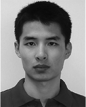 Yu Liang (梁玉)合作论文数Sichuan Provincial People's Hospital, University of Electronic Science and Technology of China3Runyang He合作论文数2Guangying Wang合作论文数2Fuyong Chen合作论文数2Gong Jinnan (巩晋南 Gong Jinnan)合作论文数2Huan Huang合作论文数2Zhangyong Li合作论文数2Mingjun Duan合作论文数2Junjie Wang合作论文数2




