Immunotherapy is effective in treating patients with a variety of cancers We developed and employed an immunotherapeutic protocol that combined intra-tumor injection of autologous immature dendritic cells , with standard of care radiation to treat patients with a variety of different advanced solid tumors. Pre and post treatment peripheral blood mononuclear cells ( PBMC) were assayed for their capacity to kill autologous tumor cell lines that had been established from patients with different advanced solid tumors. PBMC obtained post treatment were cytotoxic to autologous but not allogeneic tumor cell lines. Cytotoxic activity increased as relative numbers of CD8+-CD56+ dual positive lymphoid cells increased in PBMC obtained post treatment. The autologous tumor cell line directed cytotoxicity was shown to be mediated by both innate and adaptive immune mechanisms. Cytotoxicity of PBMC obtained post treatment was enhanced after co-culture with autologous and not allogeneic tumor cell line lysates. Antibody activity to mesothelin, a tumor associated antigen expressed in a variety of solid tumors, increased in post treatment serum samples from lung cancer patients. These sera mediated antibody directed cellular cytotoxicity ( ADCC) to mesothelin expressing tumor cell lines. SSurvival of 96 patients with a variety advanced cancers patients treated on this protocol over a 3 year period and followed for 1000+ days was significantly increased in patients with low tumor burden ( lesion at <5 sites with diameters < 3cm) (Km= <0.0001) regardless of malignancy. These findings support application of this combination of immunotherapy and radiation in the treatment of cancer patients with advanced disease
Diabetes commonly affects cancer patients. In breast cancer, it decreases patient survival. Diabetes also impacts African American women more so than European American women, potentially contributing to the breast cancer health disparity. In this study, we investigated the influence of diabetes on breast cancer biology in a diverse patient cohort using a three-pronged approach that included analysis of orthotopic human tumor xenografts, patient tumors, and breast cancer cells exposed to diabetes/hyperglycemia-like conditions. We aimed to identify shared phenotypes and molecular signatures by investigating the metabolome, transcriptome, and tumor mutational burden. Diabetes does not enhance cell proliferation but induces mesenchymal and stem cell-like phenotypes linked to increased mobility and odds of metastasis. It also promotes oxyradical formation and both transcriptome and mutational signatures of DNA repair deficiency. Moreover, food- and microbiome-derived metabolites tend to accumulate in breast tumors in presence of diabetes, potentially affecting tumor biology. Breast cancer cells cultured under hyperglycemia-like conditions acquire increased DNA damage and sensitivity to DNA repair inhibitors. Based on these observations, diabetes-associated breast tumors may show an increased drug response to DNA damage repair inhibitors that are cancer therapeutics. Key words: Breast cancer, diabetes, metabolome, transcriptome, metastasis, DNA repair, mutational signature, health disparity Citation Format: Gatikrushna Panigrahi, Julián Candia, Tiffany H. Dorsey, Wei Tang, Yuuki Ohara, Jung S. Byun, Tsion Zewdu Minas, Amy Zhang, Anuoluwapo Ajao, Ashley Cellini, Harris G. Yfantis, Dean Mann, Olga Ioffe, Xin W. Wang, Huaitian Liu, Christopher A. Loffredo, Ana Maria Napoles, Stefan Ambs. Diabetes-associated breast cancer is molecularly distinct and shows DNA repair deficiency [abstract]. In: Proceedings of the American Association for Cancer Research Annual Meeting 2023; Part 2 (Clinical Trials and Late-Breaking Research); 2023 Apr 14-19; Orlando, FL. Philadelphia (PA): AACR; Cancer Res 2023;83(8_Suppl):Abstract nr LB174.
PDF file - 40KB, ELISA Antibody Responses for the PCV (Prevnar-13(registered trademark)).
Diabetes commonly affects patients with cancer. We investigated the influence of diabetes on breast cancer biology using a 3-pronged approach that included analysis of orthotopic human tumor xenografts, patient tumors, and breast cancer cells exposed to diabetes/hyperglycemia-like conditions. We aimed to identify shared phenotypes and molecular signatures by investigating the metabolome, transcriptome, and tumor mutational burden. Diabetes and hyperglycemia did not enhance cell proliferation but induced mesenchymal and stem cell-like phenotypes linked to increased mobility and odds of metastasis. They also promoted oxyradical formation and both a transcriptome and mutational signatures of DNA repair deficiency. Moreover, food-and microbiome-derived metabolites tended to accumulate in breast tumors in the presence of diabetes, potentially affecting tumor biology. Breast cancer cells cultured under hyperglycemia-like conditions acquired increased DNA damage and sensitivity to DNA repair inhibitors. Based on these observations, we conclude that diabetes-associated breast tumors may show an increased drug response to DNA damage repair inhibitors.
Lung adenocarcinoma (LAC) is a common and aggressive form of lung cancer that is increasing in incidence among never smokers at a younger age. Current treatment of patients with LAC is insufficient and there is a need for identification of effective biomarkers and development of therapeutic targets. These demands require also improved models for in vivo and in vitro experimentation. In this study, we describe the establishment of two LAC cell lines, named LuCa-3 and LuCa-6. Both were derived from pleural effusion (PE) cells of LAC patients (L3 and L6) and readily propagated as tumor xenografts in immunodeficient mice. PE cells from the patient L6 exhibited also the capacity for in vitro growth and were cultured in two forms: (i) as a suspension growing cell population, labeled LuCa-6S, composed of non-clumping single cells; and (ii) as a monolayer-like culture, labeled LuCa-6A, exhibiting tight cell-to-cell and to culture surface adherence. Unique features of these two sublines and their cell clones are the capacity to convert from a non-clumping single-cell suspension into the adherent growth pattern and vice versa. Immunostaining of patients' tumor tissue xenografts and cultured subline cells displayed markers specific for the phenotype of human LAC. LuCa-6S and LuCa-6A cells did not reveal a noticeable disparity in quantitative growth characteristics. However, a number of differences were detected between these two cell populations manifested in detection or intensities of antigen expressions on the cell surface (CD133, SFTPC) and in the nucleus (TTF-1) including pluripotent (OCT-4, SOX-2, NANOG) genes in cancer stem-like cells (CSCs). Dissimilarities between these two sublines were also detected in N-glycan profiles and in the sensitivity to natural killer cells. Salient features of these subline cell populations are responsiveness to selective upregulation of the pluripotent genes in subsets of CSCs via conversion of their growth patterns and/or by using culture stem media with growth factors. The described in vivo/in vitro model enables broader experimental approaches in studies of lung adenocarcinoma.
Abstract Background and Aim Chronic hepatitis C virus (HCV) infection, long‐term alcohol use, cigarette smoking, and obesity are the major risk factors for hepatocellular carcinoma (HCC) in the United States, but the disease risk varies substantially among individuals with these factors, suggesting host susceptibility to and gene–environment interactions in HCC. To address genetic susceptibility to HCC, we conducted a genome‐wide association study (GWAS). Methods Two case‐control studies on HCC were conducted in the United States. DNA samples were genotyped using the Illumian microarray chip with over 710 000 single nucleotide polymorphisms (SNPs). We compared these SNPs between 705 HCC cases and 1455 population controls for their associations with HCC and verified our findings in additional studies. Results In this GWAS, we found that two SNPs were associated with HCC at P < 5E‐8 and six SNPs at P < 5E‐6 after adjusting for age, sex, and the top three principal components (PCs). Five of the SNPs in chromosome 22q13.31, three in PNPLA3 (rs2281135, rs2896019, and rs4823173) and two in SAMM50 (rs3761472, rs3827385), were replicated in a small US case‐control study and a cohort study in Singapore. The associations remained significant after adjusting for body mass index and HCV infection. Meta‐analysis of multiple datasets indicated that these SNPs were significantly associated with HCC. Conclusions SNPs in PNPLA3 and SAMM50 are known risk loci for nonalcoholic fatty liver disease (NAFLD) and are suspected to be associated with HCC. Our GWAS demonstrated the associations of these SNPs with HCC in a US population. Biological mechanisms underlying the relationship remain to be elucidated.
Hepatocellular carcinoma (HCC) is an aggressive malignancy with its global incidence and mortality rate continuing to rise, although early detection and surveillance are suboptimal. We performed serological profiling of the viral infection history in 899 individuals from an NCI-UMD case-control study using a synthetic human virome, VirScan. We developed a viral exposure signature and validated the results in a longitudinal cohort with 173 at-risk patients who had long-term follow-up for HCC development. Our viral exposure signature significantly associated with HCC status among at-risk individuals in the validation cohort (area under the curve: 0.91 [95% CI 0.87-0.96] at baseline and 0.98 [95% CI 0.97-1] at diagnosis). The signature identified cancer patients prior to a clinical diagnosis and was superior to alpha-fetoprotein. In summary, we established a viral exposure signature that can predict HCC among at-risk patients prior to a clinical diagnosis, which may be useful in HCC surveillance.
Prostate cancer is a leading cause of cancer death in US men. Yet, the etiology of prostate cancer remains poorly understood, with only older age, African ancestry, family history of the disease, and multiple germline genetic variations being established disease risk factors. Prostate cancer occurs more often in African-American men and Caribbean men of African descent than in men of other race/ethnicities. The causes of these racial and ethnic differences are multifactorial, but it has been proposed that differences in tumor biology contribute to the health disparity associated with prostate cancer. Previously, our laboratory discovered immunobiology differences between African-American and European-American men where genes involved in the inflammatory response tended to be up-regulated in tumors of African American patients. Accumulating evidence suggests that inflammation may be involved in prostate carcinogenesis. Given these observations, we hypothesized that inflammation may contribute to prostate cancer risk and the survival health disparity observed between African-Americans and European-Americans. To this end, we conducted a case-control study in the greater Baltimore area where we recruited 1000 cases and 1000 controls composed of equal populations of African-American and European-American men. In our survey, we asked about non-steroidal anti-inflammatory drugs (NSAIDs) use categorized into aspirin, Tylenol, and other pain relievers that neither contain Tylenol nor aspirin. We found a significant association between aspirin use and prostate cancer risk—where aspirin use decreased the risk of prostate cancer. Strikingly, the decreased prostate cancer risk as a result of aspirin use was most profound in African-American men. We were also able to measure the levels of C-reactive protein, a marker for inflammation, to further evaluate the relationship between aspirin use, inflammation, and prostate cancer risk. Indeed, we did observe a decrease in C-reactive protein in patients who took aspirin. Interestingly, overall levels in healthy controls and prostate cases were higher in African-American subjects and patients when compared to European-Americans. Although aspirin use decreased C-reactive protein levels in African-American and European-American cases, African-Americans had significantly higher levels of C-reactive protein. The robust association between aspirin consumption and prostate cancer risk suggests that inflammation might be a driver in prostate cancer of African-American men. Additionally, the increased levels of C-reactive protein in African-American controls and cases might contribute to the excessive disease risk and mortality among these men. Citation Format: Cheryl J. Smith, Symone Jordan, Tiffany H. Dorsey, Dean Mann, Chris A. Loffredo, Elise Bowman, Wei Tang, Stefan Ambs. Aspirin use among African American men reduces prostate cancer risk: Findings from the NCI-Maryland Prostate Cancer Case-Control study. [abstract]. In: Proceedings of the 107th Annual Meeting of the American Association for Cancer Research; 2016 Apr 16-20; New Orleans, LA. Philadelphia (PA): AACR; Cancer Res 2016;76(14 Suppl):Abstract nr 1776.
Tremendous efforts have been made to develop cancer biomarkers by detecting circulating extracellular miRNAs directly released from tumors. Yet, none of the cell-free biomarkers has been accepted to be used for early detection of non-small cell lung cancer (NSCLC). Peripheral blood mononucleated cells (PBMCs) act as the first line of defense against malignancy in immune system, their dysfunction may occur as an early event in cancer immunogenicity or immune evasion. We proposed to investigate whether analysis of miRNA expressions of PBMCs has diagnostic value for NSCLC. We first used a microarray to analyze PBMCs of 16 stage I NSCLC patients and 16 cancer-free smokers, and identified seven PBMC miRNAs with a significantly altered expression level in NSCLC patients. In a training set of 84 NSCLC patients and 69 cancer-free smokers, a panel of two miRNAs (miRs-19b-3p and -29b-3p) were developed from the seven PBMC miRNAs, producing 72.62% sensitivity and 82.61% specificity in identifying NSCLC. Furthermore, the miRNAs could identify squamous cell lung carcinoma (SCC), a major type of NSCLC, with 80.00% sensitivity and 89.86% specificity. The expression levels of the miRNAs were independent of disease stage. In a testing set of 56 NSCLC patients and 46 controls, the performance of the biomarkers was reproducibly confirmed. The study presents the first in-depth analysis of PBMC miRNA profile of NSCLC patients. The assessment of PBMC miRNAs may provide a new diagnostic approach for the early detection of NSCLC.
Background: Blood alcohol is present in a third of trauma patients and has been associated with organ dysfunction. In both human studies and in animal models, it is clear that alcohol intoxication exerts immunomodulatory effects several hours to days after exposure, when blood alcohol is no longer detectable. The early immunomodulatory effects of alcohol while blood alcohol is still elevated are not well understood.Methods: Human volunteers achieved binge alcohol intoxication after high-dose alcohol consumption. Blood was collected for analysis prior to alcohol ingestion, and 20 min, 2 h, and 5 h after alcohol ingestion. Flow cytometry was performed on isolated peripheral blood mononuclear cells, and cytokine generation in whole blood was measured by enzyme-linked immunosorbent assay (ELISA) after 24-h stimulation with lipopolysaccharide (LPS) and phytohemagglutinin-M (PHA) stimulation.Results: An early pro-inflammatory state was evident at 20 min when blood alcohol levels were similar to 130 mg/dL, which was characterized by an increase in total circulating leukocytes, monocytes, and natural killer cells. During this time, a transient increase in LPS-induced tumor necrosis factor (TNF)-alpha levels and enhanced LPS sensitivity occurred. At 2 and 5 h post-alcohol binge, an anti-inflammatory state was shown with reduced numbers of circulating monocytes and natural killer cells, attenuated LPS-induced interleukin (IL)-1 beta levels, and a trend toward increased interleukin (1L)-10 levels.Conclusions: A single episode of binge alcohol intoxication exerted effects on the immune system that caused an early and transient pro-inflammatory state followed by an anti-inflammatory state. (C) 2015 Elsevier Inc. All rights reserved.
We conducted a phase I dose escalation study to evaluate the safety and immunologic response to peptide immunomodulatory vaccines GL-0810 (HPV16) and GL-0817 (MAGE-A3) in HPV16 and MAGE-A3-positive RM–SCCHN patients, respectively.
The neutralization of toxins is considered essential for protection against lethal infection with Bacillus anthracis (BA), a select agent and bioterrorism threat. However, toxin-neutralizing activity alone would not be expected to provide sterile immunity. Therefore, we hypothesized that the development of an adaptive immune response against BA is required for bacterial clearance. We found that human monocyte-derived dendritic cells (hDCs) kill germinated BA bacilli, but not nongerminated BA spores. hDCs produce IL-1β, IL-6, IL-12, and IL-23, and these cytokines are differentially regulated by germination-proficient versus germination-deficient BA spores. Moreover, the IL-23 response to BA spores is regulated by IL-1R-mediated signaling. hDCs infected with germinating BA spores stimulated autologous CD4(+) T cells to secrete IL-17A and IFN-γ in a contact-dependent and antigen-specific manner. The T-cell response to BA spores was not recapitulated by hDCs infected with germination-deficient BA spores, implying that the germination of spores into replicating bacilli triggers the proinflammatory cytokine response in hDCs. Our results provide primary evidence that hDCs can generate a BA-specific Th17 response, and help elucidate the mechanisms involved. These novel findings suggest that the IL-23/Th17 axis is involved in the immune response to anthrax in humans.
AbstractPurpose: Myeloma-directed cellular immune responses after autologous stem cell transplantation (ASCT) may reduce relapse rates. We studied whether coinjecting the TLR-3 agonist and vaccine adjuvant Poly-ICLC with a MAGE-A3 peptide vaccine was safe and would elicit a high frequency of vaccine-directed immune responses when combined with vaccine-primed and costimulated autologous T cells.Experimental Design: In a phase II clinical trial (NCT01245673), we evaluated the safety and activity of ex vivo expanded autologous T cells primed in vivo using a MAGE-A3 multipeptide vaccine (compound GL-0817) combined with Poly-ICLC (Hiltonol), granulocyte macrophage colony-stimulating factor (GM-CSF) ± montanide. Twenty-seven patients with active and/or high-risk myeloma received autografts followed by anti-CD3/anti-CD28–costimulated autologous T cells, accompanied by MAGE-A3 peptide immunizations before T-cell collection and five times after ASCT. Immune responses to the vaccine were evaluated by cytokine production (all patients), dextramer binding to CD8+ T cells, and ELISA performed serially after transplant.Results: T-cell infusions were well tolerated, whereas vaccine injection site reactions occurred in >90% of patients. Two of nine patients who received montanide developed sterile abscesses; however, this did not occur in the 18 patients who did not receive montanide. Dextramer staining demonstrated MAGE-A3–specific CD8 T cells in 7 of 8 evaluable HLA-A2+ patients (88%), whereas vaccine-specific cytokine-producing T cells were generated in 19 of 25 patients (76%). Antibody responses developed in 7 of 9 patients (78%) who received montanide and only weakly in 2 of 18 patients (11%) who did not. The 2-year overall survival was 74% [95% confidence interval (CI), 54%–100%] and 2-year event-free survival was 56% (95% CI, 37%–85%).Conclusions: A high frequency of vaccine-specific T-cell responses were generated after transplant by combining costimulated autologous T cells with a Poly-ICLC/GM-CSF–primed MAGE-A3 vaccine. Clin Cancer Res; 20(5); 1355–65. ©2013 AACR.
SummaryInteractions between natural killer (NK) cells and dendritic cells (DC) affect maturation and function of both cell populations, including NK cell killing of DC (editing), which is important for controlling the quality of immune responses. We also know that antigen‐stimulated Vγ2Vδ2 T cells co‐stimulate NK cells via 4‐1BB to enhance the killing of tumour cell lines but we do not know what regulates 4‐1BB expression or whether other NK effector functions including DC killing, might also be influenced by NK–γδ T‐cell cross‐talk. Here we show that antigen‐stimulated γδ T cells co‐stimulate NK cells through inducible T‐cell co‐stimulator (ICOS)– ICOS ligand (ICOSL) and this signal increases NK cell killing of autologous DC. Effects of NK–γδ T‐cell co‐culture, which could be reproduced with soluble ICOS‐Fc fusion protein, included increased CD69 and 4‐1BB expression, interferon‐γ, tumour necrosis factor‐α, macrophage inflammatory protein‐1β, I‐309, RANTES and sFas ligand production, as well as elevated mRNA levels for co‐stimulatory receptors OX40 (TNFRSF4) and GITR (TNFRSF18). Hence, ICOS–ICOSL co‐stimulation of NK by Vγ2Vδ2 T cells had broad effects on NK phenotype and effector functions. The NK–γδ T‐cell cross‐talk links innate and antigen‐specific lymphocyte responses in the control of cytotoxic effector function and DC killing.
 Alan S. Cross合作论文数Center for Vaccine Development, Department of Medicine, University of Maryland School of Medicine12
 Scott E. Strome合作论文数Department of Otorhinolaryngology, Mayo Clinic10
 Kristina M. Harris合作论文数9Mikulas Popovic合作论文数9Kim G. Hankey合作论文数8
 Aaron P. Rapoport合作论文数Division of Hematology/Oncology, University of Maryland School of Medicine8Kenichiro Hasumi合作论文数7Zheng Zhaohui合作论文数7
 Ashraf Z. Badros合作论文数School of Medicine, University of Maryland7
 Bruce L. Levine合作论文数Department of Pathology and Laboratory Medicine, Perelman School of Medicine, University of Pennsylvania;Center for Cellular Immunotherapies, University of Pennsylvania7
 Edward A. Stadtmauer合作论文数Division of Hematology Oncology, Department of Medicine, School of Medicine, University of Pennsylvania7
 Kathleen Ruehle合作论文数7
 Carl H. June合作论文数Department of Pathology and Laboratory Medicine, Perelman School of Medicine, University of Pennsylvania7R C Gallo合作论文数6Hong-Bin Fang合作论文数6Akpek G合作论文数6Sandra Rollins合作论文数5Andrea L Brennan合作论文数5YinYan Xu合作论文数5Ling Cai合作论文数5Ma Fernandezvina合作论文数5
 Nicole Alyse Aqui合作论文数Department of Pathology and Laboratory Medicine, Perelman School of Medicine, University of Pennsylvania5
 Michael Kalos合作论文数IpiNovyx Bio;Next Pillar Consulting, LLC;RoslinCT5Milliron Todd合作论文数5Philip Sunita合作论文数5Kerr Naseem合作论文数5
 John C. Papadimitriou合作论文数Department of Pathology, University of Maryland School of Medicine5
 John Fahey合作论文数Department of Microbiology and Immunology, UCLA School of Medicine5
 Yanovich Saul合作论文数5
 Stefan Ambs合作论文数National Cancer Institute, National Institutes of Health5
 Brendan M Weiss合作论文数Department of Medicine, Perelman School of Medicine, University of Pennsylvania;Moderna5Stephen T. Bartlett合作论文数5Patricia Tsao合作论文数5
 Dan T. Vogl合作论文数Division of Hematology/Oncology, University of Pennsylvania School of Medicine5AB EINSTEIN合作论文数4James Kodak合作论文数4K. Cao合作论文数4
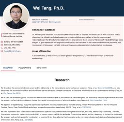 Wei Tang合作论文数Bio-X Life Science Research Center, Shanghai Jiao Tong University, 1954 Huashan Road, Shanghai 200030, PR China4Dorsey Tiffany H合作论文数4
 Alessio Fasano合作论文数Mucosal Immunology and Biology Research Center, Massachusetts General Hospital;Harvard Medical School4
 Xinwei Wang合作论文数Laboratory of Human Carcinogenesis, Center for Cancer Research, National Cancer Institute, National Institutes of Health4
 Thomas J Macvittie合作论文数4Richard B. Alexander合作论文数4Gn Rogentine合作论文数3Ryuko Watanabe合作论文数3
 Simeon E Goldblum合作论文数3As Fauci合作论文数3M Robert-Guroff合作论文数3
 Thomas Spies合作论文数Fred Hutchinson Cancer Research Center3




