Abstract Background Individualized radiopharmaceutical therapies guided by patient-specific absorbed dose assessments using imaging have the potential to improve both efficacy and safety. Understanding sources of variability in absorbed dose calculations is critical for standardization. The Society of Nuclear Medicine and Molecular Imaging Dosimetry Task Force launched the 177Lu Dosimetry Challenge to evaluate variability across different steps within the dosimetry workflow. This work aimed to assess the variability in absorbed doses due to differences in segmentation methods. Methods Anonymized datasets from two patients treated with 177Lu-DOTATATE, including serial SPECT/CT scans, were made available online. Participants were asked to segment healthy organs and lesions and perform dosimetry calculations. In a subsequent task, participants were provided with standardized segmented VOIs and asked to perform dosimetry based on these pre-defined regions. Variability in segmentation was assessed by comparing absorbed dose estimates across two scenarios: participant-generated segmentations versus predefined reference segmentations. Relative absorbed dose variability was quantified using the quartile coefficient of dispersion (QCD) and interquartile range. Results Variability in absorbed dose (measured as QCD difference between absorbed doses from participant-generated segmentations and those from reference segmentations) for kidneys was less than 5% in simple cases and 10.6% for more challenging scenarios (i.e. presence of intraparenchymal cysts, cortical defects). Lesion segmentation exhibited higher variability, with absorbed dose variability reaching up to 22.4%. Conclusions Segmentation significantly contributes to variability in absorbed dose estimates, particularly for lesions and for kidneys with anatomical complexities. Standardizing segmentation protocols and providing training on advanced segmentation methods are essential to reduce variability.
Individualized radiopharmaceutical therapies guided by patient-specific absorbed dose (AD) assessments using nuclear medicine imaging have the potential to improve both efficacy and safety. Understanding sources of variability in AD calculations is critical for standardization. The Society of Nuclear Medicine and Molecular Imaging Dosimetry Task Force launched the 177Lu Dosimetry Challenge to evaluate variability across steps within the dosimetry workflow. This work aimed to assess the variability in ADs due to different fitting and integration methods. Methods: Anonymized datasets from 2 patients treated with 177Lu-DOTATATE, including serial SPECT/CT scans, segmented organs and lesions, and time-integrated activity maps, were made available online. Participants were invited to perform dosimetry calculations and submit their results using standardized submission spreadsheets. Fitting approaches were categorized, and relative AD variability was analyzed using the quartile coefficient of dispersion and interquartile range. Results: The variability in AD due to the fitting step for patient A's kidneys was less than 1%. In contrast, patient B's kidneys showed higher variability, with values below 10%. Lesions exhibited more variability in fitting than did kidneys, with variability within 25%. Conclusion: The contribution of variability caused by fitting and integration is small for healthy organs. By following recommendations such as selection of appropriate functions, pharmacokinetic modeling, and sanity checks, this variability can be further reduced.
Image-based dosimetry-guided radiopharmaceutical therapy has the potential to personalize treatment by limiting toxicity to organs at risk and maximizing the therapeutic effect. The 177Lu dosimetry challenge of the Society of Nuclear Medicine and Molecular Imaging consisted of 5 tasks assessing the variability in the dosimetry workflow. The fifth task investigated the variability associated with the last step, dose conversion, of the dosimetry workflow on which this study is based. Methods: Reference variability was assessed by 2 medical physicists using different software, methods, and all possible combinations of input segmentation formats and time points as provided in the challenge. General descriptive statistics for absorbed dose values from the global submissions from participants were calculated, and variability was measured using the quartile coefficient of dispersion. Results: For the liver, which included lesions with high uptake, variabilities of up to 36% were found. The baseline analysis showed a variability of 29% in absorbed dose results for the liver from datasets where lesions included and excluded were grouped, indicating that variation in how lesions in normal liver were treated was a significant source of variability. For other organs and lesions, variability was within 7%, independently of software used except for the local deposition method. Conclusion: The choice of dosimetry method or software had a small contribution to the overall variability of dose estimates.
In this paper, we report the design, development, and initial experimental evaluation of the Alpha-SPECT system, a full-body clinical SPECT scanner equipped with state-of-art CZT imaging spectrometers ideally suited to simultaneous imaging of therapeutic alpha-emitters (e.g., Ac-225, Ra-223, Lu-177, etc.) and their daughters (e.g., Fr.221, At-217, Bi-213, and Po-213 from the decay of Ac-225) in patients. Besides the potential for imaging alpha-labeled radiopharmaceutical therapy (a-RPT), the Alpha-SPECT system is intended to be a general-purpose clinical SPECT scanner for a wide variety of diagnostic and therapeutic applications. The system is equipped with a dual-aperture collimator that is installed on a CT-scanner-derived rotational gantry to allow the real-time switching between the two aperture configurations, as well as micro-swing of aperture in front of the CZT detectors with a sub- 15 mm precision to provide a variable FOV of 20-50 cm diameter and facilitate super-resolution sampling for further improved imagign resolution.
BACKGROUND:Pediatric molecular imaging requires a balance between administering an activity that will yield sufficient diagnostic image quality while maintaining patient radiation exposure at acceptable levels. In current clinical practice, this balance is arrived at by the current North American Consensus Guidelines in which patient weight is used to recommend the administered activity (AA). PURPOSE:We have previously demonstrated that girth (waist circumference at the level of the kidneys) is better at equalizing image quality than patient weight for pediatric Tc-99m DMSA renal function imaging. However, the correlation between image quality (IQ), AA, and patient girth has not been rigorously and systematically developed. In this work, we generate a series of curves showing the tradeoff between AA and IQ as a function of patient girth, providing the data for standards bodies to develop the next generation of dosing guideline for pediatric DMSA SPECT. METHODS:An anthropomorphic phantom series that included variations in age (5, 10, and 15 years), gender (M, F), local body morphometry (5, 10, 50, 90, and 95th girth percentiles), and kidney size (±15% standard size), was used to generate realistic SPECT projections. A fixed and clinically challenging defect-to-organ volume percentage (0.49% of renal cortex value) was used to model a focal defect with zero uptake (i.e., full local loss of renal function). Task-based IQ assessment methods were used to rigorously measure IQ in terms of renal perfusion defect detectability. This assessment was performed at multiple count levels (corresponding to various AAs) for groups of patients that had similar girths and defect sizes. Receiver-operating characteristics (ROC) analysis was applied; the area under the ROC curve (AUC) was used as a figure-of-merit for task performance. Curves showing the tradeoff between AUC and AA were generated for these groups of phantoms. RESULTS:Overall, the girth-based dosing method suggested different amounts of AA compared to weight-based dosing for the phantoms that had a relatively large body weight but a small girth or phantoms with relatively small bodyweight but large girth. Reductions of AA to 62.9% compared to weight-based dosing guidelines can potentially be realized while maintaining a baseline (AUC = 0.80) IQ for certain 15-year-olds who have a relatively small girth and large defect size. Note that the task-based IQ results are heavily dependent on the simulated defect size for the defect detection task and the appropriate AUC value must be decided by the physicians for this diagnostic task. These results are based purely on simulation and are subject to future clinical validation. CONCLUSIONS:The study provides simulation-based IQ-AA data for a girth-based dosing method for pediatric renal SPECT, suggesting that patient waist circumference at the level of kidneys should be considered in selecting the AA needed to achieve an acceptable IQ. This data may be useful for standards bodies to develop girth-based dosing guidelines.
ObjectivesPET-based radiomic metrics are increasingly utilized as predictive image biomarkers. However, the repeatability of radiomic features on PET has not been assessed in a test-retest setting. The prostate-specific membrane antigen-targeted compound F-18-DCFPyL is a high-affinity, high-contrast PET agent that we utilized in a test-retest cohort of men with metastatic prostate cancer (PC). MethodsData of 21 patients enrolled in a prospective clinical trial with histologically proven PC underwent two F-18-DCFPyL PET scans within 7 days, using identical acquisition and reconstruction parameters. Sites of disease were segmented and a set of 29 different radiomic parameters were assessed on both scans. We determined repeatability of quantification by using Pearson's correlations, within-subject coefficient of variation (wCOV), and Bland-Altman analysis. ResultsIn total, 230 lesions (177 bone, 38 lymph nodes, 15 others) were assessed on both scans. For all investigated radiomic features, a broad range of inter-scan correlation was found (r, 0.07-0.95), with acceptable reproducibility for entropy and homogeneity (wCOV, 16.0% and 12.7%, respectively). On Bland-Altman analysis, no systematic increase or decrease between the scans was observed for either parameter (+/- 1.96 SD: 1.07/-1.30, 0.23/-0.18, respectively). The remaining 27 tested radiomic metrics, however, achieved unacceptable high wCOV (>= 21.7%). ConclusionMany common radiomic features derived from a test-retest PET study had poor repeatability. Only Entropy and homogeneity achieved good repeatability, supporting the notion that those image biomarkers may be incorporated in future clinical trials. Those radiomic features based on high frequency aspects of images appear to lack the repeatability on PET to justify further study.
Visual Abstract Dosimetry for personalized radiopharmaceutical therapy has gained considerable attention. Many methods, tools, and workflows have been developed to estimate absorbed dose (AD). However, standardization is still required to reduce variability of AD estimates across centers. One effort for standardization is the Society of Nuclear Medicine and Molecular Imaging 177Lu Dosimetry Challenge, which comprised 5 tasks (T1–T5) designed to assess dose estimate variability associated with the imaging protocol (T1 vs. T2 vs. T3), segmentation (T1 vs. T4), time integration (T4 vs. T5), and dose calculation (T5) steps of the dosimetry workflow. The aim of this work was to assess the overall variability in AD calculations for the different tasks. Methods: Anonymized datasets consisting of serial planar and quantitative SPECT/CT scans, organ and lesion contours, and time-integrated activity maps of 2 patients treated with 177Lu-DOTATATE were made available globally for participants to perform dosimetry calculations and submit their results in standardized submission spreadsheets. The data were carefully curated for formal mistakes and methodologic errors. General descriptive statistics for ADs were calculated, and statistical analysis was performed to compare the results of different tasks. Variability in ADs was measured using the quartile coefficient of dispersion. Results: ADs to organs estimated from planar imaging protocols (T2) were lower by about 60% than those from pure SPECT/CT (T1), and the differences were statistically significant. Importantly, the average differences in dose estimates when at least 1 SPECT/CT acquisition was available (T1, T3, T4, T5) were within ±10%, and the differences with respect to T1 were not statistically significant for most organs and lesions. When serial SPECT/CT images were used, the quartile coefficients of dispersion of ADs for organs and lesions were on average less than 20% and 26%, respectively, for T1; 20% and 18%, respectively, for T4 (segmentations provided); and 10% and 5%, respectively, for T5 (segmentation and time-integrated activity images provided). Conclusion: Variability in ADs was reduced as segmentation and time-integration data were provided to participants. Our results suggest that SPECT/CT-based imaging protocols generate more consistent and less variable results than planar imaging methods. Effort at standardizing segmentation and fitting should be made, as this may substantially reduce variability in ADs.
BackgroundRecent studies have shown a clear relationship between absorbed dose and tumor response to treatment after hepatic radioembolization. These findings help to create more personalized treatment planning and dosimetry. However, crucial to this goal is the ability to predict the dose distribution prior to treatment. The microsphere distribution is ultimately determined by (i) the hepatic vasculature and the resulting blood flow dynamics and (ii) the catheter position.PurposeTo show that pretreatment, intra-procedural imaging of blood flow patterns, as quantified by catheter-directed intra-arterial contrast enhancement, correlate with posttreatment microsphere accumulation and, consequently, absorbed dose.Materials and methodsPatients who participated in a clinical trial (NCT01177007) and for whom both a pretreatment dual-phase contrast-enhanced cone-beam CT (CBCT) and a posttreatment 90Y PET/CT scan were available were included in this retrospective study. Tumors and perfused volumes were manually delineated on the CBCT by an experienced radiologist. The mean, sum, and standard deviation of the voxels in each volume were recorded. The delineations were transferred to the PET-based absorbed dose maps by coregistration of the corresponding CTs. Linear multiple regression was used to correlate pretreatment CBCT enhancement to posttreatment 90Y PET/CT-based absorbed dose in each region. Leave-one-out cross-validation and Bland-Altman analyses were performed on the predicted versus measured absorbed doses.ResultsNine patients, with a total of 23 tumors were included. All presented with hepatocellular carcinoma (HCC). Visually, all patients had a clear correspondence between CBCT enhancement and absorbed dose. The correlation between CBCT enhancement and posttherapy absorbed tumor dose based was strong (R2 = 0.91), and moderate for the non-tumor liver tissue (R2 = 0.61). Limits of agreement were approximately +/- 55 Gray for tumor tissue.ConclusionThere is a linear relationship between pretreatment blood dynamics in HCC tumors and posttreatment absorbed dose, which, if shown to be generalizable, allows for pretreatment tumor absorbed dose prediction.
In the last decade, convolutional neural networks (ConvNets) have been a major focus of research in medical image analysis. However, the performances of ConvNets may be limited by a lack of explicit consideration of the long-range spatial relationships in an image. Recently Vision Transformer architectures have been proposed to address the shortcomings of ConvNets and have produced state-of-the-art performances in many medical imaging applications. Transformers may be a strong candidate for image registration because their substantially larger receptive field enables a more precise comprehension of the spatial correspondence between moving and fixed images. Here, we present TransMorph, a hybrid Transformer-ConvNet model for volumetric medical image registration. This paper also presents diffeomorphic and Bayesian variants of TransMorph: the diffeomorphic variants ensure the topology-preserving deformations, and the Bayesian variant produces a well-calibrated registration uncertainty estimate. We extensively validated the proposed models using 3D medical images from three applications: inter-patient and atlas-to-patient brain MRI registration and phantom-to-CT registration. The proposed models are evaluated in comparison to a variety of existing registration methods and Transformer architectures. Qualitative and quantitative results demonstrate that the proposed Transformer-based model leads to a substantial performance improvement over the baseline methods, confirming the effectiveness of Transformers for medical image registration.
Background In 2016, the International Commission on Radiological Protection (ICRP) published the results of Monte Carlo simulations performed using updated and anatomically realistic voxelized phantoms. The resulting specific absorbed fractions are based on more realistic human anatomy than those computed in the stylized, geometrical Cristy–Eckerman (CE) phantom. Despite this development, the ICRP-absorbed fractions have not been widely adopted for radiopharmaceutical dosimetry. To help make the transition, we have established a correspondence between source and target tissues defined in the CE phantom and those defined in the ICRP phantoms. Results The ICRP phantom has 79 source regions and 43 target regions in comparison with the 23 source and 18 target tissue regions defined in the CE phantom. The ICRP phantom provides tissue regions with greater anatomical detail. Some of this additional detail is focused on radiation protection and dosimetry of inhaled/ingested radioactivity. Some, but not all, of this detail is useful and appropriate for radiopharmaceutical therapy. We have established the correspondence between CE and ICRP phantom source and target regions and attempted to highlight the ICRP source tissues relevant to radiopharmaceutical therapy (RPT). This paper provides tables and figures highlighting the correspondences established. Conclusion The results provide assistance in transitioning from CE-stylized phantoms to the anatomically accurate voxelized ICRP phantoms. It provides specific guidance for porting the total absorbed activity for regions as defined in the CE phantom to regions within the ICRP phantoms.
In this work, we propose a learning-based framework for unsupervised and end-to-end learning of diffeomorphic image registration. Specifically, the proposed network learns to produce and integrate time-dependent velocity fields in an LDDMM setting. The proposed method guarantees a diffeomorphic transformation and allows the transformation to be easily and accurately inverted. We also showed that, without explicitly imposing a diffeomorphism, the proposed network can provide a significant performance gain while preserving the spatial smoothness in the deformation. The proposed method outperforms the state-of-the-art registration methods on two widely used publicly available datasets, indicating its effectiveness for image registration. The source code of this work is available at: https://bit.ly/3EtYUFN.
Radiopharmaceutical therapy (RPT) continues to demonstrate tremendous potential in improving the therapeutic gains in radiation therapy by specifically delivering radiation to tumors that can be well assessed in terms of dosimetry and imaging. Dosimetry in external beam radiation therapy is standard practice. This is not the case, however, in RPT. This NRG (acronym formed from the first letter of the 3 original groups: National Surgical Adjuvant Breast and Bowel Project, the Radiation Therapy Oncology Group, and the Gynecologic Oncology Group)-National Cancer Institute Working Group review describes some of the challenges to improving RPT. The main priorities for advancing the field include (1) developing and adopting best practice guidelines for incorporating patient-specific dosimetry for RPT that can be used at both large clinics with substantial resources and more modest clinics that have limited resources, (2) establishing and improving strategies for introducing new radiopharmaceuticals for clinical investigation, (3) developing approaches to address the radiophobia that is associated with the administration of radioactivity for cancer therapy, and (4) solving the financial and logistical issues of expertise and training in the developing field of RPT.
 Yong Du合作论文数Department of Otolaryngology-Head and Neck Surgery, School of Medicine, Johns Hopkins University80
 George Sgouros合作论文数Johns Hopkins University74
 Bmw Tsui合作论文数57
 Benjamin M. W. Tsui合作论文数Department of Radiology, School of Medicine, Johns Hopkins University53
 Richard L. Wahl合作论文数Precision Radiotheranostics Translation Center, Mallinckrodt Institute of Radiology, Washington University School of Medicine in St. Louis37
 Wesley E. Bolch合作论文数J. Crayton Pruitt Family Department of Biomedical Engineering, Herbert Wertheim College of Engineering, University of Florida36Robert F Hobbs合作论文数35
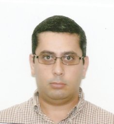 Ghaly Michael合作论文数The Russell H Morgan Department of Radiology and Radiological Science, Johns Hopkins University33Bin He合作论文数31
 Ye Li合作论文数27
 Frederic Fahey合作论文数Harvard Medical School;Department of Radiology, Boston Children’s Hospital;BCH Small Animal Imaging Laboratory, Boston Children’s Hospital27Jonathan M. Links合作论文数23Xin He (馨 賀)合作论文数21
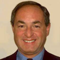 S. Ted Treves合作论文数Department of Radiology, Harvard Medical School;Boston Children's Hospital;Dana-Farber / Harvard Cancer Center, Harvard Medical School21Xinhua Cao合作论文数Children's Hospital Boston, Harvard Medical School21
 Abhinav K. Jha合作论文数19
 William Paul Segars合作论文数School of Medicine, Duke University18
 George Sgouros合作论文数Department of Otolaryngology-Head and Neck Surgery, School of Medicine, Johns Hopkins University17
 Junyu Chen合作论文数16
 Yuni Dewaraja合作论文数Department of Radiology, Michigan Medicine, University of Michigan;Center for Positron Emission Tomography, University of Michigan15
 Michael Ljungberg合作论文数Department of Medical Radiation Physics;Jubileum Institute;Lund University;Jubileum Institute, Lund University15XD Zhao合作论文数14Jingyan Xu合作论文数13
 Martin Anthony Lodge合作论文数Russell H. Morgan Department of Radiology and Radiological Science, Johns Hopkins Medicine;PET Center, Johns Hopkins University13Shannon E O'Reilly合作论文数Department of Radiation Oncology, Perelman School of Medicine, University of Pennsylvania12
 Grant T. Gullberg合作论文数Department of Radiology and Biomedical Imaging, University of California San Francisco12Katsuyuki Taguchi合作论文数12
 Xiaolan Wang合作论文数Dept. of Radiol. & Radiol. Sci., Johns Hopkins Univ.;c;Dept. of Radiol. & Radiol. Sci., Johns Hopkins Univ.11Rong Xing合作论文数11Briana Sexton-Stallone合作论文数10na song合作论文数10
 Justin Brown合作论文数Department of Paediatrics, Clinical Sciences at Monash Health, Monash University10BM Tsui合作论文数9Patt Bradley E合作论文数9Sunderland, JJ合作论文数9
 Thibault Mauxion合作论文数Inserm, France and Université Toulouse III-Paul Sabatier9
 Michael A King合作论文数Department of Radiology, University of Massachusetts Medical School9
 Dan J. Kadrmas合作论文数Department of Bioengineering CAMT, University of Utah Salt Lake City9
 David M. Loeb合作论文数The Children's Hospital at Montefiore;Montefiore Einstein9KL Gilland合作论文数8Uribe Carlos合作论文数8
 Andrew Prideaux合作论文数8
 George S. K. Fung合作论文数Laboratory for Intelligent Transportation Systems Research;Department of Electrical and Electronic Engineering;University of Hong Kong;Laboratory for Intelligent Transportation Systems Research, University of Hong Kong8David S. Lalush合作论文数Department of Biomedical Engineering
North Carolina State University8Sébastien Baechler合作论文数8Jacene Heather A合作论文数7Lishui Cheng合作论文数7Wentao Wang合作论文数7
George S. K. Fung合作论文数Laboratory for Intelligent Transportation Systems Research;Department of Electrical and Electronic Engineering;University of Hong Kong;Laboratory for Intelligent Transportation Systems Research, University of Hong Kong8David S. Lalush合作论文数Department of Biomedical Engineering
North Carolina State University8Sébastien Baechler合作论文数8Jacene Heather A合作论文数7Lishui Cheng合作论文数7Wentao Wang合作论文数7




