Focal cortical dysplasia (FCD) is one of the most prevalent and difficult to detect structural etiologies of epilepsy. Recent advancements in MRI technology have enhanced detectability of FCD, yet many remain undiagnosed by MRI. FDG-PET may increase detection rates when utilized alongside MRI, yet PET continues to be underutilized beyond temporal lobe epilepsy, likely due to challenges in identifying subtle cortical lesions. Hybrid PET/MR systems present an opportunity to improve image quality and lesion detection, but benefits of hybrid PET/MR in epilepsy beyond workflow advantages are not well established. This feasibility study assesses the potential of a prototype MR-guided PET reconstruction that integrates event-by-event motion correction, enhanced point spread function modeling, regularization techniques, and MR-guided reconstruction algorithm. We demonstrate enhancements in cortical definition and increased conspicuity of FCD in FDG-PET. The combined reconstruction approach provides a distinct advantage for PET/MR, thereby helping to realize the full potential of hybrid PET/MR systems.
Recent advances in ultrahigh-field MRI have led to accelerated adoption in clinical applications, especially after regulatory approval for 7-T MRI systems. The substantial gains in signal-to-noise ratio, tissue contrast, chemical shift, and susceptibility effects enable unprecedented image resolution and quality, resulting in more accurate diagnoses and improved treatment planning. Despite these inherent advantages of 7-T MRI, several challenges have historically limited its clinical adoption. The authors review recent technical advancements that have further enabled routine clinical implementation of 7-T MRI and highlight its applications across a diverse range of neurologic disorders. They outline key physical principles underpinning 7-T imaging, including susceptibility and chemical shift effects, and describes how innovations such as dynamic parallel transmission and deep-learning reconstructions stand to impact clinical translation by mitigating previous technical barriers. Next, the most common clinical indications are addressed, encompassing epilepsy, multiple sclerosis, pituitary microadenomas, unruptured aneurysms, cerebrovascular disease, brain tumors, neurodegenerative diseases, and applications in planning deep brain stimulation. In each of these conditions, 7-T MRI demonstrates superior lesion detection, enhanced anatomic delineation, and increased diagnostic specificity compared with MRI at lower field strengths. With an expanding body of evidence supporting its utility in both diagnosis and treatment planning, 7-T MRI is poised to play an increasingly pivotal role in clinical neuroradiology. ©RSNA, 2025 Supplemental material is available for this article.
Background: Essential and Parkinsonian tremors are common movement disorders that impair quality of life. Two incisionless ablative techniques for refractory disease are Gamma Knife radiosurgery (GKRS) and MR-guided focused ultrasound (MRgFUS); both are frame-based, and GKRS treatment times may extend to several hours. We examined a non-invasive, frameless, linear accelerator (LINAC)-based stereotactic radiosurgery (SRS) technique using a virtual cone method for thalamotomy. Methods: In this prospective, single-centre, open-label trial, 40 patients with medically refractory tremor (35 essential tremor, 4 tremor-dominant Parkinson's disease, 1 mixed) underwent unilateral LINAC virtual-cone thalamotomy with thermoplastic-mask immobilisation. The primary outcome was contralateral tremor improvement on the Fahn-Tolosa-Marin (FTM) scale at 3, 6, and ≥12 months. Secondary outcomes were safety (CTCAE), satisfaction, and quality of life. Wilcoxon signed-rank tests compared pre- versus post-treatment scores; linear mixed-effects models assessed change over time. This trial is registered with ClinicalTrials.gov, NCT03305588. Findings: At a median follow-up of 31·7 months, 38 of 40 patients (95%) achieved ≥10% contralateral FTM improvement by 6 months (p<0·001). Mean contralateral FTM reduction was 54·6% (SD 36·0) in the intention-to-treat cohort and 60·7% (24·6) among responders. Five patients (12·5%) had serious neurological adverse events (dysarthria, focal hemiparesis), typically at 6 months, generally improving with corticosteroids or bevacizumab; one died of unrelated causes. Transient paraesthesia was the most common minor event. Interpretation: Frameless, coneless, LINAC-based thalamotomy may be an effective alternative to frame-based SRS or FUS for medically refractory tremor, with comparable risk and short, mask-based treatment. Larger randomised or comparative studies with blinded assessment are warranted.
BACKGROUND AND PURPOSE:7T MRI enhances lesion detection in epilepsy but is limited by radiofrequency transmission field (B1+) inhomogeneity and long scan times. Recent advancements in dynamic parallel transmission and deep learning-based reconstructions offer promising solutions. We aimed to optimize an enhanced 7T epilepsy protocol incorporating these innovations and evaluate real-world benefits compared with standard 7T epilepsy protocol. MATERIALS AND METHODS:We retrospectively compared 40 consecutive brain MRIs acquired using a standard 7T epilepsy protocol with 40 MRIs obtained with an enhanced protocol with dynamic parallel transmission and deep learning-based k-space reconstructions. Quantitative metrics for comparison included image noise, signal homogeneity (coefficient of variation), and resolution/time trade-offs. RESULTS:The enhanced protocol demonstrated significant improvements in resolution, scan time, noise levels, and image homogeneity. The edge-enhancing gradient-echo and magnetization-prepared rapid acquisition of gradient echo with 2 inversions sequence exhibited a 57.8% reduction in voxel volume while reducing scan time by 33.0% and improving image homogeneity (P = .002) without a significant change in noise (P = .09). Deep learning-based reconstruction of coronal T2 turbo spin-echo imaging resulted in a 25.7% reduction in noise (P < .001), and patient-specific B1+ shimming achieved homogeneity comparable with dielectric pads. The sampling perfection with application-optimized contrasts using different flip angle evolutions (SPACE Sequence) FLAIR had reduced noise (P < .001), enhanced homogeneity (P < .001), and halved voxel size while maintaining similar scan times. Deep learning-based EPI SWI improved acquisition time by 56.5% with a 20.5% reduction in noise (P = .001). Despite increased resolution and parallel transmission use, the overall scan time was less than 25 minutes, one-half the duration recommended by the 7T Epilepsy Task Force. CONCLUSIONS:Integration of dynamic parallel transmission and deep learning-based reconstructions enhances image resolution, reduces scan time, and improves image homogeneity, addressing barriers to routine clinical implementation of 7T MRI. These advancements may improve lesion conspicuity and contribute to better outcomes for patients with epilepsy.
Objective This longitudinal study evaluated the long-term efficacy and safety of hematopoietic stem cell transplantation (HSCT) in slowing the progression of CSF1R-related disorder (CSF1R-RD).Methods Six symptomatic patients (mean follow-up, 6.6 years) were compared with six matched, untreated controls. The CSF1R Clinical Severity Score (CCSS), Montreal Cognitive Assessment, and Sundal radiological score were used for evaluation.Results Post-HSCT clinical progression slowed significantly from 14.1 to 3.7 CCSS/year, compared with 15.2 CCSS/year in the control group (p<0.01). Cognitive decline was substantially reduced (-1.5 points/year vs. -7.6 points/year), and radiological deterioration slowed (0.4 per year vs. 3.9 per year). Notably, during the observation period, all HSCT patients survived, whereas 50% of the patients in the control group died. No serious transplant-related complications were observed.Conclusion HSCT is a potent disease-modifying therapy for CSF1R-RD that drastically improves survival and slows deterioration. These findings underscore the necessity of early intervention during the symptomatic phase to maximize the preservation of quality of life for affected individuals.
BACKGROUND:Deep brain stimulation (DBS) has been investigated for patients with drug-resistant epilepsy who are not candidates for resective surgery. Because different types of epilepsy involve different brain networks, numerous DBS targets have been explored. METHODS:To provide a comprehensive overview of this expanding literature, we conducted a systematic review of studies for DBS in epilepsy, collecting data on surgical targets, individual disease characteristics, outcomes, and precise electrode placements. DBS electrode coordinates were gathered into a common template space and related to clinical outcomes. FINDINGS:We included 124 studies, corresponding to 1,210 patients and 20 distinct surgical targets. While the anterior (ANT) and centromedian (CM) nuclei of the thalamus remain the most studied, we also review less commonly used targets that show promise for specific forms of epilepsy and may warrant further investigation. Substantial variability in targeting strategies and electrode placement was observed within each of the target regions. Importantly, significant relationships between stimulation location and outcomes were identified for ANT- and CM-DBS. For ANT-DBS, shorter distance to the mammillothalamic tract junction was associated with greater seizure reduction on both study- and patient-level analyses (r=-0.55, p<0.001 and r=-0.51, p<0.001, respectively). For CM-DBS, localization effects may be dependent on the form of epilepsy, with stimulation of the parvocellular CM being associated with better outcomes in generalized epilepsy. INTERPRETATION:Our results emphasize the importance of accurate targeting in DBS for epilepsy. Our database and atlas of DBS targets are made publicly available, potentially serving further meta-analytical work.
BACKGROUND AND PURPOSE:Clinical adoption of 7T MRI has been limited by lengthy acquisitions. Acceleration techniques, such as controlled aliasing in parallel imaging (CAIPI) and compressed sensing (CS), can reduce scan time but are prone to artifacts and noise. This work combines CAIPI and CS within a unified, two-step deep-learning (DL) reconstruction framework to leverage their strengths: CAIPI for controlled aliasing with improved conditioning, CS for additional incoherent undersampling, and DL to reduce residual aliasing and CS-related artifacts. This hybrid approach aims to maintain image quality while enabling higher net acceleration and reducing scan time. MATERIALS AND METHODS:In this paired within-subject study, 30 patients underwent 7T sampling perfection with application-optimized contrasts using different flip angle evolutions (SPACE) FLAIR and 30 patients underwent 7T SPACE T2 acquisitions. Each scan included a reference CAIPI-DL protocol (acceleration factor=6; ∼7 minutes) and a CS-CAIPI-DL protocol (acceleration factor=14; ∼3.5 minutes). Image quality was assessed quantitatively using structural similarity index, peak signal-to-noise ratio, gradient-domain mean squared error, contrast-to-noise ratio, noise, ghosting ratio, and Natural Image Quality Evaluator (NIQE), as well as blinded qualitative assessments. Statistical comparison used linear mixed-effects models with false discovery rate correction. RESULTS:CS-CAIPI-DL reduced scan time by approximately 50% without significant differences in contrast-to-noise ratio or noise for either sequence. Ghosting ratios were significantly lower with CS-CAIPI-DL for both FLAIR (∼10% reduction; q<.001) and T2 (∼30% reduction; q<.001), consistent with reduced motion-related artifact. NIQE scores were significantly improved for T2 (q<.001) and showed a favorable trend for FLAIR (q=.06). There was no significant difference in diagnostic adequacy and no DL-specific artifacts. CONCLUSIONS:Combining CS with CAIPI and DL reconstruction enables over two-fold increase in acceleration with no statistically significant differences in key image quality metrics. Shorter acquisitions were associated with significantly lower ghosting ratios, consistent with reduced motion-related artifact, supporting the feasibility of rapid, high-resolution 7T brain MRI.
Background:High-resolution MRI is essential for accurate diagnosis and treatment planning, but its clinical acquisition is often constrained by long scanning times, which increase patient discomfort and reduce scanner throughput. While super-resolution (SR) techniques offer a post-acquisition solution to enhance resolution, existing deep learning approaches face trade-offs between reconstruction fidelity and computational efficiency, limiting their clinical applicability. Purpose:This study aims to develop an efficient and accurate deep learning framework for MRI super-resolution that preserves fine anatomical detail while maintaining low computational overhead, enabling practical integration into clinical workflows. Materials and Methods:We propose a novel SR framework based on multi-head selective state-space models (MHSSM) integrated with a lightweight channel multilayer perceptron (MLP). The model employs 2D patch extraction with hybrid scanning strategies (vertical, horizontal, and diagonal) to capture long-range dependencies while mitigating pixel forgetting. Each MambaFormer block combines MHSSM, depthwise convolutions, and gated channel mixing to balance local and global feature representation. The framework was trained and evaluated on two distinct datasets: 7T brain T1 MP2RAGE maps (142 subjects) and 1.5T prostate T2w MRI (334 subjects). Performance was compared against multiple baselines including Bicubic interpolation, GAN-based (CycleGAN, Pix2pix, SPSR), transformer-based (SwinIR), Mamba-based (MambaIR), and diffusion-based (I2SB, Res-SRDiff) methods. Results:The proposed model demonstrated superior performance across all evaluation metrics while maintaining exceptional computational efficiency. On the 7T brain dataset, our method achieved the highest structural similarity (SSIM: 0.951±0.021) and peak signal-to-noise ratio (PSNR: 26.90±1.41 dB), along with the best perceptual quality scores (LPIPS: 0.076±0.022; GMSD: 0.083±0.017). These results represented statistically significant improvements over all baselines (p < 0.001), including a 2.1% SSIM gain over SPSR and a 2.4% PSNR improvement over Res-SRDiff. For the prostate dataset, the model similarly outperformed competing approaches, achieving SSIM of 0.770±0.049, PSNR of 27.15±2.19 dB, LPIPS of 0.190±0.095, and GMSD of 0.087±0.013. Notably, our framework accomplished these results with only 0.9 million parameters and 57 GFLOPs, representing reductions of 99.8% in parameters and 97.5% in computational operations compared to Res-SRDiff, while also substantially outperforming SwinIR and MambaIR in both accuracy and efficiency metrics. Conclusion:The proposed framework provides a computationally efficient yet accurate solution for MRI super-resolution, delivering well-defined anatomical details and improved perceptual fidelity across anatomically distinct datasets. By significantly reducing computational demands while maintaining state-of-the-art performance, the model offers strong potential for feasibility toward clinical translation and scalable integration into future imaging workflows.
OBJECTIVE:Precise targeting of subcortical structures is crucial for deep brain stimulation (DBS). Although 7T MRI provides superior resolution and contrast, its clinical adoption remains limited by B1+ transmit inhomogeneity, prolonged scan times, and motion sensitivity. This study applied deep learning (DL)-based image reconstruction and dynamic parallel transmission (pTx) to optimize DBS protocols and improve image quality. METHODS:Thirteen patients scanned using a conventional 7T DBS protocol were compared to 13 imaged after implementing DL reconstruction and dynamic pTx. Two readers scored image quality, motion artifact, and target conspicuity on 5-point Likert scales. Ordinal logistic regression was used to calculate odds ratios (OR) for improvements with the enhanced protocol, adjusted for multiple comparisons. RESULTS:Enhanced MP2RAGE reduced voxel volume by 65.8% and scan time by 32.9%, with improved image quality (OR = 4.4;p = 0.003), target conspicuity (OR = 3.4;p = 0.011), and reduced motion artifacts (OR = 3.8;p = 0.006). Fast gray matter acquisition T1 inversion recovery (FGATIR) scan time decreased by 45.2% with improved target delineation of both globus pallidus interna (OR = 22.9;p < 0.001) and dentato-rubro-thalamic tract (OR = 8.8;p < 0.001). T2-weighted sampling perfection with application-optimized contrasts using different flip angle evolutions (SPACE) improved subthalamic nucleus (STN) delineation (OR = 25.3;p < 0.001). Susceptibility-weighted imaging (SWI) improved image quality (OR = 17.4;p < 0.001), STN delineation (OR = 16.9;p < 0.001), and reduced scan time by 42.6%. Enhanced 3D spoiled gradient recall echo improved image quality (OR = 17.4;p < 0.001) and vessel visualization (OR = 26.1;p < 0.001) with reduced motion artifact (OR = 8.8;p < 0.001). Scan time decreased from 4:33 to 1:35, reducing protocol duration from 42:16 to 26:40 (36.9%). CONCLUSIONS:DL reconstruction and dynamic pTx improved image quality, target definition, and motion robustness while shortening 7T DBS protocol time.
This study aimed to develop high-resolution, photorealistic three-dimensional (3D) neuroanatomical models via photogrammetry-based cadaver imaging combined with magnetic resonance imaging (MRI) tractography and 3D printing integrated into augmented reality (AR) and virtual reality (VR) platforms to support neurosurgical education. White matter dissections were performed on ten human cadaveric brains preserved via the Klingler method. Each dissection stage was photographed in a photographic studio using a DSLR camera and turntable system. Images were processed via Adobe Photoshop and reconstructed into 3D models via Agisoft Metashape. Postprocessing and optimization were carried out in Blender. The models were visualized via AR/VR devices and shared via Sketchfab. The selected models were 3D printed with a PLA filament. Representative MRI tractography data were incorporated into the models with Blender to visualize key white matter pathways. Twenty-eight detailed 3D models were created, capturing step-by-step dissections of the cerebral hemispheres, brainstem and cerebellum. On average, each model was generated from 442 photographs, producing more than six million surface triangles in the point cloud. The selected models were 3D-printed at life size using a PLA filament, and MRI tractography data were incorporated to visualize key white matter tracts. The models were successfully implemented in both the AR and VR platforms, enabling immersive neuroanatomical exploration. This study introduces a systematically produced collection of 28 high-resolution photogrammetry-based cadaveric neuroanatomical 360-degree models integrating representative tractography, AR/VR visualization, and 3D printing. This collection provides a refined and accessible resource for neuroanatomical and neurosurgical education. These models offer an interactive and accessible alternative to cadaver-based training, overcoming financial, ethical, and logistical barriers, and serve as effective educational tools to enhance visuospatial neuroanatomical understanding and improve neurosurgical training.
BACKGROUND The authors aimed to evaluate the feasibility/safety of stereo-electroencephalography (SEEG) and SEEG-guided radiofrequency thermocoagulation (RFTC) in patients with drug-resistant epilepsy previously implanted with neuromodulation devices, using a retrospective cohort design evaluating patients treated at the Mayo Clinic Florida Epilepsy Center. OBSERVATIONS Nine patients (mean age 36.3 ± 10.9 years, 33% female) with prior implantation of neuromodulation devices were included. A vagus nerve stimulator was present in 8 of 9 patients, and 4 patients had undergone previous intracranial neuromodulation. Five patients underwent SEEG-RFTC. The mean SEEG monitoring duration was 6.5 ± 2.9 days. No intraoperative or procedural complications occurred, including hardware-related issues. No permanent or significant neurological/cognitive deficits occurred. One patient experienced transient postoperative neurological symptoms attributed to perilesional edema. Seizure reduction ranged from 50% to 75% following SEEG-RFTC. LESSONS SEEG and SEEG-RFTC are feasible in patients who have implanted neuromodulation devices, including intracranial implants. These procedures may be performed without interference with existing hardware and provide meaningful seizure reduction in highly refractory populations. Further studies with larger cohorts and longer follow-up are needed to better define efficacy and patient selection. https://thejns.org/doi/10.3171/CASE26376
The pineal region presents considerable challenges for neurosurgical interventions owing to its deep anatomical location and the intricate network of critical neurovascular structures that encase it. The capacity to conduct effective and safe neurosurgical procedures in this area necessitates a comprehensive understanding of anatomical variations. This study aims to conduct an in-depth analysis of radiologic measurements pertinent to accessing the pineal region, alongside their supported by qualitative cadaveric dissections. The supracerebellar infratentorial (SCIT) approach, occipital transtentorial (OT) approach, and posterior interhemispheric (PIH) approach were performed on four cadaveric heads (eight sides). All dissections were performed with the head positioned to simulate the orientation employed in the operating room. Magnetic resonance imaging (MRI) scans from 50 male to 50 female patients were also retrospectively assessed. Descriptive statistics were presented for the quantitative data. MRI analysis of 100 adult subjects revealed a mean tentorial angle of 31.5° (range: 14.4–47.7°), indicating substantial anatomical variability. Measurements from external landmarks to the pineal gland and tentorium were quantified to assist in surgical planning. The Torcular Herophili was found to exhibit variable vertical positioning, and its distance from the inion was measured radiologically. These findings were categorized into distinct morphometric types based on tentorial inclination and torcular location. Cadaveric dissections were performed in surgical positions simulating SCIT, OT, and PIH approaches, and were used to qualitatively illustrate key anatomical corridors. Each dissection demonstrated the spatial relationships among the tentorium, splenium, deep venous system, and surrounding neurovascular structures, consistent with the anatomical context of the radiologic measurements. In neurosurgical practice, a lesion’s location is a critical determinant in selecting the approach for addressing pineal region lesions, given the complex microsurgical anatomy and deep positioning of these structures. Additionally, conducting thorough radiologic examinations during the preoperative phase is essential in informing the choice of surgical approach. We posit that employing radiologic measurements and identifying vascular variations will lead to reduced complication rates.
BACKGROUND AND OBJECTIVES:Access to the orbit, cavernous sinus, Meckel cave (MC), and infratemporal fossa (ITF) remains challenging due to the complex 3-dimensional anatomy and proximity to critical neurovascular structures. An extradural endoscopic orbitozygomatic (OZ) approach has the potential to provide a direct surgical corridor across these compartments while minimizing the limitations of compartment-specific corridors. METHODS:Five formalin-fixed, silicone-injected cadaveric heads (10 sides) were dissected using an extradural OZ approach under operative microscopy supplemented with 0° and 30° rigid endoscopes. After a two-piece OZ craniotomy, extradural dissection was performed toward the superior orbital fissure and foramen rotundum/ovale, combined with anterior petrosectomy and opening of MC. Anatomic exposure, corridor continuity, and visualization of key neurovascular structures were qualitatively assessed. RESULTS:The extradural endoscopic OZ approach created a direct working corridor from the lateral orbit to the ITF and petroclival region. Endoscopic augmentation enhanced visualization of the lateral cavernous sinus wall, trigeminal divisions (V1-V3), cavernous internal carotid artery, medial orbital apex, deep infratemporal recesses, and the petroclival region. Anterior petrosectomy extended exposure toward the upper clivus and ventrolateral brainstem. Limitations included restricted microscopic visualization of the medial orbit and inferior ITF, which were partially mitigated by angled endoscopy. CONCLUSION:This cadaveric study demonstrates that the extradural OZ-anterior petrosectomy approach establishes a continuous anatomic corridor integrating the orbit, cavernous sinus/MC, ITF, and petroclival region, while defining its boundaries, areas of optimal exposure, and inherent limitations. As a qualitative study, the findings are descriptive in nature and do not include quantitative morphometric analysis.
Background Ordered-subset expectation-maximization (OSEM) PET reconstruction is limited by spatial resolution and partial-volume effects, contributing to interreader variability and false positives and highlighting the need for more comprehensive alternative approaches. Purpose To evaluate whether MRI-guided brain PET reconstruction improves inter-reader agreement, visual consistency, diagnostic performance, and Centiloid (CL)-based quantitative separation of amyloid status compared with OSEM reconstruction. Materials and Methods In this retrospective multireader diagnostic accuracy study conducted at a single institution, consecutive brain fluorine 18 (18F)-florbetapir PET/MRI scans (September 2019 to December 2024) were analyzed. For each scan, OSEM and MRI-guided PET reconstructions were generated. Four blinded readers independently interpreted both reconstructions in separate sessions, recording amyloid status (β-amyloid [Aβ]-/Aβ+), diagnostic confidence (three-point scale), and image quality (five-point scale). The consensus-derived reference standard (Aβ-/Aβ+) was not fully independent. Diagnostic performance was assessed using generalized linear mixed-effects models. Interreader agreement was assessed using Fleiss κ, and quantitative measures (CL values and standardized uptake value ratios) were compared between reconstructions. P values were adjusted for multiple comparisons. Results A total of 120 patients (120 scans; median age, 74.8 years [IQR, 68.7-78.3 years]; 66 males) were included. MRI-guided reconstruction improved interreader agreement for amyloid status (Fleiss κ, 0.91 vs 0.79; absolute increase, 0.12 [95% CI: 0.05, 0.20]; P < .001), with greatest improvement in the posterior cingulate cortex and precuneus (κ, 0.82 vs 0.74; P = .03). Specificity improved from 91.6% (95% CI: 85.7, 96.5) to 99.2% (95% CI: 98.4, 99.7), reducing false positives from 8.1% (14 of 166) to 0.8% (two of 168) (P < .001) relative to the reference standard. CL separation was preserved between Aβ- and Aβ+ groups (MRI-guided: -4.24 vs 89.74; OSEM: 0.84 vs 89.0; both P < .001), with lower median CL values in Aβ- scans (-4.24 vs 0.84; P = .01). Conclusion MRI-guided amyloid brain PET reconstruction showed higher interreader agreement and fewer false-positive classifications than OSEM reconstruction and preserved CL-based separation between the Aβ- and Aβ+ groups. © RSNA, 2026 Supplemental material is available for this article. See also the editorial by Bedmutha in this issue.
BACKGROUND:Functional pituitary adenomas (FPAs), especially those "MRI-occult" on 1.5T/3T and smaller than 10 mm, present diagnostic and therapeutic challenges, leading to repeated investigations, invasive procedures, and suboptimal outcomes. This review examines the potential clinical use and limitations of 7T MRI and amino-acid (AA)-PET with tracers like L-[methyl-11C]methionine (MET) or O-(2-[18F]fluoroethyl)-L-tyrosine (FET) for detecting MRI-occult FPAs. MATERIALS AND METHODS:A systematic search of Medline, Embase, and Web of Science was performed up to August 2025, following the Preferred Reporting Items for Systematic Reviews and Meta-Analyses guidelines. Studies evaluating 7T MRI and AA-PET for MRI-occult PAs were included and independently reviewed by three authors. Eighteen studies met inclusion criteria: 4 evaluated the 7T MRI, 8 MET-PET, three FET-PET, and three hybrid/multi-tracer approaches. RESULTS:7T MRI offers superior spatial resolution, increased signal-to-noise ratio, and improved tissue contrast. AA-PET provides high tumor-to-background contrast, facilitating MRI-occult PAs localization. CONCLUSIONS:Despite these advances, small cohort sizes, heterogeneous protocols, high costs, and limited accessibility limit the current evidence base. Ultra-high-field 7T MRI and AA-PET represent emerging complementary imaging techniques for MRI-occult FPAs. Current evidence is preliminary, underscoring the need to standardize protocols, conduct prospective multicenter studies, and assess cost-effectiveness before widespread clinical adoption.
OBJECTIVE:This study aims to identify both fluid and neuroimaging biomarkers for CSF1R-RD that can inform the optimal timing of treatment administration to maximize therapeutic benefit, while also providing sensitive quantitative measurements to monitor disease progression. METHODS:Our study compared neuroimaging and fluid (plasma and cerebrospinal fluid (CSF)) biomarkers across three distinct populations: asymptomatic CSF1R pathogenic variant carriers (N = 14), symptomatic CSF1R pathogenic variant carriers (N = 17), and healthy controls (N = 30). We evaluated biomarker correlations with both an established (Montreal Cognitive Assessment (MoCA)) and a novel (CSF1R Clinical Severity Score (CCSS)) clinical diagnostic scale to investigate potential clinical utility. Additionally, we tested the relationship between select biomarkers and cortical thickness using 3D T1-weighted MPRAGE scans, providing a highly valuable physiological component to our analyses. RESULTS:Our results demonstrate that while plasma glial fibrillary acidic protein (GFAP) displays a high sensitivity for distinguishing early-stage CSF1R-RD patients from healthy controls, plasma neurofilament light chain (NfL) is more effective for tracking disease progression following the onset of symptoms. INTERPRETATION:Overall, our study provides evidence for plasma NfL and GFAP as valuable biomarkers of earliest symptom onset and disease progression for CSF1R-RD.
INTRODUCTION/AIMS:There are no established biomarkers of upper motor neuron degeneration to aid in the diagnosis of motor neuron disease (MND). This study examines the diagnostic value of the motor band sign as a marker of upper motor neuron degeneration and its relationship to clinical findings in MND. METHODS:Records of consecutive patients who underwent 7T magnetic resonance imaging (MRI) between October 2021 and April 2025 for evaluation of MND or other neurologic indications were retrospectively reviewed. Clinical variables and plasma neurofilament light chain (pNfL) levels were recorded. An upper motor neuron score (Mayo UMNS) was derived from reflex scores. Blinded MRI review assessed the degree of susceptibility-weighted imaging (SWI) hypointensity in the hand, foot, and bulbar motor cortex regions. RESULTS:An MBS was observed in 100 of 117 (85.5%) MND patients and in 16 (15.5%) patients with non-MND diagnoses, corresponding to a sensitivity of 85.5% (78.0%-90.7%) and 84.5% (76.2%-90.2%) specificity. The MBS in 78 MND patients (70.9%) preferentially involved the middle and deep cortical layers, giving a trilaminar appearance, while only one non-MND patient had this finding. Mayo UMNS (β = 0.89, p < 0.001), pNfL (β = 0.63, p = 0.033), and age at evaluation (β = 0.68, p = 0.027) were independently associated with the summed SWI score. DISCUSSION:The 7T MRI MBS is a sensitive and specific marker for MND that complements established clinical evaluation. Using 7T, a trilaminar appearance of the motor cortex, reflecting known histopathological changes, can be visualized and may be specific to MND.
PURPOSE:The cerebral aqueduct (CA) is the narrowest part of the ventricular system, connecting the third and fourth ventricles. This structure is encircled by important neuroanatomical features, and with the increasing application of neuroendoscopic techniques, a comprehensive understanding of the anatomy of the CA is essential. This study aims to provide a comprehensive, multi-perspective anatomical analysis of the cerebral aqueduct and its surrounding structures, integrating microsurgical dissections with endoscopic anatomical correlations to support safer surgical planning. METHODS:Three formalin-fixed human brainstem specimens were prepared using the Klingler technique and microscopically dissected with a focus on the cerebral aqueduct. In addition, endoscopic exposure was simulated in two separate formalin-fixed cadaveric heads to correlate intraventricular landmarks with aqueductal anatomy. RESULTS:The CA is positioned ventromedially to the colliculi, which are located dorsal to the mesencephalon. The frenulum veli structure, which contains the crossing of the fourth cranial nerve, was observed inferior to the colliculi. The periaqueductal gray matter surrounds the CA dorsally and ventrolaterally on both sides. A region topographically corresponding to the oculomotor nuclear complex was approximated based on known anatomical relationships and the intramesencephalic course of cranial nerve III, without direct visualization. As the dissection progressed, the courses of the deep tracts ventral to the CA were demonstrated. The medial longitudinal fasciculus, the trigeminal mesencephalic tract, and the superior cerebellar peduncle were identified. Fibers of the central tegmental tract were observed running posterior to, and also traversing, the decussation of the superior cerebellar peduncle. CONCLUSIONS:The relationship of the CA with adjacent anatomical structures presents challenges to micro-endoscopic interventions of this region, necessitating a thorough understanding of the related anatomy.
BACKGROUND:Carotid revascularization prevents ipsilateral ischemic stroke in high-grade stenosis, but its effect on hemispheric white matter disease (WMD) progression is not known. METHODS:We conducted a longitudinal study of patients ≥40 years with carotid stenosis (moderate50-69%, high-grade 70-99% or occluded) seen at Mayo Clinic (Florida, Rochester, Arizona) between 2011 and 2015 with serial brain MRIs ≥1 year apart and clinical follow-up through 2020. Hemispheric WMD volumes were quantified from axial T2-FLAIR using automated segmentation. Patients were stratified by revascularization status. The primary outcome was annual change in WMD volume ipsilateral or contralateral to the index carotid. Hemispheric differences in the annual rate of WMD progression (cm3/yr) were compared between revascularized vs not revascularized using multivariable linear regression adjusted for demographic and vascular risk factors, with additional adjustment for baseline imbalances between groups. Because three outcomes were evaluated, a Bonferroni-corrected significance threshold of p < 0.0167 was applied. RESULTS:Among 150 patients (mean age 71 ± 10 years, 36.7% female), 37.3% had moderate stenosis, 42% high-grade, and 20.7% occlusion. Seventy-three (48.7%) underwent carotid revascularization; median follow-up was 5.1 years. Baseline WMD volume was 4.86 cm3 ipsilateral and 4.39 cm3 contralateral. Revascularized patients had a numerically slower ipsilateral WMD progression than non-revascularized patients (0.32 vs. 0.63 cm3/year, p = 0.34), with no contralateral difference. In fully adjusted models, revascularization was associated with attenuation of hemispheric difference in WMD progression (β = -0.46; 95% CI -0.84 to -0.08; p = 0.019). Although this association did not meet the Bonferroni-adjusted threshold, the effect estimate remained directionally consistent. CONCLUSIONS:Carotid revascularization was associated with numerically slower ipsilateral WMD progression (∼50% relative difference) and attenuation of hemispheric asymmetry over a median 5-year follow-up. Given the modest statistical signal and correction for multiple comparisons, these findings should be interpreted as observational and hypothesis-generating. Prospective studies should evaluate whether modulation of WMD progression translates into meaningful cognitive benefit.
PURPOSE:Layer fMRI is an increasingly utilized technique that provides insights into the laminar organization of brain activity. However, both blood-oxygen-level-dependent (BOLD) fMRI and vascular space occupancy data (VASO) have certain limitations, such as bias towards larger cortical veins in BOLD fMRI and high specific absorption rate in VASO. This study aims to explore the feasibility of whole-brain laminar functional quantitative susceptibility mapping (fQSM) compared to laminar BOLD fMRI and VASO at ultra-high field. METHODS:Data were acquired using 3D EPI techniques. Complex data were denoised with NORDIC and susceptibility maps were computed using 3D path-based unwrapping, the variable-kernel sophisticated harmonic artifact reduction, as well as the streaking artifact reduction for QSM algorithms. To assess layer-specific activation, 20 layers were segmented in the somatosensory and motor cortices, obtained from a finger tapping paradigm, and further averaged into six anatomical cortical layers. The magnitude of signal change and z-scores were compared across layers for each technique. RESULTS:fQSM showed the largest activation-dependent mean susceptibility decrease in Layers II/III in M1 and Layers I/II in S1 with up to -1.3 ppb while BOLD showed the strongest mean signal increase in Layer I. Our data suggest that fQSM demonstrates less bias towards activation in superficial layers compared to BOLD. Moreover, activation-based susceptibility change was comparable to VASO data. CONCLUSION:Studying whole-brain, layer-dependent activation with submillimeter fQSM is feasible and reduces bias towards venous drainage effects on the cortical surface compared to BOLD, thereby enabling better localization of laminar activation.
 Grewal Sanjeet S合作论文数Department of Surgery, University of Cincinnati Medical Center69
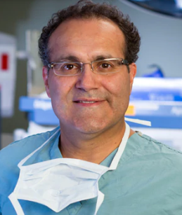 Alfredo Quinones-Hinojosa合作论文数Department of Neurosurgery, Mayo Clinic53
 Lela Okromelidze合作论文数2Radiology, and.39
 Kaisorn Chaichana合作论文数Johns Hopkins University School of Medicine35
 William Tatum合作论文数Mayo Clinic32Xiangzhi Zhou合作论文数28Shengzhen Tao合作论文数26
 Anteneh Feyissa合作论文数Department of Pharmacology and Therapeutics, NUI Galway26
 David S. Sabsevitz合作论文数Department of Neurology, Medical College of Wisconsin26Vishal Patel合作论文数20Erin Westerhold合作论文数20
 Ali Karadag合作论文数Health Science University19Jentoft Mark合作论文数18
 Vivek Gupta合作论文数17Brin Freund合作论文数Departments of1Neurology.17James F. Meschia合作论文数17
 Necmettin Tanriover合作论文数Department of Neurosurgery, Istanbul University-Cerrahpaşa16
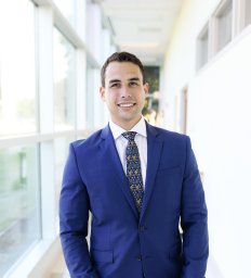 Ricardo A Domingo合作论文数Department of Neurosurgery, Mayo Clinic15
 Zbigniew K. Wszolek合作论文数Departments of Neurology, College of Medicine and Science, Mayo Clinic;College of Medicine, University of Florida Health Science Center15
 Chen Lin合作论文数Plant Dev Biol & Plant Physiol, Univ Kiel14
 Wendy J Sherman合作论文数College of Education, Kent State University14
 Elena Greco合作论文数2Radiology, and.13Alok A Bhatt合作论文数13
 Leonardo Almeida合作论文数Department of Neurology, University of Florida at Gainesville13
 Michael S. Okun合作论文数Norman Fixel Institute for Neurological Diseases, University of Florida Health;Department of Neurology, College of Medicine, University of Florida Health13
 Markus Bredel合作论文数13
 Mehmet Senoglu合作论文数Tepecik Res & Training Hosp, Univ Hlth Sci12
 Kelly D. Foote合作论文数Department of Neurosurgery, University of Florida12
 Michelle P Lin合作论文数Center for Lung Biology, University of Washington12
 Mutlu Demirer合作论文数Ohio State Univ, Coll Med, Dept Radiol, Columbus, OH 43210 USA11
 Gungor Abuzer合作论文数11
 Dhairya A. Lakhani合作论文数S.B.K.S. Medical Institute;Research Centre .;S.B.K.S. Medical Institute & Research Centre11
 Richard A Popple合作论文数11
 Harrison C Walker合作论文数11
 Hossam Youssef合作论文数10
 Joshua Wong合作论文数10
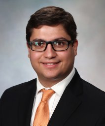 Andres Ramos-Fresnedo合作论文数Department of Neurosurgery, Mayo Clinic10
 Diogo Garcia合作论文数Department of Neurosurgery, Mayo Clinic10
 Trifiletti Daniel M合作论文数Department of Radiation Oncology, University of Virginia School of Medicine10
 Evan Marshall Thomas合作论文数Department of Radiation Oncology, University of Alabama at Birmingham;Department of Neurosurgery, University of Alabama at Birmingham;University of Alabama at Birmingham, University of Alabama at Birmingham10
 Jamie J. Van Gompel合作论文数Mayo Clinic10
 Oluwaseun Akinduro合作论文数Dept Neurosurg, Mayo Clin9
 Ayushi Jain合作论文数Mayo Clin, Dept Radiol, 4500 San Pablo Rd S, Jacksonville, FL 32224 USA9
 Philip W. Tipton合作论文数9
 Anthony Ritaccio合作论文数Department of Neurology, Albany Medical College9Paola Suarez Meade合作论文数8Neethu Gopal合作论文数Mayo Clin, Dept Radiol, Jacksonville, FL 32224 USA8
 Baran Bozkurt合作论文数School of Medicine, Acibadem Mehmet Ali Aydınlar University;School of Medicine, Acıbadem Mehmet Ali Aydinlar University;Acibadem Mehmet Ali Aydınlar University, Acıbadem Mehmet Ali Aydinlar University8




