BACKGROUND:The development of international clinical practice guidelines offers an opportunity to harmonize evidence-based care across diverse health-care systems but presents substantial logistical, methodological, and implementation challenges. These challenges are particularly pronounced for nontraumatic osteonecrosis of the femoral head, a condition characterized by heterogeneous disease biology, evolving diagnostic criteria, and limited high-certainty evidence. METHODS:This article summarizes key lessons learned during the development of the first international, evidence-based clinical practice guidelines for osteonecrosis of the femoral head led by the Association Research Circulation Osseous. RESULTS:Key lessons emerged across multiple domains, including optimization of panel structure and workflow, effective use of virtual collaboration platforms, early engagement of methodological experts, refinement of literature search and data abstraction approach, and staged formulation and approval of recommendations. Additional insights highlighted the need to anticipate global variability in resources, language, and clinical context, underscoring the value of resource-stratified recommendations, early planning for translation and cultural adaptation, incorporation of patient values and preferences, and linkage of guideline recommendations to measurable quality indicators to assess real-world uptake and impact. CONCLUSIONS:The lessons learned emphasize that international guideline development should adopt a life cycle approach extending beyond publication to promote equitable adoption, continuous refinement, and meaningful improvements in patient care worldwide.
Background/Objectives: Diagnostic evaluation and management of nontraumatic osteonecrosis of the femoral head (ONFH) vary substantially. This systematic review was conducted to inform development of the Association Research Circulation Osseous (ARCO) clinical practice guideline for diagnosis and treatment of ARCO stages I to III ONFH. Methods: We searched MEDLINE, EMBASE, Web of Science, SCOPUS, Global Index Medicus, and the Cochrane Library for studies evaluating imaging modalities and treatments for adult ONFH. We assessed risk of bias using the QUADAS-2, the ROB-2, and the ROBINS-I tools; conducted meta-analyses using random-effects regression; and evaluated certainty of evidence using GRADE methodology. Results: Among 36 included studies, 18 addressed diagnostic test accuracy and 18 addressed comparative effectiveness of treatments. Magnetic resonance imaging (MRI) demonstrated the highest pooled sensitivity (0.91; 95% confidence interval (CI), 0.87 to 0.94) and specificity (0.96; 95% CI, 0.87 to 0.99) for ONFH diagnosis. Bone marrow edema and grade 2+ joint effusion on MRI differentiated symptomatic versus asymptomatic disease. Computed tomography and MRI better detected subchondral fractures than plain radiography. Very low-grade evidence suggested lower rates of femoral head collapse with core decompression plus bone marrow concentrate compared with core decompression alone (pooled relative risk [RR], 0.55; 95% CI, 0.36 to 0.83), and with vascularized versus non-vascularized bone grafting (RR, 0.35; 95% CI, 0.14 to 0.84) over a ≤5-year follow-up. Based on three non-comparative case series, osteotomies might have a lower risk of collapse over a 10- to 20-year follow-up, but this needs to be evaluated in future comparative research. Inconsistent outcome reporting hindered treatment outcome pooling. There were no comparative studies that evaluated observation only versus intervention in asymptomatic disease or strategies for monitoring treatment response. Conclusions: Evidence supporting optimal imaging modalities and early joint-preserving interventions remains limited and predominantly observational, underscoring the need for high-quality comparative studies with consistently defined core outcomes to guide clinical decision-making.
➢ Nontraumatic osteonecrosis of the femoral head should be suspected in patients who are <50 years of age who have persistent hip pain despite normal radiographs.➢ Bilateral involvement is common; magnetic resonance imaging (MRI) of the contralateral hip is recommended.➢ MRI is the most sensitive modality for detecting early disease.➢ Femoral head sphericity is assessed on anteroposterior, lateral, and 30º view radiographs.➢ Small pre-collapse lesions have the best prognosis. Large lateral lesions and subchondral collapse predict rapid progression.➢ Systemic risk factors and multifocal disease should be actively evaluated.➢ Joint-preserving procedures are most effective before structural collapse.➢ Total hip arthroplasty remains the most reliable treatment after femoral head collapse.
BACKGROUND:Nontraumatic osteonecrosis of the femoral head (ONFH) can lead to major disability in patients of all ages. It presents at various levels of severity and can be either symptomatic or asymptomatic. There is a vast array of management strategies. Treatment is often subject to physician bias. Clinical practice guidelines that are broad-based, internationally developed, consensus-driven, and strictly evidence-based are needed. The aim of this guideline by the Association Research Circulation Osseous (ARCO) was to develop international evidence-based recommendations to assist physicians and patients in managing ONFH. METHODS:ARCO convened an international, multidisciplinary guideline panel that was balanced to minimize potential bias from conflicts of interest. The Grading of Recommendations Assessment, Development and Evaluation (GRADE) approach was utilized, including GRADE Evidence-to-Decision frameworks. The panel prioritized clinical questions, defined criteria for the systematic review of evidence, evaluated the statistical analysis, and, by consensus, approved recommendation statements, which were then subject to external review by content experts and stakeholders (a health policy-maker and a patient). RESULTS:The panel agreed on 12 recommendations for the diagnosis, evaluation, and management of ONFH. CONCLUSIONS:Key recommendations of these guidelines require accurately staging ONFH and determining when pain may be due to ONFH. They establish principles for optimal decision-making by assessing the quality of evidence backing various treatments and identifying numerous areas for additional investigation. CLINICAL RELEVANCE:This international evidence-based guideline provides standardized recommendations for the diagnosis and management of nontraumatic ONFH. It synthesizes all available evidence using GRADE methodology and offers practical, consensus-supported guidance for accurate staging, imaging selection, treatment decision-making, and the identification of patients who would benefit from joint-preserving interventions. The guideline supports clinicians in reducing practice variation, improving diagnostic accuracy, and optimizing treatment pathways for patients with ONFH.
Introduction Classical non-traumatic osteonecrosis (NTON)—most commonly affecting the femoral head—and osteonecrosis of the jaw (ONJ) represent two major forms of osteonecrosis. Ongoing debate persists regarding whether these conditions reflect manifestations of a shared underlying pathology or represent distinct clinical entities. Hypothesis We hypothesize that although NTON and ONJ share core mechanisms, their initiating events, osteoclastic responses, and reactions to anti-resorptive therapy differ sufficiently. Methods A comprehensive literature review was conducted using PubMed, Scopus, Web of Science, and the Cochrane Library. Evidence was synthesized regarding anatomical and mechanical differences, etiologic factors, pathogenic mechanisms, and therapeutic responses in NTON and ONJ. Results In NTON, compromised vascular supply is widely recognized as the primary initiating event, leading to hypoxia, increased osteoclastogenesis, and excessive bone resorption. In ONJ, impaired blood flow occurs secondarily, typically following dental trauma, infection, or anti-resorptive therapy. While both conditions involve disturbed bone homeostasis, NTON is characterized by heightened osteoclast activity, whereas ONJ features suppressed osteoclast function. This contrast results in divergent pharmacologic behavior: bisphosphonates and denosumab may be protective during the resorptive stage of NTON but significantly increase the risk of ONJ. Conclusion Although NTON and ONJ share fundamental disruptions in bone homeostasis and vascular regulation, their distinct initiating events, osteoclastic dynamics, and responses to anti-resorptive therapy suggest they are best viewed as separate clinical entities rather than variations of a single disease.
Background Nontraumatic osteonecrosis has historically been attributed to vascular insufficiency, leading to oxygen and nutrient deprivation and subsequent bone death. However, terminology used to describe this condition remains inconsistent and often fails to capture its complex pathogenesis. Terms such as “avascular necrosis” and “ischemic necrosis” may inappropriately emphasize vascular mechanisms, potentially limiting diagnostic and therapeutic approaches. Methods We performed a comprehensive review of the literature to examine the evolution of terminology for nontraumatic osteonecrosis and to evaluate the breadth of underlying pathophysiologic mechanisms. Particular attention was given to the clinical and conceptual implications of commonly used terms. Results Evidence indicates that disrupted bone homeostasis represents the final common pathway in the development of nontraumatic osteonecrosis, which may result from vascular or vascular-independent mechanisms. Continued use of terms that focus solely on vascular insufficiency may bias clinical management toward revascularization strategies, overlooking other contributory factors. Conclusions The term “osteonecrosis” more accurately reflects the multifactorial nature of this condition. Broader adoption of this terminology may foster a more comprehensive understanding of disease mechanisms and stimulate the development of innovative, multimodal treatment strategies.
Introduction Non-traumatic osteonecrosis is a frequent complication in patients with autoimmune disorders, though its prevalence varies markedly depending upon the type of disorder. Understanding the causes of this difference can help uncover the underlying pathophysiology of osteonecrosis and guide the development of effective preventive and therapeutic strategies. Areas covered In this perspective study, we reviewed available databases, including PubMed, Cochrane Library, Scopus, and Web of Science, to explore why the risk of osteonecrosis varies among different autoimmune disorders. Is this variation primarily due to the disease's pathophysiology, the use of medications such as corticosteroids, or a combination of both? If both factors are involved, what is the extent of each contribution in this context? Expert opinion Non-traumatic osteonecrosis is often induced by an interaction between disease pathophysiology and corticosteroid use. In patients with different autoimmune disorders but an identical history of corticosteroid use, the risk of osteonecrosis is influenced by how the underlying pathophysiology compromises bone health. In autoimmune disorders with multiple adverse effects on bone, such as SLE (systemic lupus erythematosus), there is a much higher risk of osteonecrosis compared to disorders with minimal impact on bone health, such as celiac disease and MS (multiple sclerosis).
BACKGROUND:Only 30%-40% of high-grade sarcomas respond to initial chemotherapy, which can have significant toxicities. Computed tomography imaging alone has limitations in evaluating treatment response. Imaging with 5-fluoro-deoxyglucose-positron emission tomography/computed tomography (FDG-PET/CT), which evaluates tumor metabolism, offers an alternative. This study examined whether early changes in the maximum standardized uptake value (SUVmax) after neoadjuvant chemotherapy can be used to predict outcomes in high-grade sarcomas. METHODS:This prospective trial assessed whether changes in the SUVmax after one or four cycles of pegylated-liposomal doxorubicin plus ifosfamide could predict progression-free survival (PFS), overall survival (OS), and histologic response. Fifty-six patients were required for 90% power. Metabolic response was defined as a reduction ≥40% in the SUVmax from baseline after either cycle 1 (delta 1) or cycle 4 (delta 2). RESULTS:Sixty-nine patients were enrolled (2006-2013) with a median follow-up of 8.1 years. The 10-year PFS rate was 74% versus 42% for delta 1 responders versus nonresponders (p = .0082), respectively; and 69% versus 33% for delta 2 responders versus nonresponder (p = .0015), respectively. The 10-year OS rate was 73% versus 58% for delta 1 responders versus nonresponder (p = .28), respectively; and 81% versus 37% for delta 2 responders versus nonresponder (p = .00026), respectively. Of 46 delta 1 nonresponders, 23 met criteria at delta 2. A positron emission tomography response was correlated with histologic necrosis. At last follow-up, 31 patients (44.9%) were alive and disease free, and 10 (14.5%) were alive with sarcoma. Five patients developed secondary malignancies. CONCLUSIONS:An SUVmax reduction verified on FDG-PET/CT imaging after neoadjuvant chemotherapy was a strong predictor of long-term outcomes. Metabolic imaging at treatment completion identified responders, supporting continued therapy in initially nonresponsive patients and guiding personalized treatment strategies.
Introduction:An increased incidence of non-traumatic osteonecrosis has been reported during the COVID-19 pandemic. Corticosteroid therapy, particularly dexamethasone, has often been implicated as a major risk factor. However, emerging evidence suggests that the pathogenesis of osteonecrosis in COVID-19 patients may extend beyond corticosteroid exposure. Hypothesis:COVID-19 infection itself may serve as an independent etiologic factor in osteonecrosis, with virus-induced pathogenic mechanisms synergizing with corticosteroid exposure to heighten risk, even at lower doses and shorter treatment durations. Methods:This review synthesizes available literature on COVID-19, corticosteroid therapy, and osteonecrosis pathogenesis. Evidence from clinical observations, mechanistic studies, and prior models of corticosteroid-induced osteonecrosis were examined to identify overlapping and distinct pathways contributing to disease development. Results:Findings indicate that COVID-19 and corticosteroids converge on common pathogenic pathways-lipid dysregulation, impaired bone homeostasis, endothelial dysfunction, and coagulopathy. COVID-19 additionally promotes osteonecrosis through cytokine storm-driven inflammation. The combined effects of viral infection and corticosteroid therapy amplify disease risk, explaining reported cases of osteonecrosis even under reduced corticosteroid exposure. Conclusion:COVID-19 may represent an independent etiologic factor for osteonecrosis, with intrinsic viral effects potentiating the impact of corticosteroids. Recognition of this dual risk underscores the need for preventive and therapeutic strategies tailored to COVID-19-associated osteonecrosis.
BACKGROUND:Historically, osteonecrosis of the femoral head (ONFH) was a major problem following kidney transplantation, occurring in up to 5%-11% of recipients. The development of new immunosuppressive agents permitted steroid minimization protocols, and consequently, the incidence of ONFH has decreased. We studied ONFH trends over four decades and evaluated risk factors in two immunosuppressive eras, pre- and post-2001. PATIENTS AND METHODS:Steroid minimization protocols at our center started in 2000. We reviewed records of 1st kidney transplants from January 1985 to May 2024 and compared ONFH incidence and risk factors between the two eras: Era 1 (January 1985-December 31, 2000) and Era 2 (January 2001-May 2024). Cox regression was used to assess for independent factors associated with a higher or lower incidence of ONFH. RESULTS:ONFH incidence in Era 1 was 7.2%; Era 2, 1.1% (p < 0.001). In Era 1, increased risk was associated with heavier weight (HR: 1.017, 95% CI: 1.010-1.023, p < 0.001) and mTOR inhibitors (HR: 4.258, 95% CI: 1.726-10.506, p = 0.002); and decreased risk with diabetes (HR: 0.362, 95% CI: 0.270-0.486, p < 0.001), and statins (HR: 0.452, 95% CI: 0.327-0.625, p < 0.001). In Era 2, increased risk was associated with steroid use (HR: 2.096, 95% CI: 1.071-4.100, p = 0.031) and decreased risk with mycophenolate (HR: 0.471, 95% CI: 0.237-0.935, p = 0.032). CONCLUSION:The incidence of ONFH in KTRs has dramatically decreased in the modern immunosuppressive era. Diabetes, statin use, and immunosuppressive medications, specifically mTOR inhibitors and mycophenolate, appear to have varying impacts depending on the immunosuppressive era.
BACKGROUND:Identifying the factors associated with the risk of osteonecrosis of the femoral head (ONFH) is crucial for implementing effective preventive measures. However, there is currently no consensus on whether a correlation exists between the incidence of ONFH and diabetes, statins, or immunosuppressive medications. We studied the associations between these factors and ONFH risk in a large cohort of kidney transplant recipients (KTRs). METHODS:Medical records of KTRs who underwent transplantation between 1985 and 2024 were gathered from a prospectively collected database. Collected data included demographic variables, immunosuppressive regimens, diabetes statuses, transplantation details, smoking histories, and statin usages. In total, 262 recipients who had ONFH were recorded in 6,926 KTRs (3.8%). Variables with a P-value < 0.1 in the univariate analyses or those considered clinically significant were included in a multivariate Cox regression model. RESULTS:In the multivariate analyses, the following factors were significantly associated with an increased risk of ONFH: older age (hazard ratio [HR]: 1.01, 95% confidence interval [CI]: 1.00 to 1.02, P = 0.018), men (HR: 1.40, 95% CI: 1.08 to 1.82, P = 0.010), mammalian target of rapamycin inhibitors (HR: 4.42, 95% CI: 2.10 to 9.32, P < 0.001), corticosteroids for maintenance immunosuppression (HR: 3.54, 95% CI: 2.02 to 6.23, P < 0.001), and cyclosporine (HR = 2.53, 95% CI: 1.12 to 5.71, P = 0.025). The following factors were significantly associated with a reduced risk of ONFH: diabetes (HR: 0.49, 95% CI: 0.38 to 0.63, P < 0.001) and statin use (HR: 0.59, 95% CI: 0.45 to 0.78). CONCLUSIONS:In addition to recipient characteristics, diabetes or statin use reduces the risk of ONFH, whereas certain immunosuppressive medications increase the risk of ONFH. These findings underscore the importance of thorough patient evaluation and the implementation of immunosuppressive strategies to reduce the risk of post-transplant ONFH. LEVEL OF EVIDENCE:III.
In keeping with our tradition of recognizing the best articles each year, JBJS Essential Surgical Techniques (EST) is pleased to announce the 2022 award winners: Key Procedures Series Award: Drs. Carlos Gamba and Albert Ginés-Cespedosa for “Proximal Medial Gastrocnemius Release: Surgical Technique”1 Subspecialty Series Award: Drs. Suhas Dasari and Nikhil Verma for “Subacromial Balloon Spacer, Indications, Rationale, and Technique”2 For our Key Procedures article series, Gamba and Ginés-Cespedosa aptly demonstrate a release of the medial gastrocnemius to reduce tension in the Achilles-calcaneus-plantar system associated with conditions such as recalcitrant plantar fasciitis. Although some may consider this to be a minor procedure from a technical standpoint, the authors emphasize that a strong understanding of the local anatomy allows surgeons to perform the procedure with minimal dissection while avoiding complications. They provide high-quality diagrams and videos and list several important tips for surgeons to consider before, during, and after surgery in order to achieve favorable outcomes. For our Subspecialty Procedures article series, Dasari and Verma describe the appropriate use of a novel, saline-solution-filled balloon implant for the treatment of one of the most challenging rotator cuff pathologies: the massive, irreparable tear. Outcomes of the procedure have been reported in randomized clinical trials3 performed in both the U.K. by the START:REACTS team and the U.S. by the SPACE group. The authors provide video demonstration of the proper insertion and positioning of the balloon. Simultaneous split-screen video of both the surgeon’s view and the arthroscope view help viewers to understand the nuances of the procedure. We congratulate the winners of the 2022 Editor’s Choice Award for Key Procedures and Subspecialty Procedures, and look forward to another year of exemplary contributions to the orthopaedic literature in 2023. JBJS EST is dedicated to optimizing the educational experience and improving patient care worldwide by providing accessible, concise video articles backed by evidence-based outcomes and JBJS peer review.
OBJECTIVE Sacroiliac joint (SIJ) fusion utilizing intraoperative navigation requires a standard reference frame, which is often placed using a percutaneous pin. Proper placement ensures the correct positioning of SIJ fusion implants. There is currently no grading scheme for evaluation of pin placement into the pelvis. The purpose of this study was to evaluate the occurrence of ideal percutaneous pin placement into the posterior ilium during navigated SIJ fusion. METHODS After IRB approval was obtained, electronic medical records and intraoperative computed tomography images of patients who underwent navigated SIJ fusion by the senior author between October 2013 and January 2020 were reviewed. A pin placement grading scheme and the definition of "ideal" placement were developed by the authors and deemed acceptable by fellow attending surgeons. Six attending surgeons completed two rounds of pin placement grading, and statistical analysis was conducted. RESULTS Of 90 eligible patients, 73.3% had ideal pin placement, 17.8% medial/lateral breach, and 8.9% complete miss. Male patients were 3.7 times more likely to have ideal placement than females (p < 0.05). There was no relationship between BMI, SIJ fusion laterality, or pin placement laterality and ideal placement. Interobserver reliability was 0.72 and 0.70 in the first and second rounds, respectively, and defined as "substantial agreement." Intraobserver reliability ranged from 0.74 (substantial agreement) to 0.92 (almost perfect agreement). CONCLUSIONS Nonideal pin placement occurred in 26.7% of cases, but a true "miss" into the sacrum was rare. Ideal pin placement was more likely in males and was not associated with BMI, SIJ fusion laterality, or pin placement laterality. The grading scheme developed has high intraobserver and interobserver reliability, indicating that it is reproducible and can be used for future studies. When placing percutaneous pins, surgeons must be aware of factors that can decrease placement accuracy, regardless of location.
Several studies have reported an association between levels of circulating blood cells, in particular the neutrophil to lymphocyte ratio (absolute neutrophil count (ANC)/absolute lymphocyte count (ALC)) and outcomes in patients with cancer. In the current study, the association between lymphocyte, neutrophil, monocyte, and platelet counts and survival was examined in a prospective trial of preoperative pegylated-liposomal doxorubicin and ifosfamide for high-grade soft-tissue sarcomas. A statistically significant association between overall survival, but not progression free-survival, was observed with the ANC/ALC ratio at a cutoff value of ≥2 and a statistically significant trend using a cutoff of ≥5. Our results suggest that a balance between the lymphocyte count and the number of circulating myeloid cells that can suppress lymphocyte function may be predictive of survival in patients with soft-tissue sarcomas. Future research should therefore examine the role of lymphocyte-myeloid cell balance in sarcoma biology.
For stable intertrochanteric hip fractures, treatment commonly involves the use of a sliding hip screw. Intertrochanteric hip fractures are increasingly common as the population ages and lives longer. More than 250,000 hip fractures occur per year in the United States1. The mortality rate within the first year following operative treatment ranges from 14% to 27.3%2,3. Early surgical repair within 48 hours of injury is associated with a lower risk of mortality2,4,5. The goals of surgical treatment are restoration of coronal plane alignment without varus angulation and early patient mobilization. Description The sliding hip screw procedure can be divided into (1) preoperative planning; (2) patient positioning; (3) C-arm setup; (4) closed reduction of fracture; (5) sterile preparation and draping; (6) lateral hip approach; (7) guide pin insertion; (8) triple-reaming the proximal aspect of the femur; (9) sliding hip screw insertion into the femoral neck and head; (10) side plate insertion, engaging the sliding hip screw, and fixation to the femur; (11) lag compression screw insertion (if appropriate); and (12) final fluoroscopic images and wound closure. Alternatives Intertrochanteric hip fractures must be surgically treated to avoid morbidity and increased risk of mortality. Nonoperative treatment is occasionally indicated in nonambulatory patients or those with high perioperative risk. If treated surgically, a common alternative implant option includes the intramedullary nail. Finally, for severely comminuted fractures or failed internal fixation, total hip arthroplasty may be necessary. Rationale Sliding hip screws are as effective as intramedullary nails and often less costly6. In general, the quality of fracture reduction is more critical than the choice of implant7. A prospective study found no significant difference in walking ability with either sliding hip screws or intramedullary nails for stable intertrochanteric fractures8. Expected Outcomes By 6 months, the majority of fractures will have healed; according to a prospective randomized study, 91% of stable fractures and 85% of unstable fractures had achieved radiographic union by that time9. Another study showed radiographically healed fractures in all 106 patients treated with sliding hip screws at median follow-up of 13.6 months8. Important Tips Watch out for comminution of the greater or lesser trochanter, which may require supplemental fixation.Prior to completely reflecting the vastus lateralis muscle, control the bleeding from any perforators with use of 2-0 silk ties. This prevents recurrent bleeding, which often occurs if only cautery is utilized to coagulate these vessels.Utilize a 4.5-mm drill hole in the lateral cortex of the femur in order to allow for minor adjustments of the anterior femoral neck guide pin; otherwise, the pin will be held tightly and continue to be bound in the same direction by the lateral cortex on repeated attempts.If the guide pin is inadvertently withdrawn along with the reamer after reaming, a lag screw may be placed backward in the newly reamed hole and the guide pin passed back through the lag screw to reposition it.Extracapsular hip fractures should be carefully scrutinized for signs of instability, such as lateral wall comminution or reverse obliquity. The fracture may displace posteriorly when the patient is supine on the fracture table.While placing the guidewire, multiple entry attempts can weaken the lateral cortex and propagate the fracture into the subtrochanteric region.Superior placement of the lag screw results in poor tip-apex distance and a higher chance of screw cut-out.Be careful to prevent guidewire penetration into the hip joint.Loss of reduction or femoral head malrotation may occur during lag screw insertion. Acronyms & Abbreviations AP = anteroposteriorfx's = fracturesIMN = intramedullary nailIV = intravenousPDS = polydioxanone sutureSHS = sliding hip screwTFL = tensor fascia lata.
The capability of the orthopaedic community to learn, teach, and demonstrate remotely has evolved over many decades, yet the recent COVID-19 pandemic has highlighted the importance of these skills. JBJS EST has kept pace, as demonstrated by the continued refinements in video presentation, quality, and efficient communication of key concepts evident across all of the articles published in 2021. Our constant attention to maintaining a high standard of quality through peer review has been integral to accomplishing these goals and staying on the leading edge of surgeon education. This quality standard was best exemplified by each of the 2021 JBJS EST Editor’s Choice Award winners. In the Key Procedures articles series, which focuses on basic techniques that most general orthopaedic surgeons perform, Lo et al.,1 paid homage to the giants of shoulder surgery, aptly summarizing and demonstrating the modified Latarjet technique for glenohumeral dislocations and instability. In the Subspecialty Procedures article series, which focuses on advanced procedures performed most often by subspecialists, Debbi et al.,2 tackled the difficult problem of chronic knee arthrofibrosis following total knee arthroplasty, exhibiting how a rotating hinge arthroplasty can address knee stiffness. Although this aggressive solution is hopefully seldom employed, it is effective when performed properly, and their presentation enables surgeons to accomplish this goal. We congratulate both teams of authors on their scholarly work and contribution to our profession. Their efforts will have an impact by improving patient care worldwide. Furthermore, we thank the larger family of surgeons in our profession who provide the foundation whereby these achievements in our field can be accomplished and encourage those surgeons on the sidelines observing to get engaged and join our endeavor.
It’s a privilege to introduce this special supplement containing research papers presented at the most recent biennial meeting of the Association Research Circulation Osseous (ARCO). This meeting was held in Dalian, China in 2019, at the Affiliated Zhongshan Hospital of Dalian University, hosted by Professor Dewei Zhao, who has led a departmental faculty with a long track record of research contributions in osteonecrosis . Physicians and scientists gathered to present and discuss the most recent findings related to this challenging disease. ARCO was founded in 1988 with the mission of studying bone circulation and associated disorders and has become the only professional organization focusing expressly on osteonecrosis. It was the hope of the founders, led by Jacques Arlet and Paul Ficat, that it would advance the science and understanding of this debilitating disease. Osteonecrosis is a bone disease that is often results in end stage arthritic joints. Unfortunately, given its relationship to steroid exposure and ethanol usage, it frequently affects adolescents or young adults. As such, it is especially tragic as joint arthroplasty is often the only means to eliminate pain and restore function, yet patients undergoing joint arthroplasties in this age range are destined to be plagued by numerous subsequent revision surgeries and are at a lifelong risk for other implant related complications such as prosthetic joint infections, particulate wear debris osteolysis, adverse local tissue reactions, and periprosthetic fractures. Advances in the understanding and treatment of this disease have unfortunately come slowly despite the cadre of experts trying to make progress. Some of these recent advances are reported in this special supplement. Organized into four major groups (basic research, clinical research, outcomes of joint preserving procedures, and outcomes of joint replacing procedures) these papers extend our knowledge and present new ideas for the scientific community to debate. Time will tell whether these new concepts will become more broadly accepted or stimulate new areas of investigation. In addition, the clinical outcomes data will provide useful information that surgeons can provide to patients, thereby enhancing their ability to make the most informed decisions possible. ARCO thanks The Journal, along with the editors and reviewers, for providing a conduit to disseminate important new information. The membership hopes that this collection of papers will expand our foundation of knowledge, be useful for the broader orthopaedic surgical community, and lead to future improvements in the care of our patients with osteonecrosis. Only then will the long-term goals and mission that the founders originally envisioned be fully realized.
 Roby C. Thompson合作论文数15
 Quanjun Cui合作论文数Department of Orthopaedic Surgery, University of Virginia Health System13
 Stuart Goodman合作论文数Department of Orthopaedic Surgery, Stanford University;Department of Bioengineering, Stanford University12
 Keith Skubitz合作论文数Division of Hematology, Oncology and Transplantation, Department of Medicine, Medical School, University of Minnesota11
 Lynne C. Jones合作论文数Department of Orthopaedic Surgery, School of Medicine, Johns Hopkins University;Center for Osteonecrosis Research and Education, Johns Hopkins University;Center for Metabolism & Obesity Research, Institute for Basic Biomedical Sciences, Johns Hopkins University10JC MANIVEL合作论文数8
 Michael Mont合作论文数Northwell Orthopedics;Lenox Hill Hospital8Philippe Hernigou合作论文数8Yong-Chan Ha合作论文数7Nobuhiko Sugano合作论文数Osaka University Graduate School of Medicine7
 Wolf Drescher合作论文数7
 Kyung-Hoi Koo合作论文数Department of Orthopaedic Surgery, Seoul National University Bundang Hospital;Department of Orthopaedic Surgery, College of Medicine, Seoul National University7Takuaki Yamamoto (山本卓明)合作论文数School of Medicine, Fukuoka University7Clohisy Denis R合作论文数6
 Yoon Byung-Ho合作论文数6
 Dewei Zhao (赵德伟)合作论文数Affiliated Zhongshan Hospital of Dalian University6Shin-Yoon Kim合作论文数6
 Takashi Sakai合作论文数6
 Mel S. Lee合作论文数Taipei Medical University;Department of Orthopaedic Surgery, Paochien Hospital;Department of Orthopaedic Surgery, Kaohsiung Chang Gung Memorial Hospital6
 Youngkyun Lee合作论文数5
 Amy Skubitz合作论文数Department of Laboratory Medicine and Pathology, University of Minnesota Medical School5Lee Yun Jong合作论文数5
 Christian M Ogilvie合作论文数5
 Tae-Young Kim合作论文数4Jo Woo-Lam合作论文数4Mark C. Gebhardt合作论文数4Denis R. Clohisy合作论文数4Laurence K Cutkomp合作论文数4
 Kathryn Dusenbery合作论文数Department of Radiation Oncology, University of Minnesota4
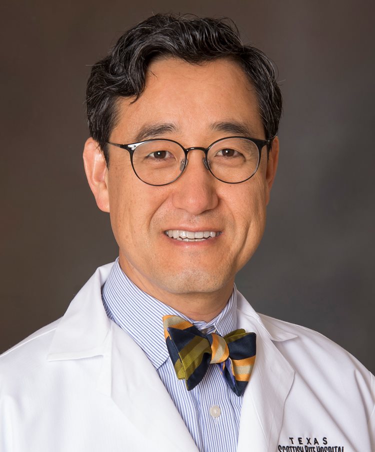 Harry Kim合作论文数Center for Excellence in Hip, Scottish Rite for Children;Department of Orthopaedic Surgery, UT Southwestern Medical Center4
 Richard Iorio合作论文数3
 Masaki Takao合作论文数3megan e anderson合作论文数3
 Robert Turcotte合作论文数Division of Orthopaedic Surgery, Department of Surgery, Faculty of Medicine and Health Sciences, McGill University3Damron Timothy A合作论文数3Remzi a Ozerdemoglu合作论文数3
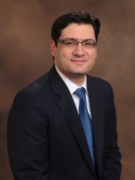 Entezari Vahid合作论文数Department of Orthopedic Surgery.3Maheshwari Aditya合作论文数Department of Orthopaedic Surgery, DownState Medical Institute3
 Vikas Khanduja合作论文数University of Cambridge3
 Sameer Naranje合作论文数3
 Ensor E Transfeldt合作论文数3
 Albert J Aboulafia合作论文数3Valérie Gangji合作论文数3
 Ara Nazarian合作论文数Nazarian Laboratory, Beth Israel Deaconess Medical Center;Harvard Medical School;Boston University;Verispellis Skis + Snowboards;Ortholevo, Inc.3Bruce R. Lindgren合作论文数3Richard M. Terek合作论文数Department of Orthopedic Surgery, Brown University3
 Marc Swiontkowski合作论文数Department of Orthopedic Surgery, Medical School, University of Minnesota3
 Arthur Matas合作论文数Division of Transplantation, Department of Surgery, Medical School, University of Minnesota3
 Brian D. Snyder合作论文数Beth Israel Deaconess Medical Center, Harvard Medical School3




