Background Glenoid bone loss during revision to reverse total shoulder arthroplasty (rTSA) presents a technical challenge, particularly following removal of central post, peg, or keel-based components. While structural allografts are commonly used to improve fixation and structural support of revision components, they often require significant intraoperative preparation. Technique This technical report describes the use of preformed cylindrical cancellous allograft bone dowels—originally designed for anterior cruciate ligament revision surgery—as a structural grafting option during revision to rTSA. Two types of contained defects are addressed: (1) cylindrical voids following removal of center posts or pegs, and (2) longitudinal defects after removal of keeled glenoid components. In each case, the dowel is prepared and impacted into the defect, followed by standard baseplate placement. Preoperative CT planning and intraoperative sizing are emphasized. Discussion Compared to traditional femoral head allografts, cancellous dowels offer a streamlined, reproducible alternative with minimal intraoperative preparation, reliable press-fit fixation, and strong graft-host contact. This approach facilitates baseplate stability and may promote early graft incorporation. Conclusion The dowel technique provides a practical and efficient solution for structural glenoid reconstruction in revision rTSA, expanding the options available to address contained glenoid defects.
Background Subscapularis healing is a critical determinant of successful anatomic total shoulder arthroplasty (aTSA) outcomes. Common subscapularis management techniques include tenotomy, peel, and lesser tuberosity osteotomy (LTO). However, data on subscapularis management in stemless aTSA remain limited. The purpose of this study was to evaluate clinical and functional outcomes at two-year follow-up in patients undergoing stemless aTSA with either subscapularis peel or LTO. Methods A retrospective review was performed of a prospectively maintained, multi-center database of primary RSAs performed by multiple surgeons between 2019 and 2023 with minimum two-year follow-up. A total of 122 patients met the study criteria, 88 in the LTO approach group versus 34 in the peel group. Patient-reported outcome measures (PROMs), range of motion (ROM), and strength were evaluated at baseline and at two-year follow-up, with comparisons made between groups. Results There were no significant differences between BMI, history of tobacco use, and diabetes between LTO and peel groups. There were no significant differences in PROMs, ROM, and strength testing at preoperative evaluation. Significant differences were observed in age (66.8 years old in LTO versus 62.7 in peel, p = 0.021), sex (52% of the LTO group was female versus 6% in peel, p = <0.0001), and Walch classification (p = 0.0004). At two-year follow-up, outcomes were similar between groups, with the exception of greater active external rotation at 90 degrees in the peel group, (84° vs 71°, p = <0.0001). No significant differences were observed in the magnitude of change from preoperative to two-year postoperative outcomes between the LTO and peel groups. Conclusions Both subscapularis peel and lesser tuberosity osteotomy (LTO) techniques in stemless anatomic TSA resulted in favorable outcomes at two-year follow-up. Postoperative outcomes were largely similar between groups, although a difference was observed in active external rotation at 90°. Despite baseline demographic differences, both techniques demonstrated favorable clinical and functional results. Level of Evidence Level III; Retrospective Cohort Comparison Study
Background Subscapularis management with lesser tuberosity osteotomy repair in stemless total shoulder arthroplasty is particularly different in terms of suture fixation as opposed to stemmed humeral implants. Surgical techniques to achieve optimal soft tissue balance in lesser tuberosity osteotomy repairs differ based on the surgeon. The purpose of this study is to determine whether there is a difference in 2 years functional or clinical outcomes in stemless total shoulder arthroplasty patients treated with lesser tuberosity osteotomy repairs conducted with bone tunnels versus suture anchors. Methods A retrospective review was performed of a prospectively maintained, multi-center database of primary TSAs performed by multiple surgeons between 2019 and 2023 with minimum 2-year follow-up. A total of 81 patients met the study criteria, including 33 in the suture anchor cohort versus 48 in the bone tunnel cohort. Patient-reported outcomes (PRO) as well as range of motion (ROM) measurements were compared between the two groups for baseline results as well as at two year follow up. Results There was no statistically significant difference in age, sex, BMI, tobacco usage, or diabetes prevalence in either cohort (p=0.610, p=0.687, p=0.980), p=0.153, p=0.355, respectively). There was no statistically significant difference in PRO, ROM, or belly press test at baseline between patients treated with suture anchors and those treated with bone tunnels. At two years, there were no statistical differences in outcomes (PROs, ROM, or strength), other than worse FF in the suture anchor group (145 degrees vs 156 degrees, p=0.026). Conclusions The current study suggests equivalent two year functional and clinical outcomes for patients treated with suture anchors versus the conventional bone tunnels in LTO repair for stemless TSA patients. The two groups had similar baseline demographics, ROM, and PRO scores and performed similarly at two years follow up. Level of Evidence Level III; Retrospective Cohort Comparison Study
BACKGROUND:Osteoporosis increases fracture risk and adversely affects outcomes after shoulder arthroplasty. Preoperative bisphosphonate use has been linked to higher rates of periprosthetic fracture after lower extremity total joint arthroplasty, but data in shoulder arthroplasty are limited. The purpose of this study was to evaluate whether anti-remodeling agents such as bisphosphonates or anabolic osteoporotic therapy influenced 1-year outcomes, including periprosthetic humeral fracture, acromial stress fracture, and all-cause revision, following anatomic total shoulder arthroplasty (aTSA) and reverse total shoulder arthroplasty (rTSA). METHODS:A retrospective cohort study was performed using the PearlDiver Mariner Patient Claims Database (2010-2025). Adult patients with a documented diagnosis of osteoporosis undergoing first-time aTSA or rTSA with at least 12 months of preoperative and postoperative continuous enrollment were included. Patients were stratified into anti-remodeling therapy, anabolic therapy, or no osteoporosis medication groups. Demographics, Charlson Comorbidity Index, and 1-year postoperative outcomes were analyzed. Univariate and multivariate logistic regressions were used to adjust for age, sex, and comorbidities. RESULTS:A total of 18,922 patients were included (13,123 rTSA; 5,817 aTSA). In the combined aTSA/rTSA cohort, anti-remodeling therapy was independently associated with increased odds of 1-year periprosthetic humeral fracture (odds ratio [OR] 1.25, 95% confidence interval [CI] 1.06-1.46, P = .006), whereas anabolic therapy was not. In the rTSA cohort, no independent association was observed between anti-remodeling therapy and 1-year periprosthetic humeral fracture after multivariate adjustment (OR 1.11, 95% CI 0.94-1.32, P = .220), and anabolic therapy was not independently associated with fracture. In the aTSA cohort, anti-remodeling therapy was independently associated with increased odds of 1-year periprosthetic humeral fracture (OR 1.85, 95% CI 1.20-2.83, P = .005), while anabolic therapy was not independently associated with fracture after adjustment. DISCUSSION:Preoperative anti-remodeling osteoporotic therapy is independently associated with increased odds of 1-year periprosthetic humeral fracture following shoulder arthroplasty. These findings may inform preoperative counseling and postoperative surveillance in osteoporotic patients undergoing shoulder arthroplasty.
Background: Ulnar collateral ligament (UCL) reconstruction (UCLR) of the elbow is common in throwing athletes and reliably allows return to play. The literature supports stress imaging of the elbow to evaluate the integrity of the UCL. The FEVER view (flexion, external rotation, and valgus stress) was developed to improve the diagnostic accuracy of UCL pathology in throwing athletes with stress magnetic resonance imaging (MRI). Hypothesis/Purpose: The present study sought to evaluate FEVER MRI imaging findings, primarily ulnohumeral joint space widening, in patients with and without a history of UCLR. The authors hypothesize that on the FEVER view there would be no difference in joint space widening between pitchers with a history of UCLR and those without. Study Design: Cross-sectional study; Level of evidence, 3. Methods: Pitchers in 2 Major League Baseball franchises who underwent preseason screening MRIs with standard and FEVER views were included. All images were read by 2 fellowship-trained musculoskeletal radiologists. Findings included gross UCL appearance, UCL edema, location of edema, type of signal, grade of injury, ligament retraction, and amount of ulnar-sided joint space opening (absolute opening, the joint space widening in the FEVER stress view; relative opening, the difference between joint space widening in the FEVER view and standard view joint space widening). Time from surgery was evaluated for impact on imaging findings. Results: There was a statistically significant increase in absolute medial-sided joint space opening in players with a history of UCLR (n = 18; mean, 4.5 ± 3.4 years between UCLR and MRI) versus no UCLR (n = 76) (4.3 vs 3.8 mm; P = .04) and increased grade of UCL signal ( P = .034) in patients with a history of UCLR. There was no difference in the other imaging parameters, including relative joint space opening (2.2 vs 1.9 mm; P = 0.11). Time from surgery was not associated with any outcome variables. Conclusion: Valgus stress MRI (the FEVER view) showed a slight, but significant increase in absolute joint space opening after UCLR and an increased grade of intrasubstance UCL signal. However, there was no difference in relative joint space opening, and the clinical significance of these findings is unclear and warrants further study.
Background:Conference narratives and publications frequently portray widespread utilization of advanced imaging, 3D-based planning, and enabling technologies in shoulder arthroplasty, but surgeon adoption in day-to-day practice is not well characterized. This study sought to characterize surgeon variation in the use of these tools, delineate perceived benefits and barriers to 3D planning, and identify surgeon characteristics associated with lower utilization. Methods:A 16-question survey was created using Google Forms and distributed to 161 shoulder surgeons members of the PacWest Shoulder and Elbow Society. The survey asked questions regarding fellowship training, years in practice, practice type, surgical volume, and patterns of pre-operative advanced imaging (computed tomography [CT], magnetic resonance imaging), 3D CT-based planning for primary and revision arthroplasty, and intraoperative enabling technologies (patient-specific instrumentation, navigation, mixed/augmented reality, robotics). Routine use was defined as ≥75% of cases. Associations between surgeon characteristics and low use of 3D planning (<25% of cases) were evaluated. Results:In total, 53% of surgeons completed the survey (85 of 161). Pre-operative CT was routinely obtained by 71% of surgeons. Routine acquisition of both CT and magnetic resonance imaging was uncommon (9.5%). Routine 3D planning was reported by 60% for primary arthroplasty, with 25% never or rarely using it. Routine 3D planning for revision cases was reported by 47%. Routine use of patient-specific instrumentation guides was infrequent (15%), and there was low adoption of navigation (13%), mixed reality (7%), and robotics (6%). Interest in robotics was high (62%), most often motivated by perceived gains in implant accuracy. There was no consensus on whether 3D planning is standard of practice (55% yes, 45% no). Reported benefits of 3D planning included implant accuracy (91%), fewer outliers (67%), education (33%), and operative time savings (32%). Barriers included imaging acquisition/protocol issues (68%) and time required to plan (47%). Surgeons in practice >10 years were more likely to report low utilization of 3D planning (P = .034). Conclusion:Pre-operative CT and 3D-based planning are common but not universal, with about 30% and 40% of surgeons not routinely using them, respectively, highlighting practice heterogeneity and a disconnect between scholarly discourse and day-to-day practice. Use of CT-based enabling technology is low, but there is interest in the nascent field of robotics. Practice variation appears shaped by workflow constraints, evidentiary uncertainty, and surgeon-specific beliefs and experience. Research that connects planning and its execution to patient outcomes is warranted, as it may change the value proposition.
Background Total shoulder arthroplasty (TSA) is increasingly performed for glenohumeral osteoarthritis (GHOA), yet the impact of nonoperative treatments on delaying surgery remains unclear. The purpose of this study was to assess the association between initial conservative treatments and time to TSA. Methods This retrospective cohort study included adults with primary GHOA who underwent either reverse or anatomic TSA between 2020 and 2023. Patients were stratified by initial nonoperative treatment: hyaluronic acid (HA), one corticosteroid injection (CS1), two corticosteroid injections (CS2), six weeks (PT6) or eight weeks (PT8) of physical therapy, platelet-rich plasma (PRP), or no treatment. Time to TSA was analyzed using Cox proportional hazards models adjusting for age, sex, BMI, and Kellgren–Lawrence grade. Secondary outcomes included passive ROM and PROMs. Results Eighty-three patients were included. Time to TSA differed significantly across groups (P = 0.016), ranging from a median of 3.00 (IQR, 2.00–6.00) months in the no-treatment cohort to 13.00 (IQR, 7.00–20.50) months in the HA cohort. On multivariable analysis, CS2 (HR 0.32; 95% CI, 0.11–0.97), HA (HR 0.31; 95% CI, 0.12–0.76), PT8 (HR 0.27; 95% CI, 0.11–0.64), and PRP (HR 0.11; 95% CI, 0.03–0.51) were associated with delayed TSA compared to no treatment. PROMs and ROM were similar across groups at initial presentation and decision for surgery, except for initial total elevation (P = 0.008). Conclusion Repeat corticosteroid injections, HA, extended PT, and PRP were each associated with delayed time to TSA in patients presenting with primary GHOA, without observed compromise in function or ROM at the time of surgical decision-making. These findings are exploratory in nature and underscore the need for larger, prospective studies to confirm whether specific nonoperative modalities meaningfully delay the decision for TSA.
Glenoid bone loss remains a significant challenge in both primary and revision reverse shoulder arthroplasty (RSA), particularly following infection, erosion, or component explantation. Adequate reconstruction is critical to restore the native joint line, inclination, and version while ensuring baseplate fixation and long-term stability. Although smaller defects can be addressed with augmented baseplates or humeral head autograft, massive bone loss often requires structural allografting. Femoral head allograft offers a viable and reproducible solution in complex revision cases where autograft is unavailable or unsuitable. This technical note details a 2-stage revision RSA approach utilizing femoral head allograft to address severe glenoid bone loss. In the first stage, the patient is positioned in the beach chair position, and previous components and cement spacers are meticulously removed. The glenoid is debrided and contoured to accept a size-matched femoral head allograft. The graft is prepared using a coring reamer and oscillating saw, trialed for optimal fit, and fixed with cannulated screws. Demineralized bone matrix is applied around the graft to promote incorporation. Importantly, the humeral component is deferred to minimize shear stress across the graft-host interface and enhance graft healing. Once incorporation is confirmed radiographically and cultures return negative, definitive RSA with a lateralized glenosphere and 135° humeral component is performed. This technique reliably restores the joint line and allows for stable baseplate fixation, even in cases with significant medial wear. The femoral head allograft provides robust structural support, facilitates biologic incorporation, and avoids donor site morbidity associated with iliac crest harvest. Our approach shows that femoral head allograft, combined with a lateralized prosthesis, can yield stable, functional outcomes in revision RSA for massive glenoid deficiency.
Background:Reverse total shoulder arthroplasty (RTSA) is an effective treatment option for multiple shoulder conditions. It is unclear if seasonal timing of surgery affects outcomes. Methods:Patients who underwent RTSA between 2015-2021 and were enrolled in a multicenter registry were eligible for inclusion. Date of surgery was divided into winter, spring, summer and fall. Patient reported outcomes, range of motion and complications at 2 years' follow up were assessed and compared between RTSA groups for each season. Results:863 patients were included. Breakdown by season was: winter (N = 214); spring (N = 183); summer (N = 178); fall (N = 288). There were no differences in 2-year clinical outcomes, range of motion, complications or strength between groups with the exception that patients who underwent RTSA in the fall had a higher Constant-Murley score and better active internal rotation at 90 degrees than in other seasons (p = 0.036 and p = 0.013, respectively). Conclusion:There were very few differences in clinical outcomes based on seasonal timing for patients who undergo RTSA. Patients should feel confident that their outcomes will not vary based on the season in which they undergo RTSA.
Background: Optimal placement of the glenosphere in reverse shoulder arthroplasty (rTSA) is a key component affecting postoperative range of motion (ROM) but remains a subject of ongoing research. The purpose of this study was to evaluate the relationship between three-dimensional (3D) glenosphere position and orientation relative to anatomic scapular landmarks and postoperative patient-reported outcomes and ROM following rTSA. Methods: A retrospective multicenter cohort study was conducted on primary rTSAs performed with a 135° humeral inlay component and a lateralized glenoid component between November 2016 and March 2022. Surgeries performed with a 3D plan and patient-specific transfer instrumentation with minimum 2-year clinical follow-up were included. Implant position was extracted from preoperative planning software, focusing on pin position (center of the glenosphere) and glenosphere diameter, version, and overhang relative to scapular anatomic landmarks. ROM and American Shoulder and Elbow Surgeons (ASES) scores were assessed at 2-year follow-up, with linear regression models utilized to analyze the relationships between preoperative and intraoperative variables and postoperative outcomes while adjusting for confounding variables. Results: A total of 75 rTSAs met the study criteria. For every 1 millimeter increase in glenosphere diameter, there was a 0.5 spinal level decrease in internal rotation (IR) spine (P ≤ .005) and a 2.5° decrease in forward flexion (P ≤ .005). For every 4° increase in baseplate retroversion, there was a 1 spinal level improvement in IR spine (P = .009). Superior tilt of the baseplate was associated with a decrease in internal rotation at 90° of abduction (3° decrease per 1° of increased superior tilt, P ≤ .001). ASES scores were also significantly affected, with a 3.5 point decrease per millimeter increase in glenosphere diameter (P ≤ .001), but improved by a 1 point per millimeter increase in pin-to-coracoid distance (P = .015). Conclusion: In patients with 3D planning and patient-specific instrumentation, smaller glenosphere diameter, increased baseplate retroversion, and avoidance of superior tilt improve IR after rTSA performed with a 135° humeral component and lateralized glenoid. A smaller glenosphere diameter and increased distance from the coracoid also improved ASES scores. This data suggests that with the use of a lateralized glenoid in rTSA, efforts should be made to increase the glenosphere distance from the coracoid, avoid a superior tilted positioning of the baseplate, and consider a smaller glenosphere when in between sizes.
BACKGROUND:Despite advances in surgical technique and implant design, internal rotation (IR) after reverse total shoulder arthroplasty (rTSA) continues to be relatively unpredictable. The purpose of this study was to compare patient characteristics, use of 3-dimensional computed tomography (3D CT)-based preoperative planning, and postoperative implant position between patients with high or low IR after rTSA. METHODS:A retrospective review was performed of a multicenter prospectively collected database on patients who underwent primary rTSA (Univers Revers; Arthrex, Inc.) from 2016-2021 with a minimum 2-year follow-up. Patients were selected for a comparative analysis who either achieved high IR (T12 or better) or low IR (below the hip) postoperatively. Baseline demographics and postoperative radiographs were evaluated for association with IR. Implant position was assessed on radiographs for lateralization shoulder angle (LSA), distalization shoulder angle (DSA), inferior glenosphere overhang, and coracoid-to-glenosphere distance. Regression analyses were performed on component and clinical variables to assess for factors predictive of high vs. low IR. RESULTS:A total of 344 rTSAs were eligible, of which 98 patients met criteria for the high-IR group and 50 met criteria for the low-IR group. Decreased body mass index (BMI) (odds ratio [OR] 1.14, 95% confidence interval [CI] 1.01-1.30, P = .044), high preoperative IR (OR 1.30, 95% CI 1.02-1.66, P = .034), and surgery on the dominant arm (OR 5.38, 95% CI 1.31-22.1, P = .019) correlated with an increased odds of high IR. The use of 3D CT-based preoperative planning was associated with having high IR (OR 9.69, 95% CI 1.83-51.3, P = .008). Radiographically, increased DSA (OR 1.09, 95% CI 1.02-1.16, P = .012) and increased inferior glenoid overhang (OR 1.39, 95% CI 1.07-1.80, P = .013) were associated with a greater chance of being in the high-IR group. CONCLUSION:Although specific baseline patient characteristics influence the ability to obtain high IR after rTSA including increased preoperative IR, decreased BMI, and surgery on the dominant arm, there are several factors within the surgeon's control. The use of 3D CT-based preoperative planning greatly increases the odds of obtaining increased postoperative IR. More precisely, mindful implant positioning including inferior glenosphere overhang and slight distalization increased postoperative IR. Therefore, the use of 3D CT-based preoperative planning may be considered in order to carefully and consciously position the glenosphere to slightly increase distalization and inferior overhang in order to optimize IR. However, further evaluation with regard to 3D planned position and postoperative outcomes are required.
Background:Between 1.7 and 3 million sports-related concussions occur every year. While most concussion symptoms resolve within 3 weeks, the absence of biomarker-based tests makes evaluating return to sport difficult. Existing guidelines primarily focus on cognitive and executive functions and often overlook proprioceptive abilities. This omission may increase athletes' risk of subsequent lower extremity injuries during competition. Purpose/Hypothesis:The purpose of this study was to assess the rate of lower extremity injuries in athletes in the year following their first concussion. It was hypothesized that athletes with a concussion would be at an increased risk for a subsequent lower extremity injury within 1 year compared with nonconcussed counterparts. Study Design:Cohort study; Level of evidence, 3. Methods:This retrospective cohort study utilized the PearlDiver database and was queried on March 29, 2024. International Classification of Diseases, Tenth Revision (ICD-10) codes for sports physicals, activity in sports, and concussions were used to create concussed and nonconcussed athlete cohorts. ICD-10 codes for acute lower extremity injuries were used to identify specific injuries within the concussed and nonconcussed populations at 3, 6, 9, and 12-month intervals. Relative risks (RRs) were then calculated for each observed injury. Results:Of the 899,269 athletes, 58,569 (6.5%) suffered a concussion, and of these concussed athletes, 6051 (10.3%) suffered a subsequent lower extremity injury within 1 year. Compared with nonconcussed athletes, there was a significantly increased risk of an unspecified ankle injury (RR, 1.40; P < .05), unspecified knee sprain (RR, 1.36; P < .05), unspecified foot injury (RR, 1.23; P < .001), medial collateral ligament sprain (RR, 1.23; P < .05), ankle sprain (RR, 1.18; P < .001), and foot sprain (RR, 1.10; P < .05). There was no significantly increased risk of sustaining lateral collateral ligament sprains, medial malleolar fractures, and lower extremity muscle strain ≤1 year after a concussion. Conclusion:This study found a significant increase in several lower extremity injuries ≤1 year after a concussion. Additional studies are warranted to better understand the different factors affecting concussion risk, which could include inadequate return-to-sport (RTS) protocols and prolonged vestibular dysfunction.
Introduction: Previous literature has shown a high revision rate after a SLAP repair operation, which led to a decreased number of SLAP repairs being performed and an increased emphasis on patient selection. The transition to the ICD-10 coding system has allowed for more descriptive coding, enhancing the validity of the data mined from large databases. This study aims to assess the surgical revision rate, risk factors for revision, and subsequent procedures after a superior labrum anterior and posterior (SLAP) repair surgery. Materials & methods: Data for the study were obtained from the national private insurance database Pearl Diver (years 2007 - 2020). All included patients underwent their first SLAP repair surgery with at least two years of follow-up. Patients were queried using Current Procedural Terminology (CPT) code 29807 and concurrent ICD-10 shoulder pathology diagnosis codes. The database was used to determine the revision rate of Subsequent Shoulder Procedures (SSP). The time between index surgery and subsequent procedures was noted. Finally, a logistic regression analysis with a significance cut-off of p ≤ 0.05 was conducted to determine the risk factors for a subsequent procedure after a SLAP repair. Results: Out of 91 million patients, 15,958 patients (16,611 shoulders) met the criteria for the study. 1,389 (8.36%) repaired shoulders underwent a subsequent shoulder procedure on average 1.13 years after their initial SLAP repair. Younger age (OR = 1.01), elevated CCI (OR = 1.07), female gender (OR = 1.3), tobacco use (OR = 1.37), and obesity (OR = 1.21) significantly increased the risk of a subsequent shoulder procedure. Subacromial decompression, debridement, rotator cuff repair, manipulation under anesthesia, and revision SLAP repair were the most common subsequent procedures. Discussion: An additional procedure may be necessary for approximately 1 in 12 patients a year after SLAP repair surgery. Patients who are younger, female, obese, have an increased CCI, and use tobacco are at increased risk of undergoing a revision procedure. Overall, the rate of revision procedures patients experience after a SLAP repair is less than initially reported.
BACKGROUND:The incidence of scapular notching has dramatically reduced with the shift from 155° to 135° humeral inclination in reverse shoulder arthroplasty (rTSA). However, humeral inclination is not the only factor as notching persists with medialized glenospheres. The purpose of this study was to assess the effect of glenoid-sided lateralization on scapular notching with a 135° rTSA. METHODS:A retrospective review was performed from a multicenter prospectively collected database to identify patients who underwent primary rTSA and had a minimum 2-year follow-up. All rTSAs were performed with the Arthrex system using a 135° inlay humeral component. Varying amounts of glenoid-sided lateralization were used from 0 to 8 mm in 2-mm increments based on surgeon preference. Postoperative radiographs were evaluated for scapular notching, distalization shoulder angle, and lateralization shoulder angle. The lateralization shoulder angle was further subdivided into humeral and glenoid contributions. Regression analysis was performed on component and clinical variables to assess for factors predictive of notching and the effect on clinical outcomes. RESULTS:Overall, scapular notching was observed in 56 of 517 shoulders (10.8%). Metallic glenoid lateralization and glenosphere overhang were protective of notching (P = .030 and P = < .001, respectively). For women, 6 mm of lateralization reduced notching compared to 4 mm or less (5.3% vs. 15.6%; P = .016). For men, 8 mm of lateralization reduced notching compared to 6 mm or less but the difference did not meet statistical significance (7.6% vs. 13.6%; P = .161). Notching also was associated with decreased forward flexion by 13° (β -12.79, 95% confidence interval: -19.63 to 5.96 P = < .001). DISCUSSION:With a 135° inlay humerus and lateralized glenoid rTSA, scapular notching decreases with metallic lateralization of 6 mm or more. Additionally, inferior glenosphere positioning is protective of notching. Notching is associated with worse clinical outcomes and lower postoperative range of motion.
BACKGROUND:The purpose of this study was to compare short- to midterm signs of stress shielding with an inlay press-fit reverse shoulder arthroplasty (rTSA) humeral component. METHODS:A retrospective review was conducted on primary rTSAs with a minimum 5-year follow-up that were implanted with a press-fit 135° inlay humeral component (Univers Revers and Revers Apex; Arthrex, Inc.) designed to achieve metaphyseal fixation. Patient-reported outcomes (PROs) and range of motion (ROM) values were collected preoperatively and compared with 2 and 5 years postoperative values. Radiographic signs of stress shielding, including cortical thinning, radiolucent lines, calcar osteolysis, and greater tuberosity (GT) resorption, were assessed and compared between 2 and 5 years postoperatively. RESULTS:One hundred thirteen patients with a mean age of 68 ± 6.6 years at the time of surgery met the study criteria. There were no differences in ROM or PROs between the 2- and 5-year postoperative time points (P > .05). Partial calcar osteolysis was observed in 15% of the patients at 2 years and 24% at 5 years, with no statistical difference (P = .093) between the time points. No cases of complete calcar osteolysis were observed at either time point. Partial GT resorption was observed in 5.4% of the cases at 2 years, which increased to 9.8% at 5 years (P = .207). There was no case of complete GT resorption at either time point. Bony adaptive changes were classified as low in 91% of cases at the final follow-up, and no stems were considered as "at risk for loosening." CONCLUSION:Stress shielding appears to be minimal at midterm follow-up after press-fit fixation of the Univers Revers, a 135° rTSA humeral component designed for metaphyseal fixation. Partial proximal bony resorption was uncommon at short-term follow-up and may progress slightly at midterm follow-up, but complete resorption was not observed. Furthermore, ROM and PROs reached at short-term follow-up are maintained at midterm follow-up, suggesting that the impact of these bony changes is not clinically important.
Background While platelet-rich plasma (PRP) is widely used for managing shoulder pathology, its implications when administered shortly before total shoulder arthroplasty (TSA) are unclear. This study investigates postoperative outcomes associated with PRP use within one year of TSA, with a focus on infection and revision rates. Methods Using the PearlDiver Mariner database (2010–2022), we identified 83 patients who received PRP injections within one year before undergoing TSA. These patients were propensity-score matched 1:4 to 332 controls based on the following: age, Charlson Comorbidity Index (CCI), gender, tobacco use, obesity, and diabetes. Postoperative outcomes, including 3-month and 3-year infection rates and revision TSA, were compared. A subgroup analysis was performed based on timing of PRP injection: 0–6 months versus 6–12 months before surgery. Statistical analysis included unpaired t-tests and chi-square tests to compare baseline demographics. Postoperative outcomes were analyzed using odds ratios (ORs) with 95% confidence intervals and chi-square tests. Statistical significance was set at p<0.05. Results Baseline characteristics were well-matched between groups (p > 0.9 for all variables). While not statistically significant, patients who received PRP within one year prior to TSA experienced numerically higher rates of 3-month infection (2.4% vs. 0.3%, OR 8.17; p = 0.384), 3-year infection (3.6% vs. 1.5%, OR 2.45; p = 0.517), and revision TSA (4.8% vs. 1.2%, OR 4.15; p = 0.270). Subgroup analysis revealed a significantly increased risk of 3-month infection in patients who received PRP within 0–6 months before TSA (6.1% vs. 0.3%; OR 21.4, 95% CI 1.88–242.21; p = 0.001). No infections occurred in the 6–12 month PRP subgroup. Conclusion PRP administration within six months of TSA may be associated with a significantly increased risk of early postoperative infection. These findings suggest that the timing of PRP injections prior to TSA may have clinical implications, but further prospective studies are necessary to determine whether PRP plays a role in postoperative infection risk. Level of Evidence Level III; Retrospective Cohort Study
Background/Objectives: Testosterone replacement therapy (TRT) has become increasingly common, particularly for patients with symptomatic hypogonadism or individuals undergoing gender-affirming therapy. The current literature is inconclusive on the association between TRT and orthopedic surgery. This study sought to examine outcomes of reverse total shoulder arthroplasty (RSA) in patients receiving TRT. Methods: A retrospective cohort of RSA patients from 2010 to 2022 was queried using the PearlDiver database. Patients were included if they underwent RSA with at least 2 years of follow-up. Patients who underwent at least 90 days of TRT prior to their surgery were matched by Charlson Comorbidity Index, age, and gender to a control cohort. Univariate analysis using chi-squared tests and Student's t-tests were used to compare demographics outcomes between groups. Results: A total of 1906 patients were identified who used TRT within 90 days of undergoing RSA, and these patients were matched to a control cohort of 1906 patients. Patients who used TRT within 90 days did not have significantly different rates of revision RSA (12.01%) compared to those without use (11.02%) (p = 0.335). Furthermore, between the TRT group and the control group, PJI rates (1.42% vs. 1.63%; p = 0.597) and periprosthetic fracture rates (0.58% vs. 1.05%, p = 0.105) were not significantly different. Conclusions: This study demonstrated that TRT use within 90 days of RSA does not increase the rates of revision, fracture, or infection. These results can assist surgeons when evaluating patients on TRT who also may be candidates for RSA.
Background While prior pneumonia (PNA) is a known risk factor for complications in surgery, its influence on outcomes following total shoulder arthroplasty (TSA) remains unclear. This study aimed to evaluate how the timing and history of PNA affect postoperative outcomes after TSA. Methods A large insurance claims database was queried to identify patients undergoing primary TSA, anatomic and reverse, with a minimum of 2 years of follow-up. Patients with a diagnosis of PNA within 2 years before TSA were propensity-score matched 1:1 to controls without prior PNA based on demographics, Elixhauser comorbidity index (ECI), and individual ECI components. Outcomes of interest included medical and surgical complications. Subgroup analyses were performed based on PNA timing (<3, 3–6, 6–12, 12–18, and 18–24 months), PNA type (bacterial vs. viral), and further stratified by causative organism. Results A total of 8,619 patients with prior PNA within 2 years of surgery were matched to 8,619 controls. Patients with prior PNA had increased odds of postoperative PNA (OR 3.06, 95% CI 2.40–3.91), acute myocardial infarction (MI) (OR 1.50, 95% CI 1.05–2.15), deep venous thrombosis (OR 1.60, 95% CI 1.15–2.22), and urinary tract infection (OR 1.18, 95% CI 1.03–1.34). PNA occurring within 3 months prior to TSA was associated with the highest odds for postoperative PNA (OR 5.21, 95% CI 3.66–7.42) and MI (2.28, 95% CI 1.23-4.21). Additionally, patients with prior PNA had increased odds of aseptic loosening (OR 1.25, 95% CI 1.04-1.51) and periprosthetic fracture (OR 1.88, 95% CI 1.21–2.91), but no significant associations were seen in time-dependent analyses. Viral PNA was associated with increased rates of postoperative MI (OR 3.40, 95% CI 1.45-8.00), compared to controls. Streptococcal PNA was notably associated with postoperative PNA (OR 7.26, 95% CI 3.90–13.55), while Mycoplasma PNA was associated with wound dehiscence (OR 10.10, 95% CI 1.33–76.76) compared to controls. Discussion A history of PNA is associated with higher risk of complications after TSA, especially when within 3 months or 6 months before surgery. Both timing and organism influence outcomes, highlighting the importance of pulmonary optimization prior to surgery. Level of Evidence Level III; Retrospective Cohort Study
 Patrick Denard合作论文数62
 Reuben Gobezie合作论文数54
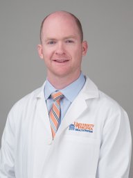 Werner Brian C合作论文数Department of Orthopaedic Surgery, University of Virginia Health System37Anthony A. Romeo合作论文数23Anup A Shah合作论文数16
 Justin W. Griffin合作论文数14Benjamin W. Sears合作论文数14
 Midhat Patel合作论文数12
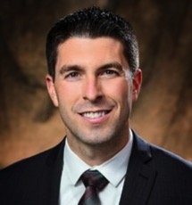 Erickson Brandon J合作论文数Rush University Medical Center, Rush University11
 Yousef Shishani合作论文数11Harmsen Samuel合作论文数8
 Youssef Galal合作论文数7Clayton H Hui合作论文数6Michael David McKee合作论文数6J Brock Walker合作论文数5
 M. Tyrrell Burrus合作论文数5Arjun Vohra合作论文数4Pamela Lund合作论文数4
 R. Alexander Creighton合作论文数4Theresa Pak合作论文数3Meghan E Bishop合作论文数Rothman Orthopaedic Institute3Javier Ardebol合作论文数3Mazzocca Augustus D合作论文数3
 Hustedt Joshua W合作论文数Department of Orthopaedics and Rehabilitation, Yale School of Medicine3
 Menendez Mariano E合作论文数Orthopaedic Hand and Upper Extremity Service, Massachusetts General Hospital3Noyes Matthew P合作论文数3
 Hartzler Robert U合作论文数The San Antonio Orthopaedic Group3
 Chalmers Peter N合作论文数Department of Orthopaedic Surgery, Rush University Medical Center3Bradford O. Parsons合作论文数3
 Patric Raiss合作论文数Stiftung Orthopädische Universitätsklinik Heidelberg3Sven Lichtenberg合作论文数3Armodios M. Hatzidakis合作论文数2Elliott W Cole合作论文数2Pierce Johnson合作论文数2anikar chhabra合作论文数2
 Mihir Sheth合作论文数2Brian S. Cohen合作论文数2Easton James Bents合作论文数2Patrick E Saunders合作论文数2Samuel G Moulton合作论文数2Richard L Auran合作论文数2
 Daichi Morikawa合作论文数2
 Georges Haidamous合作论文数2J Brett Goodloe合作论文数2
 Asheesh Bedi合作论文数Department of Orthopaedic Surgery, Medical School, University of Michigan-Ann Arbor2Provencher Matthew T合作论文数2Hanypsiak Bryan T合作论文数2Tokish John M合作论文数Steadman Hawkins Clinic of the Carolinas, Greenville Health System2
 Michael J Bercik合作论文数Temple University Hospital2




