According to the current WHO classification, noninvasive germ cell neoplasia of the testis comprises germ cell neoplasia in situ (GCNIS), specific forms of intratubular germ cell neoplasia, and gonadoblastoma. Because type II germ cell tumors (GCT, type II) arise from GCNIS, accurate detection of precursor lesions is diagnostically important. In preparation for the 2024 International Society of Urological Pathology (ISUP) Consensus Conference on genitourinary precursor lesions, which took place in Florence, Italy, an anonymous survey was distributed to ISUP members to assess current diagnostic practices regarding testicular precursor lesions. The literature and current WHO classification affirm the significance of precursor lesions in testicular tumours. Working Group 4-Precursor Lesions of the Testis-focused on their practical application by individual pathologists rather than establishing consensus from scientific data. There is strong agreement that GCNIS is the preferred and appropriate term for GCT precursor lesions and that its presence should be reported in cases of invasive GCT. Respondents also agree that "seminoma with intratubular nonseminoma" is the appropriate terminology for seminoma with an associated noninvasive nonseminomatous (embryonal carcinoma, yolk sac tumor, trophoblasts, or teratoma) component. Most pathologists prefer to use OCT3/4 as the primary immunohistochemical marker, and a panel was generally not considered necessary. No consensus is reached regarding the requirement for immunohistochemistry to confirm GCT precursor lesions in the testis. Three questions remain open: the value of subtyping intratubular lesions, the immunohistochemical approach to gonadoblastoma, and the criteria distinguishing Sertoli cell nodules from Sertoli cell tumors.
Birt-Hogg-Dubé (BHD) syndrome is an autosomal dominant disorder caused by germline inactivation of the folliculin gene (FLCN). Approximately 25
According to the current WHO classification, noninvasive germ cell neoplasia of the testis comprises germ cell neoplasia in situ (GCNIS), specific forms of intratubular germ cell neoplasia, and gonadoblastoma. Because type II germ cell tumors (GCT, type II) arise from GCNIS, accurate detection of precursor lesions is diagnostically important. In preparation for the 2024 International Society of Urological Pathology (ISUP) Consensus Conference on genitourinary precursor lesions, which took place in Florence, Italy, an anonymous survey was distributed to ISUP members to assess current diagnostic practices regarding testicular precursor lesions. The literature and current WHO classification affirm the significance of precursor lesions in testicular tumours. Working Group 4—Precursor Lesions of the Testis—focused on their practical application by individual pathologists rather than establishing consensus from scientific data. There is strong agreement that GCNIS is the preferred and appropriate term for GCT precursor lesions and that its presence should be reported in cases of invasive GCT. Respondents also agree that “seminoma with intratubular nonseminoma” is the appropriate terminology for seminoma with an associated noninvasive nonseminomatous (embryonal carcinoma, yolk sac tumor, trophoblasts, or teratoma) component. Most pathologists prefer to use OCT3/4 as the primary immunohistochemical marker, and a panel was generally not considered necessary. No consensus is reached regarding the requirement for immunohistochemistry to confirm GCT precursor lesions in the testis. Three questions remain open: the value of subtyping intratubular lesions, the immunohistochemical approach to gonadoblastoma, and the criteria distinguishing Sertoli cell nodules from Sertoli cell tumors.
AIMS:Germ cell neoplasia in situ (GCNIS) is the precursor of GCNIS-derived testicular germ cell tumours, including seminomas and non-seminomas. Most non-seminomas show mixed histology, often including components of seminoma. A small subset presents as pure non-seminomatous germ cell tumors. In these neoplasms, the absence of invasive seminoma. raises the possibility that reprogramming to non-seminoma may occur at an early in-situ stage (i.e., within the spermatogonial niche). This study examined GCNIS associated with pure embryonal carcinoma (EC) and postpubertal-type yolk sac tumour (YST) using immunohistochemistry for transcription factors characteristic of GCNIS/seminoma, EC, and YST phenotypes to determine if reprogramming to non-seminoma occurs within the spermatogonial niche. METHODS:GCNIS associated with pure testicular YST and EC was assessed using OCT4, FOXA2 (only YST), CD30 (only EC), SOX2 (only EC), SOX17, and NANOG immunohistochemistry. Adjacent foci of intratubular and invasive tumour were also evaluated, if present. RESULTS:Sixty-seven pure non-seminomas (63 EC, 4 YST) were evaluated. In all cases, GCNIS expressed OCT4, SOX17, and NANOG. GCNIS associated with EC was consistently negative for CD30 and SOX2, and GCNIS associated with YST was consistently negative for FOXA2. Adjacent invasive EC (62/67 cases) and intratubular EC (19/67) showed the classic EC immunophenotype: OCT4+/NANOG+/SOX17-/SOX2+/CD30+. Similarly, invasive YST adjacent to GCNIS was OCT4-/NANOG-/SOX17+/FOXA2+. CONCLUSIONS:Assessment of transcription factors involved in the induction and maintenance of EC and YST phenotypes suggests that, in pure non-seminomas, reprogramming occurs outside the spermatogonial niche.
A rare spindle cell tumor with a skeletal muscle phenotype, male predilection, exclusive involvement of the head and neck region, particularly the tongue, and a suggested indolent course has been previously reported as vestigial-like 3 (VGLL3)-rearranged spindle cell rhabdomyosarcoma (SRMS). We report 18 cases with extended clinical follow-up, detailed molecular results, and methylation profiling. Tumors occurred in 5 females, 12 males, and 1 patient of unknown sex with a median age of 58 years (range, 22-71). Tumors involved the tongue (n = 13), lower lip (2), retropharyngeal region (n = 1), thyroid/parathyroid (n = 1), and palatine tonsil (1), with a median size of 1.2 cm. Treatment details (15 patients) revealed that 13 patients underwent excision only, whereas 1 patient underwent adjuvant chemotherapy and 1 patient underwent adjuvant radiation therapy. Follow-up (11 patients) showed no local recurrence or metastases. At the last follow-up (median, 45 months; range, 1-326 months), all patients were alive without evidence of disease. Histologically, tumors showed spindled to histiocytoid cells arranged in a fascicular, storiform, or haphazard architecture with variably collagenous stroma, rounded to infiltrative borders, and often diffusely growing through skeletal muscle, adipose tissue, and entrapped nerves. Necrosis was consistently absent with a median mitotic rate of 1/10 high-power fields (range, 0-7/10). By immunohistochemistry, tumors diffusely expressed desmin (n = 18), multifocal MyoD1 (n = 15), and/or myogenin (n = 12), and sometimes smooth muscle actin (n = 7). Molecular testing revealed EP300::VGLL3 (n = 6), TCF12::VGLL3 (n = 5), and PPARGC1A::VGLL3 (n = 1) fusions with VGLL3 rearrangement by fluorescence in situ hybridization in 3 cases. One archival tumor failed molecular and methylation testing despite multiple attempts, likely due to decreased DNA/RNA integrity, yet was included given classic morphologic features. Methylation data (n = 12) revealed that all but 1 tumor formed a distinct group separate from other fusion-driven rhabdomyosarcomas, including 5 infantile/congenital SRMS. We describe a well-characterized series of these rare tumors with extended follow-up data confirming lack of progression or recurrence. Furthermore, our DNA methylation profiling data support the view that these tumors form a distinct cluster, regardless of VGLL3 fusion partner and are separate from morphologic mimics and other fusion-driven SRMS, including 5 cases of congenital SRMS. We propose that these neoplasms may be better classified as VGLL3-rearranged spindle cell rhabdomyoblastic tumors to reflect their indolent behavior and to prevent overtreatment.
Aims Testicular sex cord-stromal tumours (TSCSTs) are rare neoplasms that arise from (or show differentiation to) elements derived from the sex cords or intertubular stroma. The current classification (World Health Organization, 2022) is based almost entirely on morphology, with many tumour types such as granulosa cell tumours being defined based on their resemblance to ovarian counterparts. Recent studies have uncovered new clinicopathological, molecular and biological nuances within this group of tumours, underscoring differences with ovarian homologues.Methods and results Considering the new data, a group of genitourinary pathology experts (Testicular Sex Cord-Stromal Tumour [TESST] group) has proposed a revised 'TESST classification' that incorporates the best available evidence. This classification is structured and hierarchical, composed of the following tiers: (1) group, (2) category, (3) entity and (4) histological pattern. 'Group' refers to the histological compartment a specific tumour belongs to (sex cords, stroma or both), whereas 'category' is defined by the phenotype of the tumour, taking non-neoplastic elements of the ovary and testis as a reference. The third tier, 'entity', refers to neoplasms with shared clinicopathological and/or molecular features that justify classifying them as distinct types. Lastly, 'histological patterns' are recurrent morphological variations within entities that are not linked to distinct clinicopathological or molecular features.Conclusions We propose a classification that will provide a framework to improve current clinical management and guide future research in the field.
Synovial sarcoma is a rare malignant mesenchymal neoplasm that typically occurs in soft tissue sites, and the kidney is an uncommon primary location. Previous studies of primary renal synovial sarcoma are limited by small sample sizes, and our understanding remains incomplete. Here, we present the largest multi-institutional case series to date of primary renal synovial sarcoma, with a focus on novel and molecular findings. A total of 70 cases were contributed by multiple institutions. Comprehensive clinical and histopathologic data were collected and analyzed. The mean patient age was 40 years, with a male-to-female ratio of 1.9:1. The most common presenting signs and symptoms were pain and hematuria. The mean tumor size was 11.6 cm (range: 2.3 to 26 cm), with frequent cystic change and necrosis. The mean follow-up was 29 months (range: 2 to 129 mo), and the rates of metastasis, recurrence, and death due to disease were 62.7%, 33.3%, and 50.0%, respectively. Histologically, 56.1% were monophasic synovial sarcoma, 15.1% were biphasic, and 28.8% were poorly differentiated or showed round cell features. Molecularly, SS18::SSX2 fusion was detected in the majority of cases assessed (n=19), followed by SS18::SSX1 fusion (n=6) and a rare SS18::NEDD4 fusion (n=1). Given the morphologic and immunohistochemical overlaps with many other neoplasms, accurate diagnosis with preferably molecular techniques is crucial for appropriate prognostication and treatment of this aggressive tumor.
Testicular adult granulosa cell tumors (AGCTs) are rare and show several clinical, pathological, and molecular differences with their ovarian counterparts. FOXL2 p.Cys134Trp, the ubiquitous molecular driver of ovarian AGCTs, is infrequent ( 7
BACKGROUND:Solitary fibrous tumor (SFT) is a mesenchymal neoplasm characterized by NAB2::STAT6 fusion and nuclear STAT6 expression. Primary SFT of the prostate is rare, and its biological behavior remains incompletely defined. We present a multi-institutional cohort and comprehensive literature review to better characterize clinicopathologic features, management, and outcomes. DESIGN:We analyzed 33 patients with prostatic SFTs from multiple institutions, integrating clinical, radiologic, histopathologic, immunohistochemical, and follow-up data. A systematic review of published cases was performed, and findings were contextualized using established risk models. RESULTS:Patients ranged from 30 to 93 years (mean, 59; median, 61). Tumor sizes ranged from 0.6 to 19.5 cm (mean, 6.8; median, 5.75). Presenting symptoms included urinary obstruction, pelvic fullness, elevated PSA, or incidental detection. In two patients, the tumor caused urinary obstruction requiring transurethral resection. Location adjacent to the seminal vesicles was noted in several patients with one growing as a large mass into the pelvis. In several patients, the SFT was an incidental finding on needle biopsy performed for elevated PSA. Histologically, all tumors demonstrated classic SFT morphology with a spindle cell proliferation and patternless architecture. Tumor borders varied, with both well-circumscribed and infiltrative patterns observed. Cellularity ranged from low to high, and nuclear atypia was most often mild to moderate. Mitotic activity was generally low (<5 mitoses/10 HPF in most cases), although two tumors showed elevated indices (12 and 15/10 HPF). Necrosis was identified in 5 tumors and was focal in 3. STAT6 was positive in 27/27 tested cases (100%), and CD34 was positive in all cases with available results (31/31, 100%). Management was primarily surgical. Only three tumors in this cohort developed metastatic disease, limiting the ability to confirm or refute the applicability of the Demicco risk stratification model. Metastatic cases occurred in relatively older patients, two of the three were >5 cm with focal necrosis, and two had increased mitotic activity (>4/10 HPF), while the third was 4.5 cm without necrosis but showed a high mitotic count. No overtly high-grade morphology or consistent morphologic predictors of metastasis were identified in this series. Follow-up was available in 16 patients (10-171 months; mean, 38; median, 29). Aside from the three patients who developed metastatic disease, all others were alive and without recurrence or metastasis at last contact. CONCLUSIONS:This is among the largest reported cohorts of prostatic SFTs. These tumors, though rare in the prostate, appear to display similar morphologic and immunophenotypic features to SFTs in other anatomic sites. While generally indolent, rare cases may metastasize. Accurate diagnosis relies on a combination of morphologic evaluation and STAT6 IHC. This study expands the clinicopathologic spectrum of SFTs and emphasizes the need for long-term follow-up due to the potential for late recurrence or metastasis.
AIMS:Standardizing pathology reporting protocols through peer consensus review is critical for the best quality of care metrics. Reporting heterogeneity due to discrepancies among professional societies and practice patterns may lead to heterogeneous management and treatment approaches. This issue prompted a multi-institutional survey of pathologists to address potential similarities or differences in trends and practice patterns in prostate pathology reporting worldwide. METHODS AND RESULTS:A REDCap survey was distributed among 175 pathologists worldwide, recruited through invitations and social media. The response rate among invited pathologists was 83%. The practice locations were as follows: North America (USA, Canada, and Mexico, 62%), Europe (17%), Australia/New Zealand (3%), Central/South America (2%), Asia (13%), and Africa (2%). Most pathologists practiced for <5 years (28%). A genitourinary (GU) pathology fellowship was completed by 37%, 58% practiced in a subspecialized setting, and 43% in academia. Reporting includes (63%) or subtracts (37%) intervening benign tissue. Both Gleason score and Grade Groups (GG)s were reported by 96% of responders, whereas 94% report percent pattern 4 (%4). Aggregate grading and volume estimation in undesignated cores with different grades in the same jar are reported by 73% and 54% for systematic biopsies, and 83% and 62% for targeted biopsies, respectively. Cribriform morphology was reported by 81%. For presumed intraductal carcinoma (IDC), 89% use basal cell markers when isolated (iIDC), 82% with GG1 cancer, and 37% with ≥GG2. iIDC or IDC associated with GG1 or with ≥GG2 was not graded by 90%, 78%, and 70%, respectively. In radical prostatectomies, 90% report %4, but only 53% report it if the overall grade is ≥7. A tumour with Gleason 3 + 3 = 6 and <5% pattern 4 was graded as GG2 by 64%. A <5% cutoff for defining tertiary pattern was used by 74%, and 80% report >5% pattern 4 or 5 as a secondary pattern. Grading was assigned based on the dominant nodule by 59%. Finally, reporting practices were significantly associated with demographic characteristics. CONCLUSIONS:Although most issues are agreed upon, significant discordance is identified among societies and pathologists in different practice settings. We hope this survey will serve as the basis for future studies and new collaborative approaches to more standardized reporting practices.
Somatic‐type malignancies (SMs) arising in germ cell tumors (GCTs) are aggressive neoplasms resistant to systemic treatment. Most are diagnosed in metastatic sites after chemotherapy; however, they have also been well‐documented in primary testicular GCTs. Historically, SMs were thought to originate in components of teratoma that acquire molecular alterations equivalent to those that characterize their true somatic counterparts. However, recent studies have shown that SMs typically lack the hallmark molecular alterations seen in similar somatic tumors. Additionally, clinicopathologic and molecular data suggest that a subset may derive from yolk sac tumor (YST) rather than teratoma. In this study, we evaluated the relationship between conventional histological types of GCTs and SMs by comparing expression of microRNA (miR)‐371–373 and genomic methylation profiles. A total of 96 samples (including multiple paired conventional GCT–SM samples from individual tumors) were assessed for miR‐371–373 expression by RT‐qPCR and genomic DNA methylation using a clinically validated assay. Expression of miR‐371–373 was higher in conventional GCTs than in SMs (considered as a single category encompassing all histological subtypes). However, miR‐371–373 expression was heterogeneous among SMs, with significantly higher levels in sarcomatoid YST (SYST) and glandular neoplasms than in other SMs. Genomic DNA methylation analysis showed that SMs (considered as a single category) did not form a distinct cluster. Instead, they grouped into multiple clusters that did not show perfect correspondence with histology and often included conventional GCTs. Genome‐wide methylation assessment showed a higher abundance of hypermethylated regions in SMs than in conventional GCTs. Analysis of paired conventional GCT and ‘somatic‐type’ components that did not meet size criteria for SMs dissected from individual tumors demonstrated separation according to histology, suggesting that epigenetic processes play a role in the transition from conventional GCT to ‘somatic‐type’ phenotypes. Gene‐level and pathway‐level analyses identified MAPK/RAS signaling, mitosis/proliferation, differentiation towards neural tissue/neuroectoderm, epithelial‐to‐mesenchymal transition, and DNA repair as key differentially regulated processes in components with somatic‐type histology, suggesting mechanisms of progression from conventional to ‘somatic’ phenotypes in GCT. These results support the hypothesis that a subset of SMs derive from YST and suggest that some subtypes (such as SYST) may represent ‘intermediate’ phenotypes. Additionally, analysis of differentially methylated promoter regions in SM identified genes and biologic processess that may underlie 'somatic tranformation' in GCTs. © 2025 The Author(s). The Journal of Pathology published by John Wiley & Sons Ltd on behalf of The Pathological Society of Great Britain and Ireland.
Renal mucinous cystadenoma (RMC) is an exceptionally rare finding with a poorly understood pathogenesis. Links between RMC and other malignancies are not well described, nor are there known associations with familial cancer disorders. We present the first case of RMC associated with Lynch syndrome (LS) and neuroendocrine neoplasm. A 56-year-old woman presented with iron deficiency leading to a diagnosis of a colonic mixed neuroendocrine-non-neuroendocrine carcinoma. After initial surgery, she experienced local recurrences at 6 and 12 months, treated with resection and adjuvant chemotherapy. Tumours displayed deficient MMR proteins with BRAF positivity, and germline testing confirmed LS. Surveillance subsequently revealed a complex cyst arising from a horseshoe kidney, for which she underwent a partial nephrectomy. Histopathology confirmed this to be renal mucinous cystadenoma arising from the pelvicalyceal system. This case underscores the need for further investigation into RMC pathogenesis and its potential association with LS.
Ewing sarcoma (ES) is an undifferentiated round cell sarcoma arising in skeletal and extraskeletal locations usually diagnosed in the second decade of life. It is typically characterized by a monomorphic round blue cell morphology, positive membranous CD99 immunohistochemical staining, and EWSR1 gene fusion with a partner gene from the ETS family. Less frequently, other genes from the FET family of genes substitute for EWSR1. We report a case of oligometastatic Ewing sarcoma with FUS::ERG fusion with an unusual primary location in an older patient. This case highlights the application of ancillary tests, especially next generation sequencing, as an adjunct to the morphological assessment of undifferentiated round cell tumours. We also discuss potential diagnostic pitfalls and literature review of Ewing sarcomas with FUS::ERG fusion.
 Anthony Gill合作论文数Department of Anatomical Pathology, Royal North Shore Hospital;Faculty of Medicine and Health, University of Sydney42
 Kiril Trpkov合作论文数Department of Pathology and Laboratory Medicine, Cuming School of Medicine, University of Calgary;Southern Alberta Institute of Urology;Tom Baker Cancer Center34
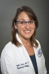 Michelle S. Hirsch合作论文数Department of Pathology, Division of Women's and Perinatal Pathology, Brigham and Women's Hospital, Boston, MA, USA34
 Sean Williamson合作论文数Department of Pathology, Cleveland Clinic33
 Ana Cristina Vargas合作论文数33
 Liang Cheng合作论文数Department of Surgery, Warren Alpert Medical School of Brown University;Department of Pathology and Laboratory Medicine, Warren Alpert Medical School of Brown University27Fiona Bonar合作论文数26
 Katrina Collins合作论文数Department of Pathology & Laboratory Medicine, Hartford Hospital26Sangoi Ankur R合作论文数25
 Chia Sui Kao合作论文数Department of Pathology, School of Medicine, Stanford University;Stanford Health Care23
 Osunkoya Adeboye O合作论文数20
 Thomas M. Ulbright合作论文数School of Medicine, Indiana University19
 Maria Tretiakova合作论文数Department of Pathology, University of Chicago19Cristina Magi-Galluzzi合作论文数19
 Maurizio Colecchia合作论文数Pathology, Fondazione IRCCS Istituto Nazionale dei Tumori of Milan19
 Rohit Mehra合作论文数Michigan Center for Translational Pathology;Department of Pathology, University of Michigan Medical School;Rogel Cancer Center, Michigan Medicine19
 Muhammad Idrees合作论文数17
 Acosta Andres合作论文数17
 Jonathan Epstein合作论文数Johns Hopkins University School of Medicine;Johns Hopkins Hospital17Manju Aron合作论文数17Annabelle Mahar合作论文数17Eva Comperat合作论文数16Ondrej Hes合作论文数16
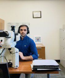 Mahmut Akgul, MD合作论文数Albany Med Ctr15
 Abbas Agaimy合作论文数Pathologisches Institut, Universitätsklinikum Erlangen15
 Kristine Cornejo合作论文数Massachusetts General Hospital/Harvard Medical School/Department of Pathology, Massachusetts General Hospital15
 Mahul B. Amin合作论文数Labcorp;Keck School of Medicine of the University of Southern California;The University of Tennessee Health Science Center15Jennifer B Gordetsky合作论文数Univ Alabama Birmingham15
 Andres Matoso合作论文数Department of Otolaryngology-Head and Neck Surgery, School of Medicine, Johns Hopkins University14
 Satish K. Tickoo合作论文数Department of Pathology, Memorial Sloan Kettering Cancer Center14
 Steven C. Smith合作论文数Pacific Northwest National Laboratory14Kvetoslava Michalova合作论文数13Cheah Alison合作论文数13
 Santosh Menon合作论文数13Ming Zhou合作论文数Department of Anatomic Pathology, Cleveland Clinic12
 Maria M Picken合作论文数Stritch School of Medicine, Loyola University12
 Sambit K. Mohanty合作论文数Department of Biomedical Informatics, University of Pittsburgh School of Medicine12
 Joao Lobo合作论文数11
 Gupta Sounak合作论文数11
 Lynette M Sholl合作论文数Department of Pathology, Brigham and Women's Hospital;Harvard Medical School11Rao Priya合作论文数11
 Priti Lal合作论文数Department of Pathology and Laboratory Medicine, Perelman School of Medicine, University of Pennsylvania11Jesse K. Mckenney合作论文数11
 Isabela Werneck da Cunha合作论文数Instituto D'Or de Pesquisa e Ensino, Rede D´Or São Luiz11
 Sara Elizabeth Wobker合作论文数Department of Pathology and Laboratory Medicine, School of Medicine, The University of North Carolina at Chapel Hil11Seema Kaushal合作论文数Dept Pathol, All India Inst Med Sci10
 Fadi Brimo合作论文数Department of Pathology, Faculty of Medicine and Health Sciences, McGill University10Sioson Loretta合作论文数10
 Przybycin Christopher合作论文数10




