BACKGROUND:The existing classifications of congenital ossicular chain usually take the motility of the stapes footplate for differentiation, with a lack of attention on the concurrent malformations of facial nerve. OBJECTIVES:To explore the distribution of different types of congenital ossicular chain malformations and to propose a new classification for the guidance of clinical treatment. MATERIAL AND METHODS:Preoperative examinations and intraoperative findings of patients with congenital ossicular chain malformations from 2014 to 2025 were retrospectively collected. Based on the traditional Teunissen and Yang classification, a new classification was further proposed according to the key anatomical structures concerned in clinical diagnosis and treatment. RESULTS:A total of 119 patients with ossicular chain malformations were included in this study, 135 ears in total. The classification of patients in this study based on this new classification was as follows: 19 ears (14.1%) of type Ia, 60 ears (44.4%) of type Ib, 7 ears (5.2%) of type IIa, 33 ears (24.4%) of type IIb, 11 ears (8.1%) of type IIIa, and 5 ears (3.7%) of type IIIb. CONCLUSIONS AND SIGNIFICANCE:Based on a large cohort study, this innovative classification that centering on abnormal stapes and facial nerve contributes much to clinical diagnosis and treatment of non-syndrome congenital ossicular chain malformations.
OBJECTIVES:This study sought to explore the association between MRI-detected inner ear hemorrhage and the prognosis and symptom of SSNHL. METHODS:A total of220 enrolled patients were asked to receive MRI and hearing test. RESULTS:Patients with MRI evidence of inner ear hemorrhage experienced significantly more severe hearing loss, differences in hearing before and after treatment, and subjective perceptions compared to individuals without these signs. The hearing recovery at speech frequencies (0.5, 1, 2 kHz) was better in the non-hemorrhage group compared to the hemorrhage group. Additionally, the non-hemorrhage group exhibited fewer symptoms of dizziness and a better prognosis for dizziness. CONCLUSION:MRI played a crucial role in both identifying the location of hemorrhage and accurately predicting symptoms and prognosis.
Background Temporal bone computed tomography (CT) helps diagnose chronic otitis media (COM). However, its interpretation requires training and expertise. Artificial intelligence (AI) can help clinicians evaluate COM through CT scans, but existing models lack transparency and may not fully leverage multidimensional diagnostic information. Objective We aimed to develop an explainable AI system based on 3D convolutional neural networks (CNNs) for automatic CT-based evaluation of COM. Methods Temporal bone CT scans were retrospectively obtained from patients operated for COM between December 2015 and July 2021 at 2 independent institutes. A region of interest encompassing the middle ear was automatically segmented, and 3D CNNs were subsequently trained to identify pathological ears and cholesteatoma. An ablation study was performed to refine model architecture. Benchmark tests were conducted against a baseline 2D model and 7 clinical experts. Model performance was measured through cross-validation and external validation. Heat maps, generated using Gradient-Weighted Class Activation Mapping, were used to highlight critical decision-making regions. Finally, the AI system was assessed with a prospective cohort to aid clinicians in preoperative COM assessment. Results Internal and external data sets contained 1661 and 108 patients (3153 and 211 eligible ears), respectively. The 3D model exhibited decent performance with mean areas under the receiver operating characteristic curves of 0.96 (SD 0.01) and 0.93 (SD 0.01), and mean accuracies of 0.878 (SD 0.017) and 0.843 (SD 0.015), respectively, for detecting pathological ears on the 2 data sets. Similar outcomes were observed for cholesteatoma identification (mean area under the receiver operating characteristic curve 0.85, SD 0.03 and 0.83, SD 0.05; mean accuracies 0.783, SD 0.04 and 0.813, SD 0.033, respectively). The proposed 3D model achieved a commendable balance between performance and network size relative to alternative models. It significantly outperformed the 2D approach in detecting COM (P≤.05) and exhibited a substantial gain in identifying cholesteatoma (P<.001). The model also demonstrated superior diagnostic capabilities over resident fellows and the attending otologist (P<.05), rivaling all senior clinicians in both tasks. The generated heat maps properly highlighted the middle ear and mastoid regions, aligning with human knowledge in interpreting temporal bone CT. The resulting AI system achieved an accuracy of 81.8% in generating preoperative diagnoses for 121 patients and contributed to clinical decision-making in 90.1% cases. Conclusions We present a 3D CNN model trained to detect pathological changes and identify cholesteatoma via temporal bone CT scans. In both tasks, this model significantly outperforms the baseline 2D approach, achieving levels comparable with or surpassing those of human experts. The model also exhibits decent generalizability and enhanced comprehensibility. This AI system facilitates automatic COM assessment and shows promising viability in real-world clinical settings. These findings underscore AI’s potential as a valuable aid for clinicians in COM evaluation. Trial Registration Chinese Clinical Trial Registry ChiCTR2000036300; https://www.chictr.org.cn/showprojEN.html?proj=58685
Intense noise poses a threat to spiral ganglion neurons (SGNs) in the inner ear, often resulting in limited axonal regeneration during noise injury and leading to noise-induced hearing loss (NIHL). Here, we propose an ultrasound-triggered nitric oxide (NO) release to enhance the sprouting and regeneration of injured axons in SGNs. We developed hollow silicon nanoparticles to load nitrosylated N-acetylcysteine, producing HMSN-SNO, which effectively protects NO from external interferences. Utilizing low-intensity ultrasound stimulation with bone penetration, we achieve the controlled release of NO from HMSN-SNO within the cochlea. In mice with NIHL, a rapid and extensive loss of synaptic connections between hair cells and SGNs is observed within 24 h after exposure to excessive noise. However, this loss could be reversed with the combined treatment, resulting in a hearing functional recovery from 83.57 to 65.00 dB SPL. This positive outcome is attributed to the multifunctional effects of HMSN-SNO, wherein they scavenge reactive oxygen species (ROS) to reverse the pathological microenvironment and simultaneously upregulate the CREB/BDNF/EGR1 signaling pathway, thereby enhancing neuroplasticity and promoting the regeneration of neuronal axons. These findings underscore the potential of nanomedicine for neuroplasticity modulation, which holds promise for advancing both basic research and the further treatment of neurological diseases.
A large-scale outbreak of Severe Acute Respiratory Syndrome Coronavirus 2 (SARS-CoV-2) occurred in Shanghai, China, in early December 2022. To study the incidence and characteristics of otitis media with effusion (OME) complicating SARS-CoV-2, we collected 267 middle ear effusion (MEE) samples and 172 nasopharyngeal (NP) swabs from patients. The SARS-CoV-2 virus was detected by RT-PCR targeting. The SARS-CoV-2 virus, angiotensin-converting enzyme 2 (ACE2), and transmembrane serine protease 2 (TMPRSS2) expression in human samples was examined via immunofluorescence. During the COVID-19 epidemic in 2022, the incidence of OME (3%) significantly increased compared to the same period from 2020 to 2022. Ear symptoms in patients with SARS-CoV-2 complicated by OME generally appeared late, even after a negative NP swab, an average of 9.33 +/- 6.272 days after COVID-19 infection. The SARS-CoV-2 virus was detected in MEE, which had a higher viral load than NP swabs. The insertion rate of tympanostomy tubes was not significantly higher than in OME patients in 2019-2022. Virus migration led to high viral loads in MEE despite negative NP swabs, indicating that OME lagged behind respiratory infections but had a favorable prognosis. Furthermore, middle ear tissue from adult humans coexpressed the ACE2 receptor for the SARS-CoV-2 virus and the TMPRSS2 cofactors required for virus entry.
Purpose: Glomus tympanicum tumors are benign primary tumors of the middle ear that can be completely removed using modern surgery. We compared endoscopic ear surgery (EES) to traditional microscopic ear surgery (MES) in terms of the removal of early-stage glomus tympanicum tumors. Methods: We retrospectively reviewed 25 cases treated from 2003 to 2021 that were of Grade I or II based on the Glasscock–Jackson classification system. Overall, 18 cases underwent MES: 8 via trans-tympanic bone and 10 via canal-wall-down or canal-wall-up tympanomastoidectomy (CWDT or CWUT) and 7 underwent EES. We compared surgery durations, the lengths and costs of hospitalization, postoperative complications, and relapse rates between the two groups and among the three specific operation ways. Results: The postoperative follow-up period ranged from 1 to 19 years. There was no between-group difference in operative time or the length or cost of hospitalization. Operative time and cost of hospitalization did not show a statistically significant correlation to the three surgical procedures, whereas it was found that the group of MES via the trans-tympanic bone had shorter length of hospitalization when compared with CWUT or CWDT group. All tumors were completely resected; pulsatile tinnitus improved in all patients, and there was no major complication. Two patients who underwent CWUT or CWDT (one each) relapsed; no patient relapsed in the EES group. Conclusion: MES via the trans-tympanic bone and EES via the ear canal safely and reliably remove early-stage tumors without excessive patient discomfort.
耳显微外科的发展与成熟依赖于显微镜及相关手术器械的辅助,随着内镜工艺和技术的逐步完善,耳内镜在耳显微外科的应用得到了众多耳科医师的广泛关注.经过近 10年的探索与研究,积累了很多临床经验,也取得了显著的临床成效.当然,耳内镜也有其不足,比如需单手操作、配套手术器械不足等,相信随着越来越多耳科医师的不断探索以及内镜工艺的不断精进,这些不足将会被逐一克服,耳内镜优势将更加突出,在耳科将有着更为广泛的应用前景.
Dentin matrix protein 1 (Dmp1) is a highly phosphorylated, extracellular matrix protein that is extensively expressed in bone and teeth but also found in soft tissues, including brain and muscle. However, the functions of Dmp1 in the mice cochlea are unknown. Our study showed that Dmp1 was expressed in auditory hair cells (HCs), with the role of Dmp1 in those cells identified using Dmp1 cKD mice. Immunostaining and scanning electron microscopy of the cochlea at P1 revealed that Dmp1 deficiency in mice resulted in an abnormal stereociliary bundle morphology and the mispositioning of the kinocilium. The following experiments further demonstrated that the cell-intrinsic polarity of HCs was affected without apparent effect on the tissue planer polarity, based on the observation that the asymmetric distribution of Vangl2 was unchanged whereas the Gαi3 expression domain was enlarged and Par6b expression was slightly altered. Then, the possible molecular mechanisms of Dmp1 involvement in inner ear development were explored via RNA-seq analysis. The study suggested that the Fgf23-Klotho endocrine axis may play a novel role in the inner ear and Dmp1 may regulate the kinocilium-stereocilia interaction via Fgf23-Klotho signaling. Together, our results proved the critical role of Dmp1 in the precise regulation of hair bundle morphogenesis in the early development of HCs.
Facial nerve tumors are rare but challenging diseases, and accurate diagnosis and effective treatment are crucial for patients′ recovery. This article aims to explore the current status, challenges, and opportunities in improving the diagnosis of facial nerve tumors and its functional surgery. Imaging techniques such as MRI and CT play a crucial role in the preoperative diagnosis and evaluation of facial nerve tumors. However, limitations still exist in the imaging diagnosis of lesions isolated within the internal auditory canal and parotid gland. Surgical treatment is the main therapeutic approach for facial nerve tumors, and with the continuous development of surgical techniques, the surgical goals for facial nerve tumors have shifted from simply completely removing the tumor to preserving facial nerve integrity as much as possible and promoting postoperative facial nerve function recovery. Challenges faced in the diagnosis of facial nerve tumors and its functional surgery include limitations in imaging diagnosis, intraoperative facial nerve protection, and postoperative improvement of facial nerve function. Future directions include the application of high-resolution imaging techniques, innovation in nerve protection and repair surgical techniques, the realization of personalized precision medicine, and multidisciplinary collaboration. Future research and technological innovations are expected to overcome diagnostic difficulties, improve facial nerve preservation, and promote postoperative facial nerve function recovery.
Tumors originating from the facial nerve are relatively rare and primarily include three types: facial nerve schwannoma, facial nerve neurofibroma, and facial nerve hemangioma. The Facial Nerve Tumor Diagnosis and Treatment Consensus Writing Group of China has established the current consensus after multiple discussions, based on a review of domestic and international literature and taking into account the domestic situation. This consensus systematically elucidates the epidemiology, pathological characteristics, and main clinical manifestations of facial nerve tumors. It establishes diagnostic criteria for facial nerve tumors, clarifies the anatomical classification of facial nerve tumors, and outlines its main differential diagnoses. Furthermore, the consensus clearly defines the principles of applying different treatment methods for facial nerve tumors and outlines a concise workflow for the diagnosis and treatment of facial nerve tumors.
Objective: To evaluate the effects of DX and HA on hearing preservation and tissue response with cochlear implantation in a cat mode. Methods: 11 cats were bilaterally implanted with Nurotron electrode. According to the different treatment, 22 ears were subdivided into four following groups: Electrode (E) only, Electrode + HA (E + H), Electrode + DX (E + DX), and Electrode + HA + DX (E + H + DX). Auditory brainstem responses (ABR) thresholds were recorded before and 3 months after surgery. After 3 months of cochlear implantation histopathological assessment of all cochleae were performed. Results: At low frequencies, the mean ABR thresholds shift in animals treated with E + H + DX demonstrated a statistically significant difference. The proportion of cochleae showing fibrous tissue and new bone formation was 50% in E + H + DX group, 66.7% in E + DX group, 83.3% in E + H group and 100% in E group. Conclusion: United HA and DX local delivery could provide better protection against hearing loss at low-frequency threshold and could more effectively reduce the trauma related to electrode insertion and the fibrous tissue formation around the electrode. (c) 2022 Japanese Society of Otorhinolaryngology-Head and Neck Surgery, Inc. Published by Elsevier B.V. All rights reserved.
Objective Our team designed a long‐lasting, well‐sealed microphone, which uses laser welding and vacuum packaging technology. This study examined the sensitivity and effectiveness of this new floating piezoelectric microphone (NFPM) designed for totally implantable cochlear implants (TICIs) in animal experiments and intraoperative testing. Methods Different NFPM frequency responses from 0.25 to 10 kHz at 90 dB SPL were analyzed using in vivo testing of cats and human patients. The NFPM was tested in different positions that were clamped to the ossicular chains or placed in the tympanic cavity of cats and human patients. Two volunteers' long incus foot and four cats' malleus neck of the ossicular chain were clamped with the NSFM. The output electrical signals from different locations were recorded, analyzed, and compared. The NFPM was removed after the test without causing any damage to the middle‐ear structure of the cats. Intraoperative tests of the NFPM were performed during the cochlear implant surgery and the cochlear implant surgery was completed after all tests. Results Compared with the results in the tympanic cavity, the NFPM could detect the vibration from the ossicular chain more sensitively in cat experiments and intraoperative testing. We also found that the signal output level of the NFPM decreased as the acoustic stimulation strength decreased in the intraoperative testing. Conclusion The NFPM is effective in the intraoperative testing, making it feasible as an implantable middle‐ear microphone for TICIs. Level of Evidence 4 Laryngoscope , 134:937–944, 2024
Ischemia-reperfusion (I/R) injury, uncommon among patients suffering from myocardial infarction, stroke, or acute kidney injury, can result in cell death and organ dysfunction. Previous studies have shown that different types of cell death, including apoptosis, necrosis, and autophagy, can occur during I/R injury. Pyroptosis, which is characterized by cell membrane pore formation, pro-inflammatory cytokine release, and cell burst, and which differentiates itself from apoptosis and necroptosis, has been found to be closely related to I/R injury. Therefore, targeting the signaling pathways and key regulators of pyroptosis may be favorable for the treatment of I/R injury, which is far from adequate at present. This review summarizes the current status of pyroptosis and its connection to I/R in different organs, as well as potential treatment strategies targeting it to combat I/R injury.
本文对2021年我国眩晕领域多个学科知名专家共同讨论和制订的国内首个前庭康复专家共识的关键内容进行了全面解读,并与美国物理治疗学会神经物理治疗分会发布的2016版外周前庭功能低下前庭康复指南和2022年更新版指南的相应内容进行比较,突出了本共识的特色和亮点,旨在帮助临床专业人员更好地理解共识的内涵和精髓,快速搭建前庭康复理论的系统框架,推动前庭康复诊治技术的规范操作和临床研究的广泛开展.
Substantial evidence suggests that pyroptosis is involved in renal, cerebral, and myocardial ischemia–reperfusion injury. However, whether pyroptosis is involved in ischemia–reperfusion injury of cochlear hair cells has not been explored. In this study, we examined the effects of melatonin on the oxygen–glucose deprivation/reperfusion (OGD/R) of hair cell-like House Ear Institute-Organ of Corti 1 (HEI-OC1) cells and cochlear hair cells in vitro to mimic cochlear ischemia–reperfusion injury in vivo. We found that melatonin treatment protected the HEI-OC1 and cochlear hair cells against OGD/R-induced cell pyroptosis and reduced the expression level of ROS in these cells. However, these effects were completely abolished by the application of luzindole (a non-selective melatonin receptor blocker) and largely offset by the use of ML385 (an nuclear factor erythroid 2-related factor 2 (Nrf2) inhibitor). These findings suggest that melatonin alleviates OGD/R-induced pyroptosis of the hair cell-like HEI-OC1 cells and cochlear hair cells via the melatonin receptor 1A (MT-1) and melatonin receptor 1B (MT-2)/Nrf2 (NFE2L2)/ROS/NLRP3 pathway, which may provide credible evidence for melatonin being used as a potential drug for the treatment of idiopathic sudden sensorineural hearing loss in the future.
To present the clinical characteristics and the misdiagnosis rate of acute coronary syndrome manifested primarily as throat discomfort, we conducted a multicentric and retrospective study in the cardiology and otorhinolaryngology departments. Records of patients with primary complaint of throat discomfort, absence of chest pain at onset, and an ultimate diagnosis of acute coronary syndrome, as well as patients with pharyngitis (as controls) were collected from May 2015 to April 2016. The patients’ main manifestations were compared. Logistic regression results showed that chest tightness, dyspnea, perspiring, and exertional throat symptoms were significantly associated with acute coronary syndrome, with odds ratios of 8.3 (95% CI 2.2–31.5), 10.9 (95% CI 1.8–66.9), 25.4 (95% CI 3.6–179.9), and 81.2 (95% CI 13.0–506.7). A total of 25 (56.82%) out of 44 acute coronary syndrome patients, who were first admitted to the otorhinolaryngology department, were misdiagnosed, with a 12% (3/25) mortality rate. Throat discomfort can be the principal manifestation of acute coronary syndrome. Such patients exhibit high misdiagnosis and mortality rates. Exertional throat symptoms, chest tightness, perspiring, and dyspnea were important indicators of acute coronary syndrome in patients whose main complaint was throat discomfort. The awareness of this condition will result in prompt diagnosis and reduce morbidity and mortality.
目的 探索重度阻塞性睡眠呼吸暂停(OSA)患者的耳石器及其传导通路功能变化情况.方法 2020年1~10月就诊于我院鼾症诊疗中心的重度OSA患者为病例组,30例(60耳);对照组为健康成年人21例(42耳),比较并分析2组眼肌前庭诱发肌源性电位(oVEMP)和颈肌前庭诱发肌源性电位(cVEMP)的引出率、潜伏期、波间期、振幅.结果 对照组的oVEMP和cVEMP引出率分别为97.6%和100.0%,病例组为73.3%和81.7%,较对照组显著下降(P=0.001、0.003).在oVEMP中,病例组的阈值为(115.68±6.52)dB SPL,显著高于对照组的(110.98±4.77)dB SPL(P<0.001);N1潜伏期为(11.85±0.85)ms,较对照组的(10.78±0.84)ms延长(P<0.001);病例组的N1-P1波间期缩短至(3.69±0.79)ms(P<0.001),N1-P1振幅下降至(9.30±8.17)μV,较对照组的(15.79±7.66)μV显著降低(P<0.001).在cVEMP中,病例组的阈值为(114.18±6.64)dB SPL,较对照组的(104.52±4.53)dB SPL显著升高;P1潜伏期由(16.71±1.84)ms显著延长至(18.02±2.53)ms(P=0.005);P1-N1波间期也因此缩短为(6.88±2.03)ms(P<0.001);P1-N1振幅和N1-P2振幅分别降至(126.73±98.55)μV和(88.71±94.99)μV,较对照组的(254.99±113.74)μV和(154.89±71.13)μV显著降低(P值均<0.001).对病例组30例患者进行分析发现,医患双方对平衡症状的关注度显著低于对打鼾症状的关注度(P<0.001).结论 在重度OSA患者中,oVEMP和cVEMP的波形出现典型异常变化,提示椭圆囊和球囊及其传导通路出现功能障碍.重度OSA患者的诊治应同时考虑耳石器功能障碍.
2022年是主办单位复旦大学附属眼耳鼻喉科医院建院70周年,在此契机下,《中国眼耳鼻喉科杂志》开设新栏目"学科前沿",为院庆献礼,是体现期刊"促进国内外眼科、耳鼻喉科领域内重要科研成果的交流"办刊宗旨的创新之举.
ObjectiveTo investigate whether otolith dysfunction is related to hearing impairment in vertigo patients with normal semicircular canal function, and to clarify the types of hearing impairment that may be related to otolith organ damage.MethodsThe demographic data, pure tone threshold audiometry (PTA) results (air-conduction), data of bithermal and video-head impulse test (vHIT), and vestibular evoked myogenic potential (VEMP) results (reaction threshold, P1–N1 amplitude) of patients with vertigo in outpatient clinic from April 2017 to January 2020 were collected. The clinical records of 51 vertigo patients with normal semicircular canal function were included in this study. Low-frequency, speech-frequency, high-frequency, full-frequency PTA were defined as the average of PTA in different frequency bands, respectively (low: 0.125, 0.25, 0.5 kHz; speech: 0.5, 1, 2 kHz, high: 4, 8 kHz, full 0.125–8 kHz). The correlations between hearing impairment in different frequency bands and otolith function impairment were analyzed.ResultsThe mean thresholds of 51 patients (102 ears) in low-PTA, speech-PTA, high-PTA, full-PTA were 20.95 ± 6.01, 21.92 ± 6.90, 40.12 ± 17.47, 26.97 ± 8.53 dB nHL, respectively. Among 102 ears, 87 ears (85.3%) could elicit c-VEMP waveforms and 65 ears (63.7%) had o-VEMP waveforms. The mean threshold and P1–N1 amplitude of c-VEMP were 83.10 ± 6.96 dB nHL and 176.79 ± 103.10 uV, while those of o-VEMP were 87.92 ± 5.99 dB nHL and 21.45 ± 32.22 uV. The mean threshold in high-PTA was significantly linearly correlated with c-VEMP threshold (P = 0.01) and P1–N1 amplitude (P = 0.028). There were not significant linear correlations between the mean threshold in each frequency band of PTA and o-VEMP threshold (low-PTA: P = 0.266, speech-PTA: P = 0.33, high-PTA: P = 0.311) or P1–N1 amplitude (low-PTA: P = 0.414, speech-PTA: P = 0.069, high-PTA: P = 0.08).ConclusionsThere is a positive linear correlation between saccule dysfunction and high-frequency hearing impairment in vertigo patients with normal semi-circular canal function. High frequency hearing loss can be expected in patients who have saccular damage. It suggests that high frequency hearing loss in PTA may act as a screening index that otolith organ function should be comprehensively evaluated.
Journal of Cellular PhysiologyVolume 236, Issue 7 p. i-i COVER Cover Image, Volume 236, Number 7, July 2021 Xiaoqing Qian, ENT Institute and Department of Otorhinolaryngology, Eye & ENT Hospital, Fudan University, Shanghai, China NHC Key Laboratory of Hearing Medicine (Fudan University), Shanghai, ChinaSearch for more papers by this authorZiyu He, ENT Institute and Department of Otorhinolaryngology, Eye & ENT Hospital, Fudan University, Shanghai, China NHC Key Laboratory of Hearing Medicine (Fudan University), Shanghai, ChinaSearch for more papers by this authorYanmei Wang, ENT Institute and Department of Otorhinolaryngology, Eye & ENT Hospital, Fudan University, Shanghai, China NHC Key Laboratory of Hearing Medicine (Fudan University), Shanghai, ChinaSearch for more papers by this authorBinjun Chen, ENT Institute and Department of Otorhinolaryngology, Eye & ENT Hospital, Fudan University, Shanghai, China NHC Key Laboratory of Hearing Medicine (Fudan University), Shanghai, ChinaSearch for more papers by this authorAlisa Hetrick, Research Service, VA Loma Linda Healthcare System, Loma Linda, California, USASearch for more papers by this authorChunfu Dai, ENT Institute and Department of Otorhinolaryngology, Eye & ENT Hospital, Fudan University, Shanghai, China NHC Key Laboratory of Hearing Medicine (Fudan University), Shanghai, ChinaSearch for more papers by this authorFanglu Chi, ENT Institute and Department of Otorhinolaryngology, Eye & ENT Hospital, Fudan University, Shanghai, China NHC Key Laboratory of Hearing Medicine (Fudan University), Shanghai, ChinaSearch for more papers by this authorHongzhe Li, Corresponding Author Hongzhe.Li@va.gov orcid.org/0000-0003-0207-5367 Research Service, VA Loma Linda Healthcare System, Loma Linda, California, USA Department of Otolaryngology—Head and Neck Surgery, Loma Linda University School of Medicine, Loma Linda, California, USA Correspondence Hongzhe Li, Research Service, VA Loma Linda Healthcare System, 11201 Benton St, Loma Linda, CA 92357, USA. Email: Hongzhe.Li@va.gov Dongdong Ren, ENT Institute and Department of Otorhinolaryngology, Eye & ENT Hospital, Fudan University, No. 83, Fenyang Rd, Xuhui, Shanghai 200031, China. Email: Dongdongren@fudan.edu.cnSearch for more papers by this authorDongdong Ren, Corresponding Author Dongdongren@fudan.edu.cn orcid.org/0000-0002-2889-9375 ENT Institute and Department of Otorhinolaryngology, Eye & ENT Hospital, Fudan University, Shanghai, China NHC Key Laboratory of Hearing Medicine (Fudan University), Shanghai, China Correspondence Hongzhe Li, Research Service, VA Loma Linda Healthcare System, 11201 Benton St, Loma Linda, CA 92357, USA. Email: Hongzhe.Li@va.gov Dongdong Ren, ENT Institute and Department of Otorhinolaryngology, Eye & ENT Hospital, Fudan University, No. 83, Fenyang Rd, Xuhui, Shanghai 200031, China. Email: Dongdongren@fudan.edu.cnSearch for more papers by this author Xiaoqing Qian, ENT Institute and Department of Otorhinolaryngology, Eye & ENT Hospital, Fudan University, Shanghai, China NHC Key Laboratory of Hearing Medicine (Fudan University), Shanghai, ChinaSearch for more papers by this authorZiyu He, ENT Institute and Department of Otorhinolaryngology, Eye & ENT Hospital, Fudan University, Shanghai, China NHC Key Laboratory of Hearing Medicine (Fudan University), Shanghai, ChinaSearch for more papers by this authorYanmei Wang, ENT Institute and Department of Otorhinolaryngology, Eye & ENT Hospital, Fudan University, Shanghai, China NHC Key Laboratory of Hearing Medicine (Fudan University), Shanghai, ChinaSearch for more papers by this authorBinjun Chen, ENT Institute and Department of Otorhinolaryngology, Eye & ENT Hospital, Fudan University, Shanghai, China NHC Key Laboratory of Hearing Medicine (Fudan University), Shanghai, ChinaSearch for more papers by this authorAlisa Hetrick, Research Service, VA Loma Linda Healthcare System, Loma Linda, California, USASearch for more papers by this authorChunfu Dai, ENT Institute and Department of Otorhinolaryngology, Eye & ENT Hospital, Fudan University, Shanghai, China NHC Key Laboratory of Hearing Medicine (Fudan University), Shanghai, ChinaSearch for more papers by this authorFanglu Chi, ENT Institute and Department of Otorhinolaryngology, Eye & ENT Hospital, Fudan University, Shanghai, China NHC Key Laboratory of Hearing Medicine (Fudan University), Shanghai, ChinaSearch for more papers by this authorHongzhe Li, Corresponding Author Hongzhe.Li@va.gov orcid.org/0000-0003-0207-5367 Research Service, VA Loma Linda Healthcare System, Loma Linda, California, USA Department of Otolaryngology—Head and Neck Surgery, Loma Linda University School of Medicine, Loma Linda, California, USA Correspondence Hongzhe Li, Research Service, VA Loma Linda Healthcare System, 11201 Benton St, Loma Linda, CA 92357, USA. Email: Hongzhe.Li@va.gov Dongdong Ren, ENT Institute and Department of Otorhinolaryngology, Eye & ENT Hospital, Fudan University, No. 83, Fenyang Rd, Xuhui, Shanghai 200031, China. Email: Dongdongren@fudan.edu.cnSearch for more papers by this authorDongdong Ren, Corresponding Author Dongdongren@fudan.edu.cn orcid.org/0000-0002-2889-9375 ENT Institute and Department of Otorhinolaryngology, Eye & ENT Hospital, Fudan University, Shanghai, China NHC Key Laboratory of Hearing Medicine (Fudan University), Shanghai, China Correspondence Hongzhe Li, Research Service, VA Loma Linda Healthcare System, 11201 Benton St, Loma Linda, CA 92357, USA. Email: Hongzhe.Li@va.gov Dongdong Ren, ENT Institute and Department of Otorhinolaryngology, Eye & ENT Hospital, Fudan University, No. 83, Fenyang Rd, Xuhui, Shanghai 200031, China. Email: Dongdongren@fudan.edu.cnSearch for more papers by this author First published: 12 April 2021 https://doi.org/10.1002/jcp.30395AboutPDF ToolsRequest permissionExport citationAdd to favoritesTrack citation ShareShare Give accessShare full text accessShare full-text accessPlease review our Terms and Conditions of Use and check box below to share full-text version of article.I have read and accept the Wiley Online Library Terms and Conditions of UseShareable LinkUse the link below to share a full-text version of this article with your friends and colleagues. Learn more.Copy URL Share a linkShare onEmailFacebookTwitterLinked InRedditWechat Abstract Front Cover: The cover image is based on the Original Research Article Hair cell uptake of gentamicin in the developing mouse utricle by Xiaoqing Qian et al., https://doi.org/10.1002/jcp.30228. Volume236, Issue7July 2021Pages i-i RelatedInformation
 Dongdong Ren (任冬冬)合作论文数Eye & ENT Hospital, Fudan University40
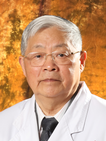 Zhengmin Wang (王正敏)合作论文数Eye & Ent Hospital of Fudan University;Institutes of Biomedical Sciences, Fudan University38
 Juanmei Yang (杨娟梅)合作论文数34
 Chunfu Dai (戴春富)合作论文数Eye & ENT Hospital of Fudan University30
 Yasheng Yuan合作论文数29Jing Wang (王璟)合作论文数24
 Ning Cong合作论文数23Yibo Huang合作论文数23Rui Ma (马锐)合作论文数19XinDa Xu (许信达)合作论文数16Na Gao合作论文数16Xian-Hao Jia合作论文数14Wenwei Luo合作论文数13Xiaoqing Qian合作论文数13Zhen Gao (高振)合作论文数13
 Tianyu Zhang (张天宇)合作论文数Eye and ENT Hospital,Fudan University,Shanghai ,China13Yongzhen Wu合作论文数12Bin-Jun Chen合作论文数11Ping Chen合作论文数11
 Yan Sha (沙炎)合作论文数Eye and Ent Hospital of Fudan University;Shanghai Institute of Medical Imaging, Shanghai Institute of Medical Imaging11
 Qin Liang (梁琴)合作论文数Eye&ENT Hospital to Fudan University10
 Huawei Li (李华伟)合作论文数The First Affiliated Hospital of Xiamen University;Department of Otorhinolaryngology, Eye & Ent Hospital of Fudan University10
 Bing Chen (陈兵)合作论文数复旦大学附属眼耳鼻喉科医院耳神经颅底外科10Xinwei Wang合作论文数9Yanmei Wang合作论文数Fudan Univ, State Key Lab Surface Phys, Shanghai 200433, Peoples R China9Jing Yu合作论文数8
 Hongjian Liu (刘宏建)合作论文数Henan Provincial People's Hospital8
 Houyong Kang (康厚墉)合作论文数重庆医科大学8
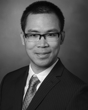 Yike Li合作论文数8
 Bin Wang (王斌)合作论文数复旦大学附属华山医院药剂科8Yu Zheng合作论文数7LiWen Wu (吴琍雯)合作论文数7Chao Han (韩朝)合作论文数7
 Xiaoyu Yang合作论文数7Jihan Lyu合作论文数6Yaoyao Fu合作论文数6Wen Li合作论文数6YanBo Yin (尹彦波)合作论文数6
 Peidong Dai (戴培东)合作论文数EYE and ENT Hospital of Fudan University6Liu Xiang合作论文数6Chen-Ru Ding合作论文数5Liang Tian合作论文数5JiangHua Jing (景江华)合作论文数5MingMin Dong (董明敏)合作论文数5LiMin Guo (郭丽敏)合作论文数5
 Shuyi Wang (王纾宜)合作论文数复旦大学附属眼耳鼻喉科医院5MeiQin Yang合作论文数5
 Aiguo Liu (刘爱国)合作论文数Tongji Hospital, Tongji Medical College of Huazhong University of Science and Technology5




