Conventional acoustoelectric imaging (AEI) is limited by the trade-off between field of view, scan efficiency, and reconstruction quality. This article presents a plane-wave acoustoelectric tomography (PW-AET) framework for sparse-angle acoustoelectric source imaging in a saline phantom. The plane-wave acquisition model is formulated and related to the Radon transform, providing the basis for tomographic reconstruction from multi-angle acoustoelectric measurements. To improve reconstruction quality under limited-angle sampling, linear back-projection (LBP) and the simultaneous algebraic reconstruction technique (SART) are used to generate physics-based initial estimates, with a simulation-trained U-Net then applied as a proof-of-concept image-domain post-processing module for sparse-angle artifact suppression. Broadband chirp excitation is further incorporated to improve demodulation robustness relative to short-pulse operation. Controlled phantom experiments demonstrate millimeter-scale source reconstruction, including dipole-pair discrimination and recovery of heterogeneous sources with distinct size scales. In dual-frequency source-emulation experiments, the framework further demonstrated frequency-specific temporal tracking, spectral discrimination, and fused spatial localization for two electrically injected sources at 7 Hz and 13 Hz. Relative to the reference focused-ultrasound scanning configuration used in this study, PW-AET provides broader instantaneous field coverage and improved task-specific source separability under the tested conditions. These results support PW-AET as a promising measurement framework for sparse-angle acoustoelectric source imaging under controlled phantom conditions and provide a basis for further development toward wider-field and faster acoustoelectric source-imaging systems.
Event-related potential (ERP)-based brain-computer interface (BCI) systems are approaching sub-microvolt-level resolution, enabling detailed decoding of sophisticated cognitive processes. This progress has increased the demand for robust classifiers. Current algorithms encounter two fundamental challenges when decoding ERPs: data scarcity and class imbalance. To address these challenges, we propose a joint-shrinkage pattern matching (JSPM) algorithm consisting of two modules. First, a novel joint-shrinkage spatial filter is constructed by integrating shrinkage-based regularization with the $\mathcal{{l}}_{2,{\bm{p}}}$-norm. This regularization approach effectively bridges the gap between complex structured regularization and implementation simplicity, which introduces automated regularization to enhance module robustness under data-scarce conditions. The $\mathcal{{l}}_{2,{\bm{p}}}$-norm provides a flexible feature distance measurement, enabling adaptation to data quality variability. Second, a weighted template matching module mitigates decision boundary shift caused by class imbalance. Using error-related potentials (ErrPs) as representative signals, we validated the algorithm through comprehensive comparisons. JSPM significantly outperformed 14 state-of-the-art classifiers on one self-collected and two public ErrP datasets. With only 40 imbalanced training samples, it achieved up to 14.84% higher average balanced accuracy (bAcc) than competing methods, maintaining a 4.88% average bAcc advantage over its nearest competitor. Notably, JSPM significantly enhanced inter-class discriminability for ErrP features with approximately 1 μV amplitude, achieving a maximum bAcc enhancement of 8.80% compared to deep learning methods. Overall, JSPM effectively addresses small-sample and imbalanced ERP decoding in BCI systems, facilitating the transition from laboratory research to real-world applications.
OBJECTIVE:Transcranial acoustoelectric brain imaging (tABI) combines focused ultrasound with electrical sensing for high-resolution current-density imaging. Its sensitivity and comparability depend on accurate measurement of the acoustoelectric (AE) interaction constant K, an intrinsic material property. Previous studies estimated K using equivalent current-source approximations under voltage-source stimulation, overlooking transient current variations. This study derives the AE amplitude-spacing relation and proposes a spacing-compensation method to eliminate spacing-induced bias, enabling quantitative estimation of current density. METHODS:Guided by finite-element modeling (FEM), a chamber with adjustable electrode spacing was constructed to ensure a uniform current density distribution. Under voltage-source conditions, AE data were acquired across multiple spacings, and a spacing-compensation curve was derived. RESULTS:With spacing compensation, K in saline stabilized at -0.024 ± 0.001%/MPa, and its coefficient of variation decreased from 44.69% to 4.41%. Application to ex vivo porcine brain tissue yielded the first reported K for biological brain: -0.022 ± 0.005%/MPa. FEM comparison showed strong agreement, with RMSEs of 9.57% for saline and 6.86% for brain tissue in the uniform current-density region. CONCLUSION:Spacing compensation corrects spacing-dependent bias in K under voltage-source stimulation, preserving its physical meaning and enabling accurate conversion of AEamplitude into local current density. SIGNIFICANCE:Accurate quantification of K enables reproducible and interpretable AE measurements, providing a quantitative foundation for current-density imaging and future applications in neural mapping and epilepsy localization.
Precise localization of deep-seated epileptic foci remains a critical challenge in preoperative assessment. Acoustoelectric (AE) imaging offers high spatial resolution and a potential route to noninvasive epileptic-source localization but remains limited by weak signal level and tissue inhomogeneity. This study aims to optimize an AE imaging system by investigating the measured AE frequency-dependent response and improving chirp-based demodulation. Specifically, we systematically characterize the measured frequency-dependent AE response over 2–4 MHz under a controlled AE measurement configuration, revealing a pronounced high-frequency roll-off. Guided by this finding, we systematically compared the performance of Tone Burst (TB) excitation with lock-in demodulation and chirp excitation with matched filtering. Furthermore, a response-corrected matched filtering strategy is proposed, incorporating the measured AE response function into the matching template to reduce spectral mismatch. Experimental results demonstrate that the chirp-based method significantly outperforms traditional TB excitation by simultaneously achieving a higher signal-to-noise ratio (SNR) and superior axial resolution. The proposed template correction further improved SNR by 1.95 dB. Validated in ex vivo porcine brain using simulated epileptiform dipoles (~500 nAm) driven by realistic ictal EEG waveforms, the system demonstrated consistent spatiotemporal imaging performance, yielding a mean localization error of 1.12 mm in cortical regions. Furthermore, it recovered the main millisecond-scale temporal pattern of the imposed ictal waveform with good temporal agreement (Global Pearson correlation r ≈ 0.79). These findings provide controlled experimental evidence for response-corrected AE weak-source imaging and support further development toward more realistic tissue and transcranial conditions.
Objective.Deep brain stimulation (DBS) is a technology employed to stimulate the central nervous system, with amplitude being the main parameter for regulating DBS. Given the effectiveness of DBS therapy depends significantly on lead placement and stimulus intensity, it is crucial to accurately map the lead field and monitor dynamic changes of stimulus amplitude. Transcranial acoustoelectric brain imaging (tABI) has been initially proved as a non-invasive method for mapping DBS currents. This study utilizes tABI to map amplitude-varying DBS currents to explore its potential for DBS monitoring.Approach.tABI was applied to six living rats' brain while DBS was delivered with 50 mV amplitude increments. The tABI images of amplitude-varying DBS currents are analyzed for spatiotemporal resolution, while the method's capability to decode DBS current is evaluated in terms of amplitude, frequency, and time domains.Main results.The results show that tABI can map the lead field of DBS with millivolt-level amplitude resolution and reveal dynamic changes with ∼2 mm spatial resolution within a single stimulus period of 7.69 ms, achieving a mean SNR of 20.4 dB. With sensitivity of 167.74μV V-1MPa-1, the acoustoelectric intensity and stimulus amplitude exhibit a strong positive correlation, with a coefficient of determination ofR2= 0.9982 for the linear fit. Additionally, the decoded acoustoelectric signal exhibits a correlation coefficient above 0.819 with the DBS current in the time domain.Significance.This study first demonstrates that tABI can reveal the spatial distribution and dynamic changes ofin vivoDBS lead currents with millivolt-level amplitude resolution. Further validation in disease-relevant animal models is warranted to assess clinical translatability.
Non-invasive neuroimaging has long faced the inherent trade-off between spatial and temporal resolution. Acoustoelectric brain imaging (ABI) holds promise to bridge this gap by combining the spatial precision of focused ultrasound (millimeter-level) with the temporal resolution of electroencephalography (EEG) signals (millisecond-level). However, its transcranial application remains fundamentally challenged by skull-induced wavefront aberration and attenuation. Here, we developed a full ABI system featuring a custom 128-element ultrasound phased array. Our system integrates developed algorithms for transcranial phase and amplitude correction, which were experimentally validated through an ex vivo human skull. We demonstrate that our system enables precise intracranial focus steering (lateral error ≤ 0.2 mm), accurate source localization (error ≤ 0.8 mm), and high-fidelity waveform reconstruction (correlation coefficient > 0.84). This work addresses the fundamental challenge of skull-induced imaging quality degradation in ABI, providing algorithmic and systemic foundations for advancing non-invasive neuroimaging techniques.
Transcranial focused ultrasound (tFUS) is a widely applied non-invasive neuromodulation technique known for high spatial resolution. However, traditional numerical simulations for tFUS are computationally intensive and time-consuming, creating a severe computational bottleneck for real-time treatment planning. To address this, we propose transcranial Focused Ultrasound Network (tFUSNet), a deep learning-based forward surrogate model for rapid acoustic field prediction. Utilizing a generative adversarial network framework, tFUSNet integrates an adversarial architecture with a multi-dimensional attention mechanism. The model was trained on simulations of 61 diverse human skulls and validated through ex vivo experiments using an independent real human skull and a 128-element phased array. Simulation results demonstrate that tFUSNet significantly outperforms traditional methods. In two-dimensional tasks, the model achieved a focal position error of 0.28 mm, a peak pressure ratio error of 7.08%, a Dice similarity coefficient of 88.21%. And in three-dimensional tasks, the model achieved a focal position error of 0.12 mm, a peak pressure ratio error of 4.37%, and a Dice similarity coefficient of 85.44%, demonstrating significantly superior performance. Experimental validation confirmed high consistency with physical measurements, showing a maximum focal localization error of 0.20 mm, an average peak deviation of 10.44%, and an average focal spot shape consistency of 75.92%. By accelerating forward acoustic evaluations from minutes to milliseconds, tFUSNet serves as a highly efficient forward surrogate conditioned on precomputed phase settings. This advancement eliminates the primary computational barrier, laying the essential groundwork for future closed-loop, real-time tFUS treatment planning systems.
Individual phase indexes in functional connectivity as biomarkers for assessing motor function in stroke patients can be affected by noise and volume conduction, resulting in unreliable and poorly correlated assessments. To explore whether multifunctional connectivity index fusion can effectively assess motor function in patients with chronic stroke. This study included 12 participants with chronic stroke and 12 healthy controls. EEG data from 14 channels near the primary motor cortex area (M1) was recorded. Fugl-Meyer scores (FMAU/FMAL) were assessed at admission. Then, correlations between PSI, PLI, WPLI differences, and FMAS between groups were analyzed. Cross-index and cross-band fusion based on validated biomarkers were performed to assess patients’ motor impairment. PSI, PLI, and WPLI of the patients with M1 were lower than those in the controls in the low-alpha. PSI and PLI were significantly correlated with FMAS and FMAU in low-alpha, and WPLI showed a strong correlation only with FMAU in low-alpha and high-beta bands. In the fusion assessment, PSI at low alpha and WPLI at low alpha showed a 13.7
It is well-established that aerobic exercise modulates mental workload (MWL) and enhances cognitive function. However, existing studies predominantly rely on indirect behavioral results, and direct neural evidence is insufficient. Therefore, it is essential to investigate how exercise modulates neural activity during cognitive tasks. In recent years, electroencephalogram (EEG) microstate analysis has emerged as a critical tool for investigating the spatiotemporal dynamics of brain electrophysiological activity. The four canonical microstate classes have been extensively studied, and their parameters are proven to correlate with MWL. In this study, participants performed N-back tasks with two MWL levels (low: 1-back; high: 3-back) following two counterbalanced exercise conditions: aerobic exercise (ES) and rest (RS). Behavioral and EEG data from 49 participants were collected during the tasks. The typical microstate parameters (Occurrences/OCC, Time Coverage/TC and Mean Duration/MD) and transition characteristics were compared. The results showed that high MWL levels increased MD of class C but decreased OCC of class D. Exercise significantly improved subjective perception of MWL during tasks and promoted transitions to class D, increasing its TC and MD. These findings suggest that exercise enhance cognitive-related microstate dynamics potentially, thereby mitigating MWL, given that class D has been associated with activations in parietal and frontal cortical regions. These findings of this study provide further insights into the potential neural mechanisms by which aerobic exercise modulates MWL.
Transcranial focused ultrasound (tFUS) technology achieves precise stimulation or treatment of the area of interest in the head by directing ultrasound beams to penetrate the human skull to form an intracranial focal point, with the advantages of eliminating the need for craniotomy and the absence of ionizing radiation. High-intensity tFUS treats brain diseases such as essential tremor or brain tumors through thermal effects, while low-intensity tFUS can safely and reversibly open the blood-brain barrier or conduct neuromodulation studies through mechanical effects. However, in practical applications, ultrasound waves undergo strong phase distortion and energy attenuation due to the strong acoustic attenuation properties and inhomogeneous structure of the skull. Acoustic simulation models the interaction between ultrasound and media based on acoustic fluctuation equations to predict the propagation properties of sound waves in different media. Therefore, acoustic simulation is commonly used to predict the intracranial acoustic field for single-element tFUS or to perform phase correction for each element of multi-element tFUS to ensure accurate focusing of intracranial ultrasound. According to the different methods of solving the acoustic fluctuation equations, the commonly used acoustic simulation methods in tFUS can be categorized into numerical and semi-analytical methods. The numerical methods include k-space pseudo-spectral method, time-domain finite difference method and finite element method, etc., and the semi- analytical methods include ray-tracing method and hybrid angular spectrum method. Simulation tools based on numerical methods synthesize various forms of wave propagation in media, such as nonlinear effects, scattering and diffraction, and are widely used in academic research. The k-Wave toolbox based on the k-space pseudo- spectral method and various programs based on the time-domain finite-difference method are the most widely used simulation tools in the current tFUS accurate simulation and experimental research. Although the finite element method has the advantage of dealing with complex boundary conditions, the excessive consumption of computational resources limits its direct application in complex 3D simulations. Compared to numerical methods, semi-analytical-based simulations cannot accurately model full-wave effects, but their computational speed makes them more suitable for clinical scenarios where simulation time is critical. Ray-tracing, developed by Insightec, is currently the only phase-correction method that has been used in clinical applications. Based on geometric acoustic principles, ray tracing enables near real-time tFUS phase correction. At the same time, the hybrid angular spectroscopy method shows higher accuracy in precise targeting than the conventional ray tracing method. In addition, the hybrid application of different simulation methods significantly improves the simulation efficiency and accuracy, e. g., the boundary element method can be coupled with the finite element method to limit the computational area to the region involving only the skull, which drastically reduces the computational load. In recent years, the acoustic simulation for tFUS has continued to make progress, but there is still a huge room for improvement in terms of computational efficiency and accuracy, and the optimal use of computational resources and the combination of multiple simulation techniques may be the direction of the future development of simulation technology. In this paper, the research on simulation techniques based on numerical, semi-analytical and hybrid methods commonly used in the field of tFUS in recent years is reviewed and sorted out, and the research and application of various simulation methods are summarized and prospected.
A 12-month cluster randomized controlled trial (RCT) demonstrated the effectiveness of an application-based education program in reducing the salt intake and systolic blood pressure (SBP) of schoolchildren’s adult family members. This study aimed to assess whether the effect at 12 months persisted at 24 months. Fifty-four schools were randomly assigned to either the intervention or control group. All participants (594 children in grade 3 and 1188 of their adult family members) who completed the baseline survey were contacted again 12 months after the trial. The primary outcome was the difference in salt intake change between the intervention and control groups at 24 months versus baseline and 12 months, measured by the mean two consecutive 24-h urinary sodium excretions. The secondary outcome was the difference in the change of blood pressure and salt-related Knowledge, Attitude, Practice (KAP) score. The difference in salt intake change in adults between the intervention and control groups after adjusting for confounding factors was − 0.38 g/day at 24 months versus baseline (95
Objective Electroencephalography (EEG) serves as a non-invasive electrophysiological monitoring technique employed to record brain electrical activity. Nonetheless, traditional EEG electrodes are susceptible to reference activation influences and exhibit limited spatial resolution. Laplacian electrodes, devoid of reference dependencies, possess the potential to amplify the spatial resolution of EEG recordings. Anchored in the utilization of bipolar concentric ring Laplacian electrodes, this study delves into the autonomous referencing attributes intrinsic to Laplacian electrodes. Furthermore, it conducts a comparison of spatial resolution disparities between Laplacian electrodes and their conventional counterparts. Methods A three-dimensional (3D) hemispherical tank experiment was conducted utilizing 21 Ag/AgCl bipolar concentric ring Laplacian electrodes to simulate whole-brain signal acquisitions. A sinusoidal signal with an amplitude of 400 mVpp@13 Hz was employed for detection. The positions of the ground electrodes in the Laplacian electrode array were varied, alongside the reference electrode positions in the case of the traditional electrodes. Subsequently, the spatial distribution of the 13 Hz source frequency component was extracted and subjected to comprehensive analysis. Results With varying ground electrode positions, the spatial distribution of the signal-to-noise ratio (SNR) among Laplacian electrodes maintains remarkable consistency, yielding a correlation coefficient of 0.94. In contrast, for traditional electrodes, the correlation coefficient for SNR distribution under distinct reference electrode positions barely reaches 0.07. While Laplacian electrodes exhibit independence from reference electrodes, traditional counterparts display a notable susceptibility to changes in reference electrode positions. Comparing amplitude's 3 dB attenuation area ratio, Laplacian electrodes showcase a mere 2.1% reduction, a significantly favorable outcome when juxtaposed with the 6.9% reduction evident in traditional electrodes. Similarly, the SNR's 3 dB attenuation area ratio for Laplacian electrodes is a mere 1.0%, contrasting with the considerably higher figure of 30.1% for traditional electrodes. Conclusion Laplacian electrodes remain impervious to reference electrode influence, displaying distinctive reference-independent attributes, in addition to boasting a heightened spatial resolution. These characteristics imbue them with the capacity to achieve heightened precision in localizing brain electrical activities, thus constituting a cornerstone for the integration of Laplacian electrodes into brain-computer interfaces (BCIs).
Motor imagery (MI) is a promising motor training method that activates the same cortical regions involved in motor execution. However, its clinical application remains limited for unpredictable outcomes. To enhance the motor modulation of MI, we applied 5 Hz repetitive transcranial magnetic stimulation (rTMS) to the right primary motor cortex (M1), synchronized with left-hand grasping MI training. The control session applied sham rTMS. TMS-evoked electromyography (TMS-EMG) and electroencephalography (TMS-EEG) data were collected pre- and post- training, offering insights into neurological effects of MI-rTMS. The results revealed a significant increase in the peak-to-peak value (PPV) of motor evoked potentials (MEP) (p = 0.017) and the N15-P30 complex of TMS-evoked potentials (TEP) (p = 0.044), supporting the positive effects of MI-rTMS. The N100 of TEP from the active group indicated bilateral suppression of GABAB-mediated inhibition (ROI2: p = 0.029, ROI5: p = 0.007). Correlations between MEP PPV and affected M1 N100 (r = -0.473, p = 0.013), as well as between MEP PPV and contralateral P180 (r = 0.314, p = 0.018), affirmed positive motor effects in both groups. Moreover, MI-rTMS increased right-to-left interhemispheric partial directed coherence (PDC) outflows (p = 0.044), suggesting bilateral recruitment of sensorimotor cortical activity. As ROI1 N45 exhibited a positive correlation with right-to-left PDC (r = 0.378, p = 0.004) and a negative correlation with MEP terminal-included duration (r = -0.376, p =0.004), activated GABAA-mediated inhibition in the contralateral hemisphere may enhance MEP efficacy. In conclusion, MI-rTMS improves MI motor outcomes and promotes bilateral sensorimotor cortical activation.
Sudden cardiac death (SCD) remains one of the leading causes of mortality worldwide, with coronary artery disease (CAD) as its predominant underlying condition. However, noninvasive and accessible screening approaches for CAD are still limited. This study aims to develop and evaluate a photoplethysmography (PPG)-based method for CAD detection using a two-dimensional Gramian angular field (GAF) transformation combined with deep learning. We enrolled 89 patients with CAD and 70 healthy controls and converted their PPG signals into two GAF representations—Gramian angular summation field (GASF) and Gramian angular difference field. The GASF representation, which preserves both magnitude and phase relationships within the PPG waveform, was found to provide superior discriminative capability. Using GASF as input, the proposed SE-ResNet model achieved an accuracy of 92.43% (95% CI: 91.51–93.36), outperforming prior work that reported 83.8% accuracy (95% CI: 82.2–85.3). These results demonstrate that the GAF transformation enhances CAD detection by encoding the temporal–phase dynamics of PPG signals, which are often overlooked in conventional one-dimensional analyses. The proposed GASF-SE-ResNet framework therefore shows strong potential as a noninvasive low-cost tool for CAD screening and SCD risk reduction.
The bimodal balance-recovery model suggests that intervention strategies should differ based on the competition or vicariation model. Current training methods are predominantly influenced by the competition model, resulting in limited benefits for motor rehabilitation. Transcranial magnetic stimulation (TMS) has the capacity to modulate neural plasticity. Motor imagery (MI) activates the same cortical areas as motor execution. Therefore, we applied 5 Hz repetitive magnetic stimulation (rTMS) synchronously with MI to investigate the combination effects. This study recruited 14 healthy subjects. 5 Hz rTMS was applied on right motor cortex, and MI was left grasping imagery. TMS-evoked electromyography (TMS-EMG) and electroencephalography (TMS-EEG) data were recorded before and after training to assess the effects. The results demonstrated a significant increase in the peak-to-peak value (PPV) of motor evoked potential (MEP) (p = 0.017) and a significant increase in the N15-P30 complex of TMS-evoked potentials (TEP) (p = 0.044) after MI-rTMS training, suggesting the enhancement in motor function. Furthermore, the N100 results of the active group exhibited bilateral suppression of GABAB-mediated inhibition (ROI2: p = 0.029, ROI5: p = 0.007), providing theoretical support for its application under the vicariation model. Correlations between MEP PPV and affected M1 N100 (r = -0.473, p = 0.013), as well as between MEP PPV and contralateral P180 (r = 0.314, p = 0.018), confirmed positive motor effects in both groups. Therefore, MI alone shows the lateralization intervention and MI-rTMS shows bilateral activation. This synchronous integration of rTMS and MI provides a novel motor training method tailored for the vicariation model of motor rehabilitation.Clinical Relevance— This synchronous integration of rTMS and MI provides a novel motor training method tailored for vicariation model of motor rehabilitation.
Deep brain stimulation (DBS) is a well-established treatment for both neurological and psychiatric disorders. Directional DBS has the potential to minimize stimulation-induced side effects and maximize clinical benefits. Many new directional leads, stimulation patterns and programming strategies have been developed in recent years. Therefore, it is necessary to review new progress in directional DBS. This paper summarizes progress for directional DBS from the perspective of directional DBS leads, stimulation patterns, and programming strategies which are three key elements of DBS systems. Directional DBS leads are reviewed in electrode design and volume of tissue activated visualization strategies. Stimulation patterns are reviewed in stimulation parameters and advances in stimulation patterns. Programming strategies are reviewed in computational modeling, monopolar review, direction indicators and adaptive DBS. This review will provide a comprehensive overview of primary directional DBS leads, stimulation patterns and programming strategies, making it helpful for those who are developing DBS systems.
High spatiotemporal resolution of noninvasive electroencephalography (EEG) signals is an important prerequisite for fine brain-computer manipulation. However, conventional scalp EEG has a low spatial resolution due to the volume conductor effect, making it difficult to accurately identify the intent of brain-computer manipulation. In recent years, transcranial focused ultrasound modulated EEG technology has increasingly become a research hotspot, which is expected to acquire noninvasive acoustoelectric coupling signals with a high spatial and temporal resolution. In view of this, this study established a transcranial focused ultrasound numerical simulation model and experimental platform based on a real brain model and a 128-array phased array, further constructed a 3-dimensional transcranial multisource dipole localization and decoding numerical simulation model and experimental platform based on the acoustic field platform, and developed a high-precision localization and decoding algorithm. The results show that the simulation-guided phased-array acoustic field experimental platform can achieve accurate focusing in both pure water and transcranial conditions within a safe threshold, with a modulation range of 10 mm, and the focal acoustic pressure can be enhanced by more than 200% compared with that of transducer self-focusing. In terms of dipole localization decoding results, the proposed algorithm in this study has a localization signal-to-noise ratio of 24.18 dB, which is 50.59% higher than that of the traditional algorithm, and the source signal decoding accuracy is greater than 0.85. This study provides a reliable experimental basis and technical support for high-spatiotemporal-resolution noninvasive EEG signal acquisition and precise brain-computer manipulation.
The steady-state visual evoked potential-based brain-computer interface (SSVEP-BCI) has gained considerable attention due to its high information transfer rate (ITR) and stable performance. However, the comfort of SSVEP-BCI still needs to be improved, as strong flickering stimuli cause users’ visual fatigue. Reducing the pixel density of the stimuli has been demonstrated as an effective method to improve its comfort. However, the signal-to-noise rate (SNR) of the SSVEP signal induced by such very weak stimuli is low, posing challenges for their decoding. Therefore, it is necessary to develop suitable strategy for better decoding the SSVEP induced by very weak stimuli. This study employed the source aliasing matrix estimation (SAME) method to enlarge the dataset and improve decoding accuracy for SSVEP induced by low-pixel density stimuli. Additionally, this study further optimized the SAME with a regularization method to achieve much higher decoding performance. A SSVEP experiment was designed with various pixel densities (100%, 90%, 80%, 70%, 60%, 50%, 40%, 30%, 20%, 10% and 1%) and frequencies (low: 7Hz, 11Hz, and 15Hz; mid-to-high: 23Hz, 31Hz, and 39Hz) to verify our methods. The results indicated SAME significantly improved the classification accuracy compared to traditional method without the SAME, especially under very weak stimulation conditions (pixel densities ≤ 50%), with the maximum increase reaching 8.6%. Besides, regularization SAME further yielded a significant enhancement, achieved maximum improvements of 4.29% compared to SAME. The regularization SAME proposed in this study significantly improves SSVEP decoding performance under low-pixel density stimuli, paving the way for the development of comfortable and effective SSVEP-BCI.
Deep brain stimulation (DBS) is a well-established neural regulation therapy whose clinical efficacy is highly dependent on the DBS current field distribution. There remains an urgent need of directly detecting DBS current field to guide the lead direction and the subsequent regulation. As an emerging neuroimaging method, acoustoelectric brain imaging (AEBI) can directly map current density distribution which provides a promising approach for noninvasively detecting DBS current field. To further promote AEBI into a specific DBS therapy, this study applies acoustoelectric signals to detect DBS current field distribution towards coma arousal targets. With a DBS current applied to the coma arousal target, the AEBI experiment is implemented on a living rat brain to detect the DBS current field. The results show that the DBS current field can be mapped by acoustoelectric (AE) signals with ~1.5 mm spatial resolution. The instant AEBI images, during one period of 7.69 ms, can describe the dynamic activation pattern of the DBS stimulus current. Besides, the DBS current field mapped by SNR values of decoded AE signals at DBS frequency closely matches the AEBI image(r=0.85). This study confirms the ability of AE signals to noninvasively detect DBS current field distribution of coma arousal target. AEBI is expected to develop into a noninvasive DBS current real-time monitoring technique.
Affective body expression recognition technology enables machines to interpret non-verbal emotional signals from human movements, which is crucial for facilitating natural and empathetic human-machine interaction (HCI). This work proposes a new framework for emotion recognition from body movements, providing a universal and effective solution for decoding the temporal-spatial mapping between emotions and body expressions. Compared with previous studies, our approach extracted interpretable temporal and spatial features by constructing a body expression energy model (BEEM) and a multi-input symmetric positive definite matrix network (MSPDnet). In particular, the temporal features extracted from the BEEM reveal the energy distribution, dynamical complexity, and frequency activity of the body expression under different emotions, while the spatial features obtained by MSPDnet capture the spatial Riemannian properties between body joints. Furthermore, this paper introduces an attentional temporal-spatial feature fusion (ATSFF) algorithm to adaptively fuse temporal and spatial features with different semantics and scales, significantly improving the discriminability and generalizability of the fused features. The proposed method achieves recognition accuracies over 90% across four public datasets, outperforming most state-of-the-art approaches.
 Dong Ming (明东)合作论文数Medical School, Tianjin University127
 Graham A MacGregor合作论文数Wolfson Institute of Population Health, Queen Mary University of London115
 Zhiqi Hong (洪之奇)合作论文数Zhejiang University72
 Minpeng Xu (许敏鹏)合作论文数Tianjin University57
 Shuang Liu (刘爽)合作论文数Medical School of Tianjin University37
 Peng Zhou (周鹏)合作论文数School of Medicine, Tianjin University;Department of Biomedical Engineering, School of Precision Instrument and Opto-electronics Engineering, Tianjin University;Hebei University of Technology32
 Zhongpeng Wang (王仲朋)合作论文数Academy of Medical Engineering and Translational Medicine, Tianjin University30
 Long Chen (陈龙)合作论文数Medical School of Tianjin University29
 Lixin Zhang (张力新)合作论文数School of Precision Instrument and Optoelectronics Engineering, Tianjin University22
 Monique Tan合作论文数Wolfson Inst Populat Hlth, Queen Mary Univ London20
 Campbell Norm R C合作论文数20
 Yijie Zhou (周伊婕)合作论文数Medical School of Tianjin University17
 Puhong Zhang (张普洪)合作论文数George Inst Global Hlth, Peking Univ17Yuan Li (李园)合作论文数16
 Yufeng Ke (柯余峰)合作论文数Medical School of Tianjin University16
 Xizi Song (宋西姊)合作论文数Medical School of Tianjin University;Department of Graduate, Tianjin University of Sport15
 Weibo Yi合作论文数15Bin Gu合作论文数12Bokun Wan (万柏坤)合作论文数Tianjin University12
 Yuan Ma合作论文数12
 Bruce Neal合作论文数School of Biomedical Sciences, University of New South Wales;Royal Prince Alfred Hospital;Imperial College London;The George Institute for Global Health,University of Sydney11
 Xin Zhao (赵欣)合作论文数School of Mechanical Engineering, Tianjin University11
 Kun Wang (王昆)合作论文数School of Mechanical Engineering, Tianjin University11Caryl Nowson合作论文数11
 Jiayuan Meng (孟嘉源)合作论文数University of Virginia11
 Jing Song (宋 菁)合作论文数Wolfson Inst Populat Hlth, Queen Mary Univ London10
 Carley A Grimes合作论文数School of Exercise and Nutrition Sciences, Deakin University10Hashem Kawther M合作论文数Wolfson Institute of Preventive Medicine, Queen Mary University of London10
 Rudi Balling合作论文数Institute of Molecular Psychiatry, University of Bonn;Luxembourg Centre for Systems Biomedicine, University of Luxembourg10GA MACGREGOR合作论文数10
 Yangfeng Wu (武阳丰)合作论文数Department of Epidemiology and Biostatistics, School of Public Health, Peking University;Peking University Clinical Research Institute10Rong Luo合作论文数9
 Jacqui Webster合作论文数The George Institute for Global Health, The University of Sydney9
 Jiajia Yang (杨佳佳)合作论文数Medical School of Tianjin University9Suckling Rebecca J合作论文数9
 Francesco P Cappuccio合作论文数WHO Collaborating Centre for Nutrition, Warwick Medical School, University of Warwick;European Centre of Excellence in Hypertension and Cardio-Metabolic Research, Warwick Medical School, University of Warwick9
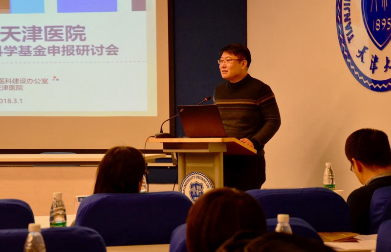 Bai-Kun Wan (万柏坤)合作论文数School of Precision Instrument and Opto-Electronics Engineering,Tianjin University9Changqiong Wang合作论文数8Li Xian合作论文数8
 Rui Xu (徐瑞)合作论文数Medical School of Tianjin University8
 Mark Woodward合作论文数Professorial Advisory Unit, The George Institute for Global Health;Faculty of Medicine, University of New South Wales, Sydney;Medical Sciences Division, University of Oxford8
 Hao Zhang (张浩)合作论文数School of Precision Instrument and Opto-Electronics Engineering, Tianjin University8
 Xingwei An (安兴伟)合作论文数Medical School, Tianjin University8
 Mhairi Brown合作论文数7XueJun Jiao (焦学军)合作论文数7
 Jiapeng Xu (徐加鹏)合作论文数Department of Automation, School of Electrical and Information Engineering, Tianjin University7
 Trieu Kathy合作论文数World Health Organization Collaborating Centre on Population Salt Reduction, The George Institute for Global Health7
 Lin Meng (孟琳)合作论文数Medical College, Tianjin University7




