BACKGROUND:Conventional envelope flap techniques for implant placement in edentulous mandibles are associated with greater postoperative morbidity, especially in elderly patients or those with anatomical limitations. Minimally invasive alternatives aim to preserve soft tissues and reduce complications, enhancing clinical predictability. METHODS:A clinical study was conducted with ten fully edentulous patients, in whom 20 regular implants were placed in the interforaminal region without surgical guides. A modified Neumann incision with a mesial relaxing cut and partial-thickness "L"-shaped flap was used. Digital planning was performed using free software. Surgical time, number of anesthetic tubes, adverse events, flap healing, peri-implant inflammation (PI), gingival bleeding index (GBI), and probing depth (PD) were evaluated at 14, 30, 60, and 90 days postoperatively. RESULTS:No surgical complications, such as dehiscence, infection, paresthesia, or early implant loss, were observed. All patients achieved primary wound healing. The mean surgical time was 44 min (range: 25-61 min), with an average of four anesthetic tubes per patient. GBI and PD remained low throughout follow-up, with few sites showing PD ≥ 4 mm. Healing scores and inflammatory parameters progressively improved over time. CONCLUSIONS:The modified Neumann incision with mesial relaxing cut, guided by digital planning, proved to be a safe, predictable, and minimally invasive approach for implant placement in edentulous mandibles. This technique demonstrated favorable healing, reduced surgical time, and effective PI control, making it suitable for patients with systemic fragility or anatomical challenges. KEY POINTS:The modified Neumann incision with mesial relaxing cut allows for safe and direct visualization of the mental foramen with limited flap elevation, reducing surgical trauma and preserving soft tissue integrity. This technique demonstrated favorable healing outcomes and low postoperative morbidity, even in elderly patients and anatomically challenging mandibles, without the need for physical surgical guides. Digital preoperative planning using free software enhances precision and predictability, making the approach reproducible and applicable in daily clinical practice. PLAIN LANGUAGE SUMMARY:This study describes a new, less invasive surgical technique for placing dental implants in the lower jaw of patients who have lost all their teeth. The method, called the modified Neumann incision with a mesial relaxing cut, was tested in 10 patients who received 2 implants to support a mandibular overdenture. The technique allows the surgeon to see important anatomical structures, such as nerves, while reducing the amount of tissue that needs to be cut or lifted. Patients were followed for 3 months after surgery, and the healing process, inflammation, and peri-implant health were monitored. The results showed that all patients healed well, with no significant complications or pain, and the soft tissues around the implants remained healthy. The approach proved to be safe, efficient, and well tolerated, particularly for older adults and individuals with health conditions that increase surgical risks. This technique may help make implant treatments more comfortable and predictable for edentulous patients, although larger studies are still needed to confirm these findings.
OBJECTIVE:Medication-related osteonecrosis of the jaw (MRONJ) is a serious complication associated with antiresorptive, and antiangiogenic therapies commonly used to treat osteoporosis, malignant bone tumors, and metastatic diseases. This review provides a comprehensive overview of the pathophysiology, clinical presentation, diagnosis, prevention, and management of MRONJ. METHODS:This narrative review examined the prevalence of MRONJ and its clinical and radiological features, particularly those relevant to disease staging, and outlined current preventive and treatment strategies, aiming to improve patient outcomes and quality of life. RESULTS:Patients with cancer receiving high-dose intravenous antiresorptive medications exhibited a higher incidence of MRONJ than that observed in patients treated for osteoporosis. Clinical staging systems, especially those developed by the American Association of Oral and Maxillofacial Surgeons, facilitate disease classification based on clinical and imaging findings, thereby guiding treatment selection and prognosis assessment. Preventive approaches, conservative management, surgical procedures, and adjunct therapies have been proposed to promote healing of both hard and soft tissues. CONCLUSION:Although emerging therapeutic options are promising, prevention and personalized treatment remain major challenges.
Accurate analysis of the bone bed is essential for surgical planning in implant dentistry and for understanding the relationship between bone parameters and primary implant stability. However, characterization of the bone bed after implant placement still presents methodological limitations, particularly in retrospective studies. This study presents a novel methodology of image superimposition between two cone-beam computed tomography (CBCT) acquisitions that retrospectively enables localization of the exact tomographic slice corresponding to the three-dimensional position of the placed implant, thus allowing detailed characterization of the bone bed. Pre- and postoperative CBCT scans of patients who received implants in the interforaminal region of the mandible were used. The Blue Sky Plan software was employed for segmentation of the mandible and implants, as well as for image superimposition based on stable anatomical landmarks. This methodology enabled direct measurements of bone thickness, implant position relative to the cortical plates, and evaluation of the buccal cortical bone. All measurements on CBCT images were performed by a single calibrated examiner, in duplicate, with a one-month interval between assessments. The intraclass correlation coefficient (ICC) was applied, and values equal to or greater than 0.80 were considered acceptable. The proposed superimposition method demonstrated high accuracy and reproducibility, allowing objective assessment of the bone bed even after implant placement. This methodology is free, accessible, and applicable to diverse clinical and research contexts, as well as adaptable to different areas of oral and maxillofacial surgery. The technique represents an innovative tool for retrospective analyses in implant dentistry, with potential to improve methodological quality and optimize digital planning protocols.
This randomized clinical trial (RCT) aimed to evaluate clinical, radiographic, functional, and patient-centered outcomes in users of mandibular two-implant overdentures (2-IMO) subjected to immediate loading (IL) and conventional loading (CL) protocols over a five-year follow-up period. Twenty participants were randomly allocated to the CL (n = 10) and IL (n = 10) groups. Clinical peri-implant health parameters, radiographic assessments of marginal bone loss (MBL) and posterior area index (PAI), functional outcomes through masticatory performance (MP), patient-reported outcomes using the Daily Impact on Daily Life (DIDL) questionnaire, and prosthetic maintenance events were evaluated at 1, 3, and 5 years post-treatment. Data were analyzed using multilevel mixed-effects linear regression models, and differences in prosthetic maintenance events over the 5-year follow-up were assessed using binary logistic regression, with the significance level set at 5
Peri-implantitis poses a persistent challenge in implant dentistry, driving interest in laser therapy as a potential treatment option. Despite encouraging outcomes, clinical applications of laser therapy differ significantly in terms of wavelength, power setting and session frequency, hindering the development of standardized protocols. This scoping review aimed to map and synthesize current clinical evidence on the efficacy of laser therapy in peri-implantitis management, identify knowledge gaps and provide a foundation for future clinical recommendations. Following the Preferred Reporting Items for Systematic reviews and Meta-Analyses extension for Scoping Reviews (PRISMA-ScR) and Joanna Briggs Institute (JBI) guidelines, a comprehensive search was conducted across 5 databases (Scopus, PubMed, Cochrane Library, Embase, and Web of Science) between May and July 2024, covering studies published from 2000 to 2024, with no language restrictions. Two independent reviewers extracted data with high inter-rater agreement (κ = 0.97). A total of 98 clinical studies were included: 56 randomized controlled trials (RCTs), 38 cohort studies and 4 retrospective studies. Diode lasers were the most frequently studied (n = 50), followed by Er:YAG, aPDT, Nd:YAG, and Er,Cr:YSGG lasers. Exposure times ranged from 10 s to 700 s, most commonly around 60 s. Key clinical outcomes included probing depth (PD) reduction, bleeding on probing (BoP) and plaque index (PI), with additional outcomes related to bone loss, clinical attachment level (CAL), gingival recession (REC), cytokine levels, microbial analysis, suppuration, and gingival index (GI). Overall, laser therapy was associated with reduced inflammation, accelerated epithelialization, improved bone parameters, fewer complications, and better patient-reported outcomes. While laser therapy shows considerable promise in the treatment of peri-implantitis, further robust and standardized clinical research is essential to confirm its efficacy, optimize treatment parameters and inform evidence-based clinical guidelines.
OBJECTIVES:The goal of this systematic review was to critically appraise the existing evidence evaluating osteoporosis' effects on dental implant osseointegration and survival rate. DATA SOURCE:A search was conducted in two databases, PubMed/MEDLINE and Scopus, until October 2024, using the keywords 'osteoporosis,' 'osteopenia,' 'osseointegration,' and 'dental implants'. The inclusion criteria were clinical studies that evaluated the implant placement, complications, and osseointegration results in patients with osteoporosis; literature reviews and clinical studies addressing the outcome were considered; and articles written in English and published since 2000. Descriptive data analysis included author, year of publication, study design, number of patients, osteoporosis assessment, follow-up, and main findings. JBI quality assessment was performed. Overall, 24 articles were included with a total of 2,102 patients; 5,954 dental implants were considered and evaluated. Most studies evaluated bone density for osteoporosis by dual-energy x-ray technology. The follow-up ranged from 1 month to 25 years. Four studies evaluated implants with over 10 years of follow-up. All studies' survival rate was higher than 90%, even for osteoporotic patients. Most studies indicated no differences between osteoporotic and healthy patients regarding marginal bone loss, bone-to-implant contact, cytokine levels, and mineral bone density. A prospective cohort study found a small marginal bone loss (-0.34 mm) in osteoporotic women, but there was insufficient evidence to prove any causal relationship between marginal bone loss and osteoporosis. Another study showed no clinical differences between implants placed in osteoporotic and healthy individuals. In contrast, other studies showed lower stability scores for implants placed in osteoporotic sites and a higher risk of failure for implant placement. Osteoporosis status was not a risk factor for dental implant failure, which was also confirmed by histologic studies. Three studies had a medium risk of bias, and 21 a low risk. CONCLUSION:Osteoporosis is not a contraindication for dental implant placement. Osseointegration in patients with osteoporosis is feasible; however, planning must be cautious and personalized for the placement of dental implants.
The global increase in the older population has led to a rise in untreated oral conditions such as edentulism. Oral rehabilitation for edentulous patients is essential to mitigate systemic health risks, including the progression of sarcopenia. This study aimed to investigate the association between age, sex, general health status, type of oral rehabilitation, sarcopenia indicators, bite force and masticatory function in individuals aged 70 years and older. This cross-sectional study assessed the masticatory function of 52 edentulous individuals aged 70 and older. Masticatory function was evaluated using the swallowing threshold test, with outcomes measured as X50 (median particle size) and B (particle homogenization). Exposure variables included age, sex, type of rehabilitation, general health status, sarcopenia indicators, and bite force. Statistical analyses were performed using Stata 14.1 software. Continuous variables (bite force and B) were tested for normality using the Kolmogorov-Smirnov test, and cutoff values were based on the mean. Other variables were dichotomized based on cutoff points established in the literature. Multivariate logistic regression with stepwise selection was performed to calculate odds ratios (OR) and 95
Objective: This longitudinal clinical study monitored annually the maxillary and mandibular bone remodeling and masticatory function in complete denture (CD) wearers rehabilitated with implant-retained mandibular overdentures (MO) over three years and combined radiographic and masticatory function data to assess the correlation between bone remodeling and masticatory function. Design: Thirty-nine MO wearers were monitored annually to assess changes in: i) residual ridge in the anterior and posterior maxillary region; ii) posterior height and posterior area index (PAI) in the mandible; and iii) masticatory function. Bone remodeling was measured through linear and angular measurements using panoramic radiographs. Masticatory function was evaluated by swallowing threshold test, to determine the median particle size (X50) and homogenization index (B). A mixed-effects multilevel regression model estimated differences between radiological and masticatory parameters over time, with associations verified using Pearson's correlation test. Results: Significant changes in mandibular bone variables (PAI and L1/R1, L2/R2, L3/R3; p <= 0.01) occurred when transitioning from CD to MO, showing a reduction in PAI values and slight reduction in mandibular linear measurements. Improvements in masticatory parameters were also observed, highlighting the benefits of transitioning to MO. Over three years of MO usage, significant changes in all radiographic bone parameters were found, with increased PAI and maxillary bone values in the first year. The correlations between masticatory function and radiological parameters indicated that mandibular morphology can significantly influence masticatory function especially in patients wearing CDs. Following the transition to MO, only one consistent significant correlation was observed between X50 and R3/L3 at the 1-year and 3-year intervals. Conclusion: Continuous changes in bone parameters were observed in MO wearers, with stabilization of maxillary ridge resorption and ongoing mandibular remodeling, alongside slight changes in masticatory function, suggesting transitory correlations between linear changes in the posterior region of mandible and masticatory function over time.
PURPOSE:This longitudinal clinical study aimed to investigate the circumferential peri‑implant bone level (CPBL), posterior mandibular resorption (PMR), and masticatory function (MF) in users of implant-retained mandibular overdentures (IMOs), comparing individuals with and without combination syndrome (CS) over a 5-year follow-up period. MATERIALS AND METHODS:A total of 39 patients were enrolled and stratified into two groups: absence of CS (ACS; n = 23) and presence of CS (PCS; n = 16). Assessments were conducted at 1, 3, and 5 years. CPBL and PMR were measured using cone beam computed tomography. PMR parameters included total bone height, cortical and medullary bone height, percentages of cortical and medullary bone and bone density at 4 standardized mandibular regions. MF was assessed via masticatory performance (MP) and swallowing threshold (ST) tests. RESULTS:The PCS group showed a significant reduction in CPBL over time (p ≤ 0.05), particularly at the mesial and distal surfaces, with average reductions of -1.09 mm (mesial) and -0.70 mm (distal) after 5 years. MF was also significantly impaired in the PCS group at 5 years, demonstrated by reduced bolus homogenization in both tests, MP and ST tests; prolonged masticatory cycles, and inferior performance in the ST (p ≤ 0.05). In contrast, the ACS group showed significantly greater PMR values (p ≤ 0.05), with increased total and medullary bone heights and higher medullary bone percentages. The ACS group also demonstrated lower cortical bone percentages at L1 and L2 distances. CONCLUSIONS:The presence of CS is associated with accelerated peri‑implant bone loss and deterioration in masticatory function over time. Conversely, greater mandibular resorption was observed in the absence of CS, particularly affecting the medullary compartment. These findings underscore the influence of CS on both structural bone remodeling and functional outcomes in IMO users. CLINICAL SIGNIFICANCE:Patients with CS would require stricter prosthetic maintenance protocols due to the impact of anterior hyperfunction. This functional overload accelerates peri‑implant bone resorption in the anterior region of mandible and can lead to maladaptive changes in the posterior mandible. As posterior occlusal support diminishes, masticatory function deteriorates, reinforcing the need for early intervention and ongoing monitoring to preserve oral function and implant stability.
OBJECTIVES:This prospective longitudinal case series investigated changes in functional, radiographic, and patient-centered outcomes in users of two narrow-diameter implant-retained mandibular overdentures (IMO) over a 5-year follow-up. MATERIALS AND METHODS:Twenty-four patients from a previous 3-year study (8 man, 16 women, 71.58 ± 7.03 years, mean mandibular edentulism duration 23.9 ± 14.4 years) were invited for a 5-year follow-up evaluation of radiographic and functional parameters. Masticatory function (MF) was assessed using the Swallowing Threshold Test (ST), with X50 and B cut-off values indicating satisfactory performance. Oral health-related quality of life (OHRQoL) was evaluated using the OHIP-Edent questionnaire. The Posterior Mandibular Area Index (PAI) was analyzed using digital panoramic radiographs. Data was analyzed using multilevel mixed-effects linear regression. RESULTS:Twenty participants completed the 5-year follow-up evaluation. ST_X50 (4.00-3.70; p ≤ 0.01; 95% CI: 0.47-1.82) reached a significant reduction along with an increased number of masticatory cycles (59.33-65.60; p ≤ 0.01; 95% CI: 0.32-1.11) between years. The number of IMO wearers presenting satisfactory mastication increased but persisted as unsatisfactory in nine participants by year 5. Declines in the Functional Limitation and Physical Disability domains of OHRQoL were noted, with unsatisfactory mastication correlating with higher pain (coef: 2.5; p: 0.03; 95% CI: 0.25-4.74) and psychological disability scores (coef: 0.94; p ≤ 0.01; 95% CI: 0.19-1.68). PAI increased significantly (1.13-1.68; p ≤ 0.01; 95% CI: 0.24-0.56), indicating ongoing bone remodeling. CONCLUSIONS:Although MF and bone parameters improved over 5 years, persistent unsatisfactory mastication continued to negatively affect quality of life. These findings underscore the interdependence between functional and patient-centered outcomes in IMO users. TRIAL REGISTRATION:The Brazilian Registry of Clinical Trials (ReBEC), Trial identification: UTN code: U1111-1259-4127.
PURPOSE:This longitudinal study investigated the relationship between secondary stability and circumferential bone level of narrow diameter implants to retain mandibular overdentures (MO) after 1 and 3 years. MATERIALS AND METHODS:Thirty patients (19 men, 11 women) with a mean age of 67.5 were monitored by the implant stability coefficient (ISQ) and the vertical (VBL) and horizontal (HBL) bone level assessed by CBCT were measured on the 4 implant faces. Two-way ANOVA with repeated measures followed by Sidak's post-hoc test was used to compare ISQ, VBL and HBL over time and between faces. RESULTS:The linearity and association between ISQ and VBL and HBL were analyzed by Spearmann's correlation and linear regression tests. ISQ, VBL and HBL did not differ significantly over time (p≥0.05). The comparison between the faces showed that only VBL presented significant differences (p≤0.05) at 1 and 3 years between the following faces: distal and mesial; lingual and mesial; and mesial and buccal. A positive and moderate correlation was observed between ISQ and HBL of the lingual (r=0.502; p<0.01) and mesial (r=0.536; p<0.01) faces at 1 year. The VBL of the mesial face, HBL of the mesial and lingual faces and the overall HBL at 1 year were associated with the ISQ values obtained on these faces. CONCLUSION:VBL and HBL remained stable across all implant faces over the 3-year follow-up period. Nevertheless, the lingual face demonstrated the greatest VBL, whereas the mesial face exhibited the highest stability, with minimal VBL observed in the third year. Early bone remodeling, particularly during the first year of OM use, may affect implant stability and appears to be associated with HBL changes on the lingual and mesial faces.
BACKGROUND:The long-term influence of mandibular atrophy on masticatory function, patient-reported outcomes, and prosthetic maintenance in users of implant-retained overdentures remains unclear. OBJECTIVE:To assess the impact of mandibular bone atrophy on masticatory function, quality of life, patient satisfaction, and prosthetic maintenance over a five-year period. METHODS:Twenty-four fully edentulous patients were rehabilitated with mandibular overdentures retained by two narrow-diameter implants and divided into two groups according to mandibular bone height: atrophic mandible (AM) and non-atrophic mandible (NAM). Masticatory performance (MP) and swallowing threshold (ST) were assessed, while quality of life and satisfaction were evaluated using the Dental Impact on Daily Living (DIDL) questionnaire. Prosthetic maintenance events were recorded over 5 years. Multilevel mixed-effects regression evaluated temporal trends and group differences, and chi-square tests were used for prosthetic events. RESULTS:Four participants were lost to follow-up, resulting in 10 individuals per group at 5 years. The AM group showed significant reductions in MP_X50 (-9.66%; p = 0.00) and ST_X50 (-1.9%; p = 0.01); and increase in ST_ME5.6 (+43.32%; p = 0.01). The eating/chewing domain of the DIDL was significantly lower in the AM group (0.35 ± 0.72) compared to the NAM group (0.73 ± 0.47). Although overall prosthetic maintenance did not differ significantly, the AM group exhibited a higher frequency of Equator dislodgement (9.24%; p = 0.00) and attachment replacement (6.02%; p = 0.00). CONCLUSION:Mandibular atrophy adversely affected masticatory function, patient-reported chewing satisfaction, and the frequency of specific prosthetic complications over 5 years.
Recent advances in 3D printing technology have significantly impacted dentistry, enabling the precise fabrication of 3D-printed denture bases. However, the effects of resin selection, printer type, and glaze application on the biological, physical, and surface properties of these denture base materials remain understudied. This study aimed to evaluate the influence of glaze on two 3D-printed resins for denture bases, using two different printers. The study utilized two printers [Flashforge Hunter (FP) and Elegoo Mars 4 (EP)], two resins [Yller Cosmos Denture (YR) and Makertech Labs priZma Bio Denture Class II (MR)], and their respective glazes. Specimens were divided into eight groups (n = 10): G1: EP + YR + YG, G2: EP + YR (no glaze), G3: EP + MR + MG, G4: EP + MR (no glaze), G5: FP + YR + YG, G6: FP + YR (no glaze), G7: FP + MR + MG, and G8: FP + MR (no glaze). Biofilm inhibition was assessed via Candida albicans adhesion, physical properties were evaluated through sorption and solubility, and surface properties were analyzed for roughness, microhardness, and surface free energy. One-way ANOVA followed by post-hoc tests (P ≤ 0.05) were applied. There was no significant influence of resin type, printer, or glaze on biofilm formation (P > 0.05). MR exhibited significantly higher sorption (P < 0.001), though solubility remained unaffected. YR showed higher hardness (P < 0.001) and reduced roughness (P = 0.044). The glaze application increased hardness (P = 0.004) and decreased roughness (P < 0.001), enhancing hydrophilicity. In conclusion, the type of resin and glaze exhibited a significant influence on the physical and surface properties, whereas the printer type demonstrated no substantial effect.
Purpose: To evaluate the circumferential bone level and morphologic changes in the posterior mandibular ridge of mandibular overdenture wearers submitted to conventional and immediate loading through CBCT at 1 and 3 years. Materials and Methods: In total, 20 participants who received two narrow-diameter implants (NDIs; Facility-Equator, Neodent; 2.9 x 10 mm) were randomly allocated to the conventional loading (CL; n = 10) or immediate loading (IL; n = 10) group. CBCT scans were performed in each group to analyze the vertical bone level (VBL) and horizontal bone level (HBL) around the implants, analyzed together with the posterior bone remodeling of the mandible measured at distances of 5, 10, 15, and 20 mm from the mental foramen (L1 to L4, respectively). Differences between groups were verified using Student t test at a significance level of 5%. Results: The IL group showed a significantly higher HBL in the first year (P = .028) and a significantly higher VBL in the third year (P = .032), with an HBL of -0.22 mm and VBL of -0.59 mm, respectively. After the third year, the percentage of cortical and medullary height indicated that the IL group presented a significantly higher percentage of cortical bone height (40.1 +/- 9.3 mm) compared to the CL group (31.8 +/- 5.8 mm) at position L1 (P = .05). No difference was found for posterior ridge remodeling (P > .05). Conclusions: The IL group showed slightly higher HBL and VBL values and locally displayed more replacement of medullary bone by cortical bone. However, circumferential bone levels in the IL group were still clinically acceptable, and the posterior resorption rate remained stable after 3 years of function, irrespective of the loading type.
STATEMENT OF PROBLEM:Rehabilitation with wide-diameter reduced-length implants has become popular for patients with minimal vertical bone. However, a consensus on the benefits of this approach is lacking. PURPOSE:The purpose of this systematic review with meta-analysis was to evaluate the influence of wide compared with regular diameter on the clinical performance of short (<10 mm) and extrashort (≤6 mm) dental implants used for rehabilitations with single crowns, fixed partial dentures, or both, in the posterior region. MATERIAL AND METHODS:A search in 6 databases was conducted to select randomized controlled trials (RCTs) and nonrandomized controlled trials (N-RCTs). Five meta-analyses were performed, where the risk ratio (RR) was evaluated. The certainty of evidence was evaluated, and the risk of bias was determined from the Joanna Briggs Institute checklist. RESULTS:Fourteen articles were included, 272 wide- and 478 regular-diameter implants. One study presented a low, 3 an unclear, and 11 a high risk of bias. Meta-analyses showed no statistical difference: implant survival, short dental implants in N-RCTs (up to 1 year - RR 1.01 [0.98; 1.03], 1 to 5 years - RR 1.01 [0.94; 1.08], more than 5 years - RR 1.01 [0.97; 1.06]), extrashort dental implants in N-RCTs (RR 1.04 [0.90; 1.20]), RCTs (RR 1.05 [0.88; 1.25]); implant success in N-RCTs (RR 1.01 [0.97; 1.05]); prosthesis success in N-RCTs (RR 1.01 [0.97; 1.05]). CONCLUSIONS:Short and extrashort dental implants with a wide and regular diameter appear to be clinically appropriate options for implant-supported posterior restorations, with high survival, success, and prosthesis success rates.
Objectives: This parallel cross-sectional study compared masticatory function and patient-centered outcomes among 3 removable treatment strategies for mandibular edentulism. Methods: Thirty-six edentulous participants rehabilitated using 3 removable treatment strategies for mandibular edentulism were allocated to one of the three groups (n=12): CCD (control), 2-IMO, and 3-IMO. Narrow diameter implants were installed using single-stage surgery and conventionally loaded with stud abutments. After 3 months of usage, the quality of life, maximum bite force, and masticatory function were evaluated. Data were analyzed with the Kruskal-Wallis test and post hoc pairwise Mann-Whitney tests with Bonferroni correction (alpha=0.05). Results: The 2-IMO and 3-IMO wearers did not show significant differences in any outcome, except for the 2 domains of 3-IMO wearers quality of life that obtained better Pain and General Performance scores. Both IMO groups presented significantly higher maximum bite force than CCD wearers. The 2-IMO wearers showed superior outcomes to the CCD group in all outcomes of the masticatory performance test and the X50, B and ME5.6 outcomes of the swallowing threshold test (p = 0.01), while 3-IMO participants only showed superior masticatory performance in terms of lower X50 values (19.86%, p = 0.02) and higher ME 2.8 (141.15%, p = 0.04) than CCD wearers. Conclusions: Compared to 2-IMO wearers, 3-IMO wearers report superior impact of treatment on daily living activities in the Pain and General Performance domains. 2-IMO and 3-IMO wearers showed no significant differences between them for all evaluated bite force and masticatory function parameters. Clinical Significance: Although overdentures retained by 3 implants (3-IMO) provide biomechanical advantages over those retained by 2 implants (2-IMO), superior clinical and functional effectiveness is not yet proven.
Background and Objective: Parkinson’s disease is a neurological disorder that affects 1% of individuals aged 60 years and older. The associated symptoms can impose limitations on the available dental treatment options. Case description: This case report follows the CARE guidelines and presents an adapted and simplified technique to fabricate complete dentures for a 74-year-old male edentulous patient with Parkinson’s disease. This modified technique enabled the fabrication of complete dentures in 4 clinical sessions of approximately 40 minutes. The first session involved manufacturing a preliminary impression with fast-setting alginate. The base plates and occlusal rims were then adjusted for artificial teeth arrangement during the second session. The final prosthesis was completed in the third session, which involved a teeth try-in and fabrication of a functional impression with low-melting thermoplastic material and polyether. Finally, denture installation was performed in the fourth session and follow-up consisted of 3 weekly sessions. Discussion: Considering that the treatment provided satisfactory aesthetics and function, mastication and socialization benefits, and improved the self-esteem and well-being of the older patient with Parkinson’s disease, the authors suggest this adapted and simplified technique for fabricating complete dentures. Keywords: Parkinson’s disease; complete denture; aged.
Objectives There is a scarcity of randomized clinical trials (RCT) that report medium- and long-term results and a lack of consensus in the literature on the predictability of immediately loaded unsplinted narrow diameter implants supporting mandibular overdentures. This RCT compared the performance of conventional (CL) and immediate loading (IL) of mandibular overdentures retained by two narrow-diameter implants for 3 years. Materials and methods Patients from an RCT treated with CL or IL were invited to attend to 2- and 3-year follow-ups. Clinical, radiographic, functional, and oral health-related quality of life parameters were evaluated. Prosthetic maintenance events, biological complications, and success and survival rates were also recorded. The data were tested by multilevel mixed-effects linear regression analysis and chi-squared tests. Results The 1-year survival rates of 90% in the CL group and 85% in the IL group were maintained as no implants were lost between 1 and 3 years. The marginal bone loss (MBL) in the IL group was significantly lower after year 3 (-0.04; p < 0.01). Significant changes were found only for the intra-group comparisons in the third year of function: (i) CL and IL presented similar progression of implant stability, MBL, and posterior bone area resorption; (ii) while CL started deteriorating of masticatory function, IL still exhibited functional evolution and (iii) oral comfort domain in the CL and pain domain in the IL were improved. Conclusion Although IL experienced the lowest MBL after 3 years, the outcomes showed that both loading protocols result in predictable medium-term rehabilitation when monitored annually. Clinical relevance It can be expected that in the third year of function, patients with immediate loading may present more complaints related to general performance even with acceptable masticatory function and self-reported improvements in oral comfort.
STATEMENT OF PROBLEM:The medium-term effect of an implant-retained mandibular overdenture on bone remodeling in the maxilla and posterior mandible of edentulous patients and the effects on quality of life have not been established.PURPOSE:The purpose of this prospective observational clinical study was to evaluate the 3-year effects of implant-retained mandibular overdentures on oral-health-related quality of life (OHRQoL) and bone remodeling in different regions of the maxilla and mandible in participants with atrophic or nonatrophic mandibles.MATERIAL AND METHODS:Twenty-six edentulous participants received 2 narrow-diameter implants in the anterior mandible. Mandibular bone atrophy was categorized from presurgical panoramic radiographs according to the Cawood and Howell criteria. OHRQoL was assessed by using the OHIP-EDENT questionnaire. Participants were evaluated annually for 3 years to measure the marginal bone loss and bone area of the posterior mandible, and the anterior and posterior regions of the maxilla were assessed annually through panoramic radiographs. The data were analyzed by using a mixed-effects linear regression to estimate time-dependent trends and a mixed-effect linear regression model to verify differences between groups. The Pearson correlation coefficients between bone variables and 3-year OHIP-EDENT outcomes were calculated.RESULTS:In the third year, atrophic participants had a significantly lower marginal bone loss (0.02 mm) than nonatrophic participants (-0.39 mm) (P=.030). Differences were also found in the functional limitation (nonatrophic=1.82 ±1.75, atrophic participants=1.92 ±1.54; P=.018) and handicap domains (nonatrophic=0.36 ±0.54, atrophic participants=0.08 ±0.27; P=.003). For nonatrophic participants, comparisons between baseline and 3-year outcomes showed significant bone resorption as indicated by the area ratio in the anterior maxilla (P=.035), posterior maxilla (P=.022), and posterior mandible (P=.009). Conversely, the bone area of the anterior maxilla (P=.019) decreased in atrophic participants between baseline and year 1, while the bone area of the anterior maxilla and posterior mandible increased (P<.001) between years 1 and 3. Higher effect sizes were observed in the OHRQoL domains of the atrophic participants.CONCLUSIONS:Bone atrophy influenced both the OHRQoL profile and bone remodeling profile in different regions of the mandible and maxilla in mandibular overdenture users. In atrophic participants, bone tissue in both jaws responded positively to overdenture use, with bone apposition after the first year and bone area preservation in the anterior maxilla, posterior mandible, and peri-implant regions after 3 years of follow-up.
Objectives:To evaluate the success rate of bone grafts and implants carried out at the Latin American Institute for Research and Dental Education (ILAPEO), considering the following: (i) the different pure bone substitutes (autogenous, xenogeneic, and alloplastic), (ii) the presurgical bone height, and (iii) how the treatment is compromised when membrane perforation occurs during sinus lift in maxillary sinus surgeries. Material and Methods. The initial sample comprised 1040 records of maxillary sinus lifting surgeries. After evaluation, the final sample retained 472 grafts performed using the lateral window technique with a total of 757 implants. The grafts were divided into 3 groups: (i) autogenous bone (n = 197), (ii) xenogenous bovine bone (n = 182), and (iii) alloplastic material (n = 93). One calibrated examiner classified the sample into two groups based on the residual bone height (<4 mm and ≥4 mm) of the area of interest measured on parasagittal sections of tomographic images. Data on membrane perforation occurrences in each group were collected; qualitative variables were described using frequency, expressed as percentages. The Chi-square test was used to analyze the success of the graft types and the survival rate of the implants as a function of the grafted material and the residual bone height. The Kaplan-Meier survival analysis was used to calculate the survival rate of bone grafts and implants according to the classifications adopted in this retrospective study.Results:The success rate of grafts and implants was 98.3% and 97.2%, respectively. There was no statistically significant difference in the success rate among the different bone substitutes (p = 0.140). Only 8 grafts (1.7%) and 21 implants (2.8%) failed. There was a greater success rate for both grafts (96.5%) and implants (97.4%) when the bone height was ≥4 mm. The success rate in the 49 sinuses in which the membrane was perforated was 97.96% for the grafts and 96.2% for the implants. The follow-up periods after rehabilitation ranged from 3 months to 13 years.Conclusions:Within the limitations of the data analyzed in this retrospective study, maxillary sinus lift was a viable surgical technique that enabled implant placement with a predictable long-term success rate, regardless of the type of material used. The presence of membrane perforation did not interfere with the success rate obtained for grafts and implants.
 Altair A Del Bel Cury合作论文数Chair Professor of Removable Partial Denture, Piracicaba Dental School51
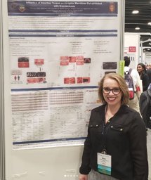 Marcello-Machado Raissa Micaella合作论文数36
 Alessandra Julie Schuster合作论文数34
 Bielemann Amália M合作论文数28Pinto Luciana de Rezende合作论文数School of Dentistry, Federal University of Pelotas-UFPEL28Possebon Anna Paula合作论文数25
 Otacilio Luiz Chagas Junior合作论文数22
 Wander José da Silva合作论文数Piracicaba Dental School, State University of Campinas (UNICAMP),16Renata Cunha Matheus Rodrigues Garcia合作论文数Department of Prosthetics and Periodontics, State University of Campinas12
 Gustavo G. Nascimento合作论文数National Dental Centre Singapore;Duke-NUS Medical School10
 Katleen Vandamme合作论文数Department of Oral Health Sciences, KU Leuven9de Mattias Sartori Ivete Aparecida合作论文数9
 Joke Duyck合作论文数Department of Oral Health Sciences, KU Leuven;Faculty of Medicine, KU Leuven8
 Marissa P Chatterjee合作论文数8Laura Lourenço Morel合作论文数6
 Ana Claudia Melo合作论文数6Rosena Santos da Rosa合作论文数5André Ribeiro Schinestsck合作论文数5Ana Paula Pinto Martins合作论文数5
 Flávia Noemy Gasparini Kiatake Fontão合作论文数Department of Postgraduation, ILAPEO College5
 Geninho Thomé合作论文数5Luiz Carlos do Carmo Filho合作论文数4Otacílio Luiz Chagas-Júnior合作论文数4Samille Biasi Miranda合作论文数4
 Shibli Jamil Awad合作论文数Dept Periodontol & Oral Implantol, Univ Guarulhos4
 William Custodio合作论文数4Lima Emilena Maria Castor Xisto合作论文数School of Dentistry, State University of Campinas4
 Dimorvan Bordin合作论文数4Corrêa Cássia Bellotto合作论文数School of Dentistry at Araraquara, São Paulo State University (UNESP),4Hermann Caio合作论文数4
 Ignace Naert合作论文数Department of Oral Health Sciences, Faculty of Medicine, KU Leuven4
 Fábio Renato Manzolli Leite合作论文数Department of Dentistry and Oral Health, Aarhus University4Juliana Silva Moura合作论文数4A CURY合作论文数3PM Senna合作论文数3RCMR Garcia合作论文数3Simone Guimarães Farias Gomes合作论文数3Hugo Eduardo de Miranda Peixoto合作论文数3
 Bassi Ana Paula Farnezi合作论文数3
 Hugo Ramalho Sarmento合作论文数3Diego Abreu Pastorino合作论文数2Marina de Matos Madruga合作论文数2Dalton Suzuki合作论文数2Vanessa Felipe Vargas-Moreno合作论文数2
 Michele Costa de Oliveira Ribeiro合作论文数2
 Gabrielle Silva合作论文数2
 Flavio Fernando Demarco合作论文数Departamento de Odontologia Restauradora, Faculdade de Odontologia, Universidade Federal de Pelotas2
 Plinio Mendes Senna合作论文数3 Department of Health Sciences Centre, Unigranrio2
 Padovan Luis Eduardo Marques合作论文数2




