AIM:To evaluate the perceived usefulness of a flipped-classroom model that integrated podcasts generated using an artificial intelligence (AI) tool in undergraduate dental education. MATERIALS AND METHODS:The present within-subject, quasi-experimental study was conducted at a Swiss dental school. A cohort of 49 clinical-year dental students participated in eight conventional seminars and two flipped-classroom seminars, each covering a distinct clinical topic. In both formats, a subset of five or six students acted as instructors under the supervision of residents or faculty members. The flipped-classroom model required students to engage with AI-generated podcast summaries of research articles before in-class discussions. These podcasts were created using NotebookLM, an online platform powered by a large language model. Surveys using Likert-type scales assessed student and student instructor satisfaction, engagement, and learning preferences. RESULTS:The surveys obtained response rates of ≥ 93.9%. The flipped-classroom model achieved high satisfaction levels among students. Compared with conventional seminars, students reported increased engagement and comprehension in the flipped-classroom format. Student instructors valued the ease of preparation and the enhanced interactivity of in-class sessions. AI-generated podcasts were rated as effective pre-class resources. CONCLUSIONS:The integration of a flipped-classroom model with AI-generated podcast resources seemed to improve student self-perception of engagement, comprehension, and satisfaction in dental education. This instructional approach may also foster critical appraisal skills for AI-generated content-an increasingly essential competence in academic and professional practice as AI tools gain prominence.
This laboratory study evaluated the effect of deep margin elevation (DME) on the marginal integrity and fracture resistance of anterior teeth with crown-root fractures. Fifty-two extracted permanent human maxillary central incisors were randomly divided into four groups. All teeth received a standardized simulated crown-root fracture, selective enamel etching followed by universal adhesive application, resin-based composite (RBC) build-up (conventional or bulk-fill), root canal treatment, and glass fiber post placement. Crown preparations were performed either entirely in natural tooth structure with subgingival finish lines (DME absent) or partially on the RBC build-up, yielding supragingival finish lines (DME present). Marginal integrity was assessed microscopically at baseline, after initial thermomechanical loading (TML), and after extended TML. Fracture resistance was determined by load-to-fracture testing. Statistical analyses included Kruskal-Wallis, Dunn’s post hoc, and Fisher’s exact tests (α = 0.05). Median marginal continuity remained 100
Large language models (LLMs) are increasingly used in clinical and educational settings. However, there is a paucity of data on LLMs' performance in specialized dental domains. This study assessed the performance of four LLMs, including two general-purpose models, ChatGPT-4o and DeepSeek-R1, and two research-focused models, Consensus and Perplexity, using a validated set of 50 multiple-choice questions in periodontology. Each LLM completed five independent trials encompassing the full question set under both primed and non-primed conditions. A validated answer key served as the benchmark. Performance was analyzed using one-way and two-way analysis of variance and independent-samples t-tests, with additional item-level analyses to identify questions that were consistently difficult. Overall accuracy across all models and trials was 65.0% (95% confidence interval: 63.4-66.6%) with a standard deviation of 3.0. There were no significant differences between models (p = 0.336). Role-specific priming, in which models were instructed to respond as board-certified periodontists, did not improve performance (p = 0.836). At the item level, four questions were never answered correctly, and several others were answered correctly in fewer than 13% of trials. These difficult items generally required detailed procedural knowledge, rare factual recall, or application of classification frameworks. Overall, these findings suggest that current LLMs demonstrate moderate domain knowledge in periodontology but fall short of the reliability required for unsupervised clinical decision support.
Artificial intelligence (AI) and large language models (LLMs) are increasingly being explored for clinical decision-making and evidence-based information retrieval in dental implantology. However, concerns remain regarding the accuracy, reliability, and clinical applicability of different AI approaches, including general-purpose, fine-tuned, and retrieval-augmented generation (RAG) models. This study aimed to compare their ability to provide accurate, evidence-based, and clinically relevant responses to implantology-related questions. Sixteen master’s-level clinical questions were presented to all four LLMs under controlled conditions. Their anonymized responses were independently evaluated by three dental implantology specialists based on three core criteria: specificity, accuracy, and clinical utility, using a 0–10 scale. Descriptive statistics were reported as medians and interquartile ranges. Differences among models were assessed primarily using Kruskal–Wallis tests, and inter-rater reliability was evaluated using Fleiss’ kappa. LLM4 consistently achieved the highest median scores across all criteria (Specificity: median 9.0, IQR 9.0–10.0; Accuracy: median 9.5, IQR 9.0–10.0; Clinical Utility: median 10.0, IQR 9.0–10.0), significantly outperforming the other models (all p < 0.001). Cohen’s d revealed large to very large effect sizes in comparisons involving LLM4, particularly against LLM2 and LLM3. LLM2 scored lowest and showed the highest variability. Inter-rater reliability was slight across all metrics (κ = 0.078–0.180), indicating moderate evaluator disagreement. Among the models tested, LLaMA 3.2 + RAG demonstrated superior and more consistent clinical performance in dental implantology. While fine-tuning provided moderate benefits, real-time access to structured guidelines significantly enhanced response quality. Despite low inter-rater agreement, these findings highlight the promise of RAG models in supporting high-complexity, evidence-based clinical domains, while emphasizing the need for standardization in AI evaluation protocols.
Algorithms, programs, and networks based on artificial intelligence (AI) are currently among the most important drivers of innovation in healthcare. In dentistry, AI-based algorithms emerged just over a decade ago, initially in dentomaxillofacial radiology, where the following applications were researched and implemented in everyday dental practice: improving radiologic image quality, automatically identifying teeth and implants (including their restorative status), diagnosing dental and oral pathologies, and localizing or segmenting anatomical landmarks and structures, for example, in the fields of orthodontics or oral surgery. Al is also a promoter of digital workflows in dentistry, particularly in oral implantology and prosthodontics, where it actively supports the planning, implementation, and monitoring of therapies. Exciting developments include speech recognition systems and visual language models (VLMs), which could significantly reduce and streamline the administrative burden in dental practices. The future use of virtual reality (VR), augmented reality (AR), and mixed reality (MR) in dental studies, or the active integration of this technology into the curriculum, also holds great promise for oral implantology, where complex surgical procedures can be trained and repeated virtually until both a suitable routine and practitioner expertise bare achieved to apply these techniques in real patients. Alongside all these possibilities, however, it is important to consider the risks associated with AI, such as deskilling and automation bias. Furthermore, the dental profession should actively participate in the discussion on the ethical use and regulation of this technology.
Periodontal debridement using ultrasonic instruments and air polishing is a standard procedure in clinical dental practice. While effective in managing periodontal health, these techniques can compromise the integrity of dental restorations. This article reviews laboratory evidence on the effects of ultrasonic instrumentation and air polishing on various restorative biomaterials, focusing on surface roughness and marginal integrity. Findings indicate that high-strength ceramics such as zirconia and lithium disilicate are more resistant to damage than feldspathic ceramics, resin-modified glass-ionomer cements, and resin-based materials, which are more susceptible to damage. The use of low-abrasive powders such as erythritol and glycine is recommended to mitigate adverse effects. Actionable recommendations are provided to inform favorable maintenance protocols for restorations during periodontal therapy.
The introduction and advancement of large language models (LLMs), such as ChatGPT, DeepSeek, and Google Gemini, present both opportunities and challenges for peer review in dental research. In this article, we propose a framework to inform the discourse on the responsible use of LLMs in dental peer review. We conducted a cross-sectional review of peer review policies from the top 50 dental journals, based on their 2024 Journal Impact Factor, to assess current guidance on LLM use. Our analysis revealed variability across dental journals: some journals permit restricted LLM use under specific conditions, while many either prohibit their use or lack explicit policies. Key concerns regarding LLM use identified by the authors include potential breaches of confidentiality, ambiguity in authorship, reduced reviewer accountability, and inherent limitations of LLMs in terms of domainspecific expertise and factual accuracy. Our proposed framework addresses confidentiality safeguards, suggested appropriate LLM applications, areas requiring caution, disclosure requirements, and accountability standards. It emphasizes that reviewers retain full responsibility for all submitted content, irrespective of LLM assistance. To protect confidentiality, the framework encourages offline or locally hosted LLMs. It also recommends regular policy reviews and reviewer training. This framework aims to support the thoughtful adoption of LLMs in dental research publishing. When employed judiciously, LLMs offer potential benefits in improving review clarity and efficiency, particularly for reviewers writing in a non-native language. However, their use must be grounded in clear ethical principles to ensure the integrity of dental peer review.
OBJECTIVE:The aim of this systematic review was to evaluate the effects of ultrasonic instrumentation and air polishing on surface roughness and marginal integrity of dental restorations, addressing their potential adverse impacts on various biomaterials. MATERIALS AND METHODS:A search of five databases, supplemented by manual searches, identified relevant laboratory studies. Forty-two studies met the inclusion criteria, with data on surface roughness and marginal quality extracted for analysis. The RoBDEMAT tool was used to assess risk of bias. RESULTS:Ultrasonic instrumentation significantly increased roughness, particularly in resin-modified glass ionomer cements and resin-based composites, whereas zirconia and lithium disilicate were less affected. Air polishing, especially with sodium bicarbonate and calcium carbonate powders, also increased roughness. Erythritol and glycine powders were the least abrasive. Both ultrasonic scaling and air polishing negatively impacted marginal quality. The RoBDEMAT assessments revealed shortcomings in randomization, sample size justification, and blinding. CONCLUSIONS:Ultrasonic instrumentation and air polishing can adversely affect dental restorations, with the degree of impact varying by biomaterial and debridement method. Low-abrasive powders, such as erythritol and glycine, are recommended. Methodological refinements and clinical studies are needed to enhance the applicability of these findings to patient care. CLINICAL SIGNIFICANCE:This review highlights the critical need to choose appropriate debridement methods to minimize iatrogenic damage to dental restorations. Low-abrasive air polishing powders, such as erythritol and glycine, are recommended for the removal of soft deposits and stains to preserve surface smoothness and marginal integrity.
OBJECTIVE:The optimal duration of phosphoric acid etching (PAE) for enamel bonding remains uncertain, particularly in the context of universal adhesives. The aim of this systematic review was to determine whether shortened etching times (< 15 s) provide comparable bond strength to conventional protocols. MATERIALS AND METHODS:Searches were conducted across four databases, including Embase, OpenGrey through DANS, PubMed, and Scopus. Laboratory studies on human or bovine enamel specimens, treated with varying PAE durations prior to the application of universal adhesives, were included. Data regarding etching protocols, adhesive types, substrate preparation (ground vs. unground enamel), and bond strength outcomes were analyzed. Risk of bias was evaluated using the RoBDEMAT tool. RESULTS:Of 762 records screened, eight laboratory studies met inclusion criteria. Etching times of 3-15 s achieved bond strengths that were statistically non-inferior to conventional durations, with no added benefit from prolonged etching. Ground enamel consistently showed enhanced bond strength with PAE, whereas unground enamel exhibited similar bonding trends. The risk of bias was most often due to inadequate sample size justification, deficiencies in randomization, and absence of blinding. CONCLUSIONS:Short-duration PAE (3-15 s) appears sufficient for effective bonding of universal adhesives to enamel, while minimizing potential risk of over-etching of adjacent dentin. Future research should focus on tailoring etching protocols based on cavity-specific characteristics and exploring alternative pretreatment methods to enhance bonding performance. CLINICAL SIGNIFICANCE:This review suggests that abbreviated phosphoric acid etching (3-15 s) provides enamel bond strengths equivalent to conventional durations when universal adhesives are used. This approach may reduce the risk of inadvertent dentin over-etching in mixed-substrate cavities while maintaining optimal adhesive performance.
To investigate the long-term tooth discoloration induced by different hydraulic calcium silicate-based cements (HCSCs), depending on blood contamination and placement method in-vitro. Eighty bovine teeth, sliced to a length of 18 mm (crown 8 mm, root 10 mm), were randomly assigned to 10 groups (n = 8), receiving ortho- or retrograde apical plug treatment (APT). Apical plugs were 4 mm in length and made of ProRoot MTA (Dentsply), Medcem MTA (Medcem), TotalFill BC RRM Fast Set Putty (Brasseler), or Medcem Medical Portland Cement (Medcem) plus bismuth oxide (Bi2O3) with and without bovine blood. Orthograde (with or without preoperative adhesive coronal dentin sealing) and retrograde APT were compared. Teeth were root-filled with gutta-percha and sealer, adhesively restored, and stored in distilled water. Tooth color was measured on apical plug, gutta-percha/sealer, and crown surface before treatment, after 24 h, and up to 72 months after treatment by spectrophotometry. Color difference (ΔE) values were calculated and analyzed by ANOVA with post-hoc-tests, Shapiro-Wilk-test, Friedman-test, Mann-Whitney-U-test, t-test, and post-hoc-tests with Bonferroni correction (α = 0.05). The increase of tooth discoloration occurred in all groups with no significant differences between HCSCs (p >.05). After six years, color changes were strongly marked on roots but insignificant on crowns. The color differences on the measuring surface of the apical plug were statistically significant between 24 and 72 months (p <.001). Blood had a more relevant impact on tooth color than Bi2O3. There were no substantial long-term effects of retrograde placement (p >.05) or preoperative dentin sealing (p >.05). Apical plugs of the tested HCSCs cause ongoing discoloration of bovine roots, but no discoloration of bovine tooth crowns within a six-year period. Apical plugs should be carefully placed. If direct contact with the coronal dentin is avoided, long-term aesthetic impairments are unlikely to occur.
Radiation therapy for head and neck cancers often leads to radiation-induced caries and microstructural dentin alterations. This laboratory study evaluated the impact of radiation on dentin microhardness and the effectiveness of various topical fluoride treatments in mitigating these effects. One hundred dentin specimens from extracted human teeth were irradiated with a cumulative dose of 60 Gy, simulating therapeutic conditions. Post-irradiation, specimens received weekly applications of silver diamine fluoride (SDF), SDF with potassium iodide, or sodium fluoride varnishes with and without additional compounds. Dentin microhardness was measured using the Knoop hardness test at three key phases: baseline, immediately after reaching a cumulative irradiation dose of 60 Gy, and weekly for five weeks following repeated fluoride applications. At baseline, the average Knoop Hardness Number (KHN) was 57.6 ± 3.9. After irradiation, the average KHN decreased significantly to 41.2 ± 8.1, reflecting an approximate 30% reduction in dentin hardness across all groups (p < 0.0001). Five weeks of weekly fluoride treatments led to partial recovery of microhardness, with average KHN values ranging from 42.4 to 54.3, but none of the groups returned to baseline hardness levels. Notably, no significant differences were found among the fluoride treatments in their ability to enhance microhardness. These findings underscore the detrimental effects of radiation therapy on dentin integrity and emphasize the need for targeted dental management strategies. While fluoride applications provide some protective benefit, their limitations highlight the necessity of comprehensive approaches to prevent radiation-related dental deterioration in head and neck cancer patients.
OBJECTIVE:Obtaining strong resin bonds to computer-aided design and computer-aided manufacturing (CAD-CAM) resin-based composites with dispersed fillers (CCRBCs) poses a challenge. Therefore, this study aimed to assess the effect of three universal adhesives and a two-component silane coupling agent on the shear bond strength to three (CCRBCs).MATERIALS AND METHODS:Eight hundred and sixty-four specimens of Brilliant Crios, Lava Ultimate, and Tetric CAD were polished or grit blasted, bonded with Adhese Universal DC, One Coat 7 Universal (OC7), and Scotchbond Universal Plus Adhesive (SBU) with or without a silane primer. Shear bond strength was measured after 24 h and 10,000 thermocycles. Linear regressions were performed (α = 0.05).RESULTS:After thermocycling, bond strengths were similar for the universal adhesives on polished Brilliant Crios and Lava Ultimate (p ≥ 0.408). Grit blasted Tetric CAD showed no significant differences (p ≥ 0.096). The silane primer had minimal impact on grit blasted Tetric CAD (p ≥ 0.384). The silane primer increased the bond strength of OC7 to Brilliant Crios (p = 0.001) but decreased the adhesion of SBU to Brilliant Crios and Lava Ultimate (p ≤ 0.018).CONCLUSIONS:Bond strength of universal adhesives varied with CCRBC type. The two-component silane coupling agent showed mixed effects on adhesive performance.CLINICAL SIGNIFICANCE:Selecting universal adhesives from the same CCRBC product line does not always guarantee superior bond strength. The efficacy of silane coupling agents differs based on the bonding substrate.
GSK-3 inhibitors, such as Tideglusib (TG) and CHIR-99021 (CHIR), show promise in stimulating reparative dentin formation. The aim of this study was to assess the discoloration potential of TG and CHIR in an established in vitro model.Enamel-dentin specimens made from bovine incisors were randomly allocated to five groups (n=15 each): group bovine blood (BB), group dimethyl sulfoxide (DMSO), group TG, group CHIR, and group mineral trioxide aggregate (MTA). Each specimen had a central cavity in which the respective material was applied and sealed with resin-based luting material. Color deter- mination was conducted using a dental spectrophotometer at t0 (before filling), t1 (immedi- ately after filling), t2 (after one week), t3 (after one month), t4 (after three months), t5 (after six months), and t6 (after one year). Statistical analysis involved descriptive statistics, Kruskal- Wallis tests, and analysis of variance (α=0.05).Group BB and group CHIR exhibited the most significant decrease in lightness (∆L*) after one year (∆L*-4.7 and ∆L* -5.7, respectively), whereas groups DMSO, TG, and MTA showed mini- mal changes (DMSO ∆L*: -0.3; TG ∆L*: 1.4; MTA ∆L*: -0.5). Group BB and CHIR exhibited the highest ∆E values (6.4±0.6 and 6.5±0.8, respectively).Unlike CHIR, TG did not result in discoloration exceeding the threshold of visual perception, defined by a ∆E value of 5.5, during the one-year observation period. This laboratory study therefore suggests that TG could be utilized for indirect or direct pulp capping without major discoloration concerns. However, additional research is required to corroborate these findings.
OBJECTIVE:This article puts forward consensus recommendations from PROSEC North America regarding single indirect restorations made from ceramic and nonmetallic biomaterials in posterior teeth. OVERVIEW:The consensus process involved a multidisciplinary panel and three consensus workshops. A systematic literature review was conducted across five databases to gather evidence. The recommendations, informed by findings from systematic reviews and formulated based on a two-phase e-Delphi survey, emphasize a comprehensive treatment strategy that includes noninvasive measures alongside restorative interventions for managing dental caries and tooth wear. The recommendations advocate for selecting between direct and indirect restorations on a case-by-case basis, favoring inlays and onlays over crowns to align with minimally invasive dentistry principles. The recommendations highlight the critical role of selecting restorative biomaterials based on clinical performance, esthetic properties, and adherence to manufacturer guidelines. They emphasize the importance of precision in restorative procedures, including tooth preparation, impression taking, contamination control, and luting. Regular follow-up and maintenance tailored to individual patient needs are crucial for the longevity of ceramic and nonmetallic restorations. CONCLUSIONS:These PROSEC recommendations provide a framework for dental practitioners to deliver high-quality restorative care, advocating for personalized treatment planning and minimally invasive approaches to optimize oral health outcomes. CLINICAL SIGNIFICANCE:The PROSEC North America recommendations highlight the importance of minimally invasive techniques in posterior tooth restorations using ceramic and non-metallic biomaterials. These principles prioritize tooth structure conservation and personalized treatment planning, essential for enhancing clinical outcomes and long-term oral health.
Objective: Syringe irrigation, commonly used for delivering sodium hypochlorite (NaOCl) during root canal treatment, requires careful handling to prevent NaOCl extrusion into periapical tissues. This randomised controlled in vitro study aimed to quantify syringe plunger forces exerted by dental undergraduates and to assess the impact of an educational intervention on adherence to safe irrigant delivery parameters. Methods: Fifty-two undergraduates performed syringe irrigation at three intervals: baseline (T1), after two weeks (T2), and after 12 weeks (T3). At T1, irrigation was conducted without prior educational intervention, while at T2, it was preceded by an intervention. The educational intervention involved a short video highlighting safe irrigation practices, including plunger force and time recommendations for syringe irrigation. At T3, the undergraduates were randomly allocated to two groups: One received a repeated intervention, while the other did not. Syringe irrigation was performed on a 3D-printed tooth using two side-vented cannulas: one of 25 Gauge (G) and another of 30 G. A syringe equipped with a force sensor recorded the plunger forces. Based on earlier research, plunger forces exceeding 10 Newtons (N) for the 25 G cannula and 40 N for the 30 G cannula were deemed critical.The data were subjected to descriptive statistical analyses. Results: Overall, the mean of maximum values of plunger forces remained under 10 N for the 25 G and below 20 N for the 30 G cannulas, with only a few measurements exceeding 40 N. Instances of surpassing the critical plunger force threshold were more common with the 25 G side-vented cannula than with the 30 G variant. At T3, the group that received the repeated educational intervention exhibited loweraverage maximum plunger forces for both cannulas than the group that did not receive the intervention. Conclusion: Integrating plunger force measurements with targeted educational interventions presents an effective approach for instructing undergraduates in the proper techniques of syringe irrigation. The findings suggest that, generally, undergraduates do not apply excessive plunger forces that could risk apical irrigant extrusion. Implementing repeated educational interventions decreased the plunger forces exerted by undergraduates, underscoring its effectiveness in fostering safe endodontic irrigation.
GSK-3 inhibitors, such as Tideglusib (TG) and CHIR-99021 (CHIR), show promise in stimulating reparative dentin formation. The aim of this study was to assess the discoloration potential of TG and CHIR in an established in vitro model. Enamel-dentin specimens made from bovine incisors were randomly allocated to five groups (n=15 each): group bovine blood (BB), group dimethyl sulfoxide (DMSO), group TG, group CHIR, and group mineral trioxide aggregate (MTA). Each specimen had a central cavity in which the respective material was applied and sealed with resin-based luting material. Color determination was conducted using a dental spectrophotometer at t0 (before filling), t1 (immediately after filling), t2 (after one week), t3 (after one month), t4 (after three months), t5 (after six months), and t6 (after one year). Statistical analysis involved descriptive statistics, Kruskal-Wallis tests, and analysis of variance (α=0.05). Group BB and group CHIR exhibited the most significant decrease in lightness (ΔL*) after one year (ΔL*-4.7 and ΔL* -5.7, respectively), whereas groups DMSO, TG, and MTA showed minimal changes (DMSO ΔL*: -0.3; TG ΔL*: 1.4; MTA ΔL*: -0.5). Group BB and CHIR exhibited the highest ΔE values (6.4Å}0.6 and 6.5Å}0.8, respectively). Unlike CHIR, TG did not result in discoloration exceeding the threshold of visual perception, defined by a ΔE value of 5.5, during the one-year observation period. This laboratory study therefore suggests that TG could be utilized for indirect or direct pulp capping without major discoloration concerns. However, additional research is required to corroborate these findings.
Intraoral scanners (IOSs) have transformed digital dentistry by enabling the capture of detailed intraoral optical impressions, resulting in high-precision virtual models. This article provides an overview of the technological advancements and emerging applications of IOSs in various dental specialties. Recent innovations have improved IOS speed, resolution, and functionality, eliminating the need for scanning powder and allowing for seamless integration with other digital tools. IOSs are widely used in restorative dentistry for computer-aided design/computer-aided manufacturing of dental restorations, offering accuracy comparable to traditional methods. Additionally, IOSs play critical roles in prosthodontics, orthodontics, and oral and maxillofacial surgery. Automated tooth segmentation and orthodontic landmarking have become more efficient due to advancements in machine learning. However, although IOSs are highly accurate for most dental procedures, they face limitations in edentulous areas and full-arch implant impressions. Efforts to expand their diagnostic capabilities-such as caries detection and the monitoring of soft-tissue levels and tooth wear-are underway, aided by advancements in imaging technologies. As research continues, future developments in IOS technology, including better integration with other digital tools and teledentistry applications, should further enhance its utility in dental care. This article highlights the pivotal role IOSs play in modern dentistry while acknowledging ongoing challenges.
Large language models (LLMs) such as ChatGPT have potential applications in healthcare, including dentistry. Priming, the practice of providing LLMs with initial, relevant information, is an approach to improve their output quality. This study aimed to evaluate the performance of ChatGPT 3 and ChatGPT 4 on self-assessment questions for dentistry, through the Swiss Federal Licensing Examination in Dental Medicine (SFLEDM), and allergy and clinical immunology, through the European Examination in Allergy and Clinical Immunology (EEAACI). The second objective was to assess the impact of priming on ChatGPT's performance. The SFLEDM and EEAACI multiple-choice questions from the University of Bern's Institute for Medical Education platform were administered to both ChatGPT versions, with and without priming. Performance was analyzed based on correct responses. The statistical analysis included Wilcoxon rank sum tests (α=0.05). The average accuracy rates in the SFLEDM and EEAACI assessments were 63.3% and 79.3%, respectively. Both ChatGPT versions performed better on EEAACI than SFLEDM, with ChatGPT 4 outperforming ChatGPT 3 across all tests. ChatGPT 3's performance exhibited a significant improvement with priming for both EEAACI (p=0.017) and SFLEDM (p=0.024) assessments. For ChatGPT 4, the priming effect was significant only in the SFLEDM assessment (p=0.038). The performance disparity between SFLEDM and EEAACI assessments underscores ChatGPT's varying proficiency across different medical domains, likely tied to the nature and amount of training data available in each field. Priming can be a tool for enhancing output, especially in earlier LLMs. Advancements from ChatGPT 3 to 4 highlight the rapid developments in LLM technology. Yet, their use in critical fields such as healthcare must remain cautious owing to LLMs' inherent limitations and risks.
Introduction: Radiation therapy is often used as the main or adjunctive treatment for cancer in the oral cavity. Unfortunately, prolonged exposure to radiation has adverse effects on tissues of the oral cavity, including a decrease in the hardness of dentin. Consequently, patients who receive radiation therapy are susceptible to tooth sensitivity and caries. Fluoride treatments have proven to be effective in restoring the hardness of dentin as it remineralizes the tooth. The aim of this study was to determine the optimal fluoride treatment for restoring the hardness of irradiated teeth. Methods: 100 teeth were extracted, cleaned, and mounted in acrylic resin. The teeth were ground and polished to expose the dentin and enamel. The initial Knoop hardness of the teeth was measured and split equally among five groups and treated as follows: (1) sodium fluoride varnish (SF), (2) MI varnish (MI), (3) silver diamine fluoride and potassium iodide (SDFKI), (4) silver diamine fluoride (SDF), and (5) no treatment (NT). All groups were subjected to 2 grays of daily radiation treatment for 6 weeks. The hardness of the dentin was measured immediately following radiation. Afterwards, the 5 treatments were applied weekly for 6 weeks. Hardness was measured 1 week after each treatment. Kruskal-Wallis one way analysis of variance on ranks was used to compare the different groups. Results: After 6 weeks of fluoride treatment, there was a significant difference (p = 0.013) between the hardness of the irradiated enamel, with Group 4 resulting in the largest median measurement (263.4). Similarly, 5 weeks of fluoride application resulted in a mean hardness of dentin that was 15.8%, 13.6%, 16.2%, and 19.4% higher than the control for groups 1, 2, 3, and 4, respectively. Again, Group 4 had the highest hardness measurement. Conclusions: Radiation therapy decreases the hardness of enamel and dentin. SDF treatment resulted in the highest hardness for both irradiated dentin and enamel, suggesting that SDF is promising as a fluoride treatment for those undergoing radiation therapy for oral cancer. Citation Format: Rachel Kulchar, Florin Eggmann, Celine Mina, Steven Szewczyk, Kakiuchi Yusuke, Jie Deng, Francis Mante. Effects of radiation therapy on the hardness of enamel and dentin [abstract]. In: Proceedings of the American Association for Cancer Research Annual Meeting 2023; Part 1 (Regular and Invited Abstracts); 2023 Apr 14-19; Orlando, FL. Philadelphia (PA): AACR; Cancer Res 2023;83(7_Suppl):Abstract nr 1109.
 Roland Weiger合作论文数26
 Markus B. Blatz合作论文数Department of Preventive and Restorative Sciences, School of Dental Medicine, University of Pennsylvania15
 Thomas Connert合作论文数Department of Periodontology, Endodontology and Cariology, University Centre for Dental Medicine, University of Basel14
 Andreas Filippi合作论文数University of Basle, University of Gießen10
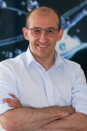 Gabriel Krastl合作论文数Dental School, University of Basel9
 Klaus W. Neuhaus合作论文数School of Dental Medicine, University of Bern7
 Julia Amato合作论文数4
 Eva Magni合作论文数4Jose Ayub合作论文数4
 Francis K. Mante合作论文数School of Dental Medicine, University of Pennsylvania4
 Nicola U. Zitzmann合作论文数Klinik für Rekonstruktive Zahnmedizin, Universitären Zentrums für Zahnmedizin Basel;Universität Basel4
 Lucia K Zaugg合作论文数4Svetlana Kapor合作论文数3
 Mila Janjic Rankovic合作论文数3Stavroula Michou合作论文数3Marie C D N J M Huysmans合作论文数3
 Julián Conejo合作论文数School of Dental Medicine, University of Pennsylvania3
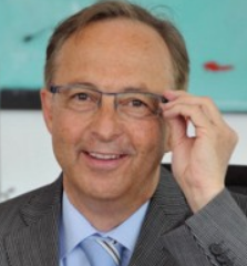 Lussi Adrian合作论文数University of Bern;University of Innsbruck3
 Felix Krause合作论文数3
 Ina Schueler合作论文数3Kühnisch Jan合作论文数Department of Operative Dentistry and Periodontology, University Hospital, Ludwig-Maximilians-Universität München, Munich, Germany.
Klinik für Zahnerhaltung und Parodontologie, Klinikum der Ludwig-Maximilians-Universität München, Goethestraße 70, 80336, Munich, Germany.3Kim Rud Ekstrand合作论文数3Ashraf Elayouti合作论文数Division of Endodontology, Department of Conservative Dentistry, School of Dental Medicine, University of Tübingen, Tübingen, Germany.3Clemens Walter合作论文数3Sebastian Kuehl合作论文数2Jelena Karacic合作论文数2
Ina Schueler合作论文数3Kühnisch Jan合作论文数Department of Operative Dentistry and Periodontology, University Hospital, Ludwig-Maximilians-Universität München, Munich, Germany.
Klinik für Zahnerhaltung und Parodontologie, Klinikum der Ludwig-Maximilians-Universität München, Goethestraße 70, 80336, Munich, Germany.3Kim Rud Ekstrand合作论文数3Ashraf Elayouti合作论文数Division of Endodontology, Department of Conservative Dentistry, School of Dental Medicine, University of Tübingen, Tübingen, Germany.3Clemens Walter合作论文数3Sebastian Kuehl合作论文数2Jelena Karacic合作论文数2 Christian Dettwiler合作论文数2
 Fabio Saccardin合作论文数2
 Khaled Mukaddam合作论文数2Christian Meller合作论文数2
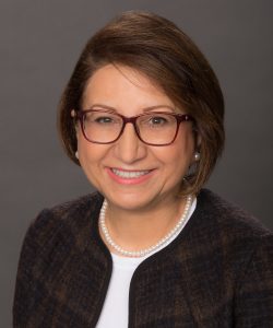 Fusun Ozer合作论文数School of Dental Medicine, University of Pennsylvania2
 Sebastian Kuhl合作论文数2
 Thomas Attin合作论文数Clinic of Conservative and Preventive Dentistry, Center of Dental Medicine, University of Zurich2
 Julia Buhler合作论文数2
 Ralf Krug合作论文数2
 Alexander Crispin合作论文数2
 Michael Marc Bornstein合作论文数Faculty of Dentistry, The University of Hong Kong;Department of Oral Health and Medicine, University Center of Dental Medicine Basel2Yusuke Kakiuchi合作论文数1Steven T Szewczyk合作论文数1Michelle Simonek合作论文数1Elisabeth Reichardt合作论文数1C. de Julián合作论文数1Michael Bergler合作论文数1Asin Ahmad Haschemi合作论文数1Theisen Catherine E. R.合作论文数1
 Awab Abdulmajeed合作论文数1Rachel J Kulchar合作论文数1Wadim Leontiev合作论文数1




