Rheumatic diseases constitute a major cause of chronic pain, functional impairment, and long-term disability worldwide. In rheumatic diseases, reliable assessment of structural damage and inflammatory activity is essential for diagnosis, disease monitoring, and treatment response evaluation, yet remains largely dependent on time-intensive expert image interpretation. In this study, we develop and evaluate a pipeline for automatic landmark detection and subsequent pathology scoring in hand magnetic resonance images for three main pathologies in rheumatic diseases, namely erosions, osteitis, and synovitis. We explicitly exploit two orthogonal acquisitions (coronal and transversal) by integrating multi-view information at different stages of the pipeline to assess their impact on automatic scoring performance. We train and compare two landmark detection models that utilize all three magnetic resonance imaging sequences to predict predefined landmarks annotated by experts. The YOLO model achieves better landmark predictions for both metrics and across all distances, with a successful detection rate of 94% for a clinically relevant distance of 6 mm and an overall mean Euclidean distance of 3 mm from ground truth landmarks to predicted landmarks. By using a super-resolution approach to fuse coronal and transversal images for the automatic scoring, we achieve an improved performance for synovitis detection. This paves the way for more fully automated precision medicine in magnetic resonance imaging, reducing the workload for physicians while enabling faster, more standardized results to support the decision-making process.
Psoriatic disease initially affects the skin and later extends to the joints. Here, we show a two-step process that orchestrates the spread of inflammation from the skin to the joints. Induction of psoriatic skin disease in photoconvertible mice, followed by sequencing and computational characterization of skin-derived cells in the joints, was used to identify a population of CD2+MHC-II+CCR2+ myeloid precursors that builds a skin-derived myeloid cell compartment in the joints. Single-cell cross-species reference mapping and mitochondrial variant tracing showed an orthologous human cell population. Interactome analysis of the joints showed that in a second step, resident regulatory CD200+ fibroblasts regulate the priming of CD2+MHC-II+CCR2+ myeloid precursors, which subsequently control IL-17 expression in T cells. Hence, the spread of inflammation requires a distinct migratory myeloid precursor population and a permissive local tissue environment, similar to tumor metastasis.
Background : The skin is a central organ involved in systemic autoimmune diseases (AID). Next to T-cell activation, the formation of pathogenic B-cells and plasma cells as well as the emergence of autoantibodies is a crucial process in AID. While some patients with AID can be managed well by conventional immunosuppressants, others develop highly-treatment resistant disease and require new therapeutic concepts that tackle pathologic B-cell and plasma cell activation. T-cell engagers are a powerful therapeutic tool to deplete B-cell and plasma cells and may be efficacious to improve skin manifestations of systemic AID. Methods : We evaluated the effect of the BCMA-directed T-cell engager teclistamab on cutaneous manifestations of highly treatment-resistant systemic AID. Skin activity was assessed using the Cutaneous Dermatomyositis Disease Area and Severity Index (CDASI), and skin-related quality of life using the Dermatology Life Quality Index (DLQI), based on clinical findings and patient interviews before and after treatment. Results : Three patients (two with dermatomyositis, one with systemic sclerosis) with treatment-resistant disease and cutaneous manifestations were treated with teclistamab. In both dermatomyositis patients, skin disease activity substantially decreased, and skin-related quality of life improved. In the patient with systemic sclerosis, digital ulcers resolved, skin fibrosis decreased, hair regrew and the skin-related quality of life normalized. Conclusion : In this small case series, BCMA-directed T-cell engager therapy was associated with improvement of skin manifestations and skin-related quality of life in patients with highly treatment-refractory AID. Larger studies are required to systematically assess efficacy, safety, and durability of cutaneous responses to T-cell engagers.
OBJECTIVES:The role of autoantibody-producing B cells in connective tissue diseases (CTD) has recently been highlighted by the successful treatment with CD19-targeting CAR T cells. Detrimental effects of autoantibodies are linked to the formation of deposited IgG complexes and the activation of immune cells via Fcγ receptors (FcγRs). The role of circulating immune complexes (cICs) as a link between adaptive and innate immunity has remained understudied. Clinical testing of cICs has been hindered by the lack of reliable detection methods. The aim of this study was to determine the potential of IgG-containing cICs to activate FcγRs (their bioactivity) using a new detection method. METHODS:A reporter cell platform was used to assess the presence and bioactivity of cICs in IgG-autoantibody-positive CTD patients (cross-section analysis) and in patients treated with CD19-CAR T cells (longitudinal analysis). RESULTS:The bioactivity of cICs in the cohort of patients with CTDs was significantly higher compared with healthy controls and patients with IgG-autoantibody-negative systemic inflammatory disease (psoriatic arthritis). Analyses of individual diseases revealed the presence of cICs in the sera of all CTDs, including systemic sclerosis (SSc) and primary Sjögren's syndrome, although there was significant heterogeneity among individuals. Within SSc, patients positive for anti-topoisomerase-I (Scl70) autoantibodies, diffuse cutaneous and lung involvement had significantly enhanced cIC bioactivity. Finally, the bioactivity of cICs was significantly reduced in CTD patients after CD19-CAR T cell therapy. CONCLUSIONS:Our study reveals the presence of FcγR-engaging cICs in CTDs and demonstrates that the bioactivity of cICs is correlated with clinical phenotypes and treatment outcomes.
Psoriatic arthritis (PsA) is characterized by heterogeneous musculoskeletal manifestations. The aim of this post-hoc cluster analysis from the ULTIMATE study was to explore whether objective assessment of inflammation, using power-Doppler ultrasound (PDUS), identified homogeneous groups of patients based on the level of severity of ultrasound-detected synovitis and then to determine the longitudinal trajectory of response with secukinumab vs. placebo for each cluster up to week 12. The ULTIMATE study enrolled patients with PsA and active clinical and PDUS synovitis. Patients were randomized to either secukinumab 150 or 300 mg, according to psoriasis severity, or placebo for the first 12 weeks. Multiple factor analysis was performed on baseline B-mode and power Doppler (PD) signal core components of the composite Global EULAR-OMERACT synovitis score (GLOESS) at joint level. Three clusters were identified from all patients enrolled in the ULTIMATE study (N = 166), with 49 (30
Chimeric antigen receptor (CAR) T cell therapy was recently proposed as a treatment for adults with B-cell-mediated autoimmune diseases (ADs) refractory to conventional immunomodulatory therapy. We present a case series of eight children with severe/refractory AD (four systemic lupus erythematosus, three dermatomyositis, one systemic sclerosis) treated at Ospedale Pediatrico Bambino Gesù, Rome, and University Hospital Erlangen with a single infusion of 1 × 10⁶ kg-1 point-of-care manufactured autologous CD19 CAR T cells (zorpocabtagene autoleucel), in a hospital exemption (HE) program. In Europe, the HE pathway offers the opportunity to treat patients with life-threatening or seriously debilitating disorders who lack valid therapeutic options, using an advanced therapy medicinal product (ATMP) authorized on a nonroutine, single-patient basis. In contrast to the 'compassionate use' pathway, the ATMP does not necessarily need to have undergone clinical trials or marketing authorization applications. Manufacturing was successful in all patients, yielding several drug product bags. Once infused after lymphodepletion, zorpocabtagene autoleucel cells expanded in vivo, promoting prompt B cell clearance. Grade 1 cytokine release syndrome was reported in six patients, and grade 1 immune effector cell-associated neurotoxicity syndrome was reported in one patient. Late-hematotoxicity was limited to grade 1 in two patients. All these adverse events were manageable and no severe infections occurred. With a median follow-up of 16.5 months (range = 9-24 months), all patients experienced a clinically substantial improvement/resolution of AD, as evidenced by reduction in disease activity scores and signs of reversal of organ damage. This improvement enabled sustained discontinuation of immunomodulators, even after B cell reconstitution. The activation of formal clinical trials enrolling children and adolescents is urgently needed to confirm these preliminary results and to assess the long-term safety of this approach.
Insufficient treatment response is common in systemic lupus erythematosus (SLE) and may be associated with progressive organ damage, yet the molecular underpinnings of treatment resistance remain elusive. RNA sequencing was performed in blood samples from 21 SLE patients who failed to achieve Lupus Low Disease Activity State after six months of treatment with cyclophosphamide (n = 9), rituximab (n = 5), or belimumab (n = 7). Molecular endotypes were identified via unsupervised clustering of Functional Analysis of Individual Microarray Expression (FAIME) scores and cluster stability was validated in an independent cohort (n = 23). Endotype-specific druggability was assessed using the L1000CDS2 platform. The pathway-based molecular stratification revealed three major endotypes among patients with inadequate treatment response: (i) a T cell-centric cluster characterized by enrichment of PD-1 signaling and DNA damage response pathways along with downregulation of CD28 co-stimulatory signaling, indicative of T cell senescence; (ii) a cytokine-driven cluster defined by elevated IL-6 and IL-17 signaling and reduced IL-2 signaling, suggesting a Th17/Treg imbalance and potential responsiveness to cytokine inhibition or low-dose IL-2 therapy; and (iii) an inflammasome-dominant cluster. A multinomial LASSO regression-derived Molecular Endotype Classification Index (MECI) - validated via 1000-fold bootstrap resampling - demonstrated potential as a surrogate marker for endotype assignment (median AUC-ROC 0.889). Transcriptome reversal analysis suggested that patients of the T cell-dominant endotype may be more responsive to CD19 CAR-T cell therapy. In summary, distinct molecular endotypes underlie insufficient response to therapy in SLE, providing a framework for personalized treatment strategies and improved clinical trial design.
OBJECTIVE:Members of the superfamily of nuclear receptors have been implicated in inflammatory processes and pathologic tissue remodeling and have emerged as attractive targets for pharmaceutical intervention. However, the role of testicular receptor 4 (TR4; or Nr2c2) in fibroblast activation and rheumatologic diseases has not yet been investigated. METHODS:TR4 expression in human skin and experimental fibrosis were determined by immunofluorescence staining and Western blot. RNA sequencing (RNAseq) was performed in transforming growth factor β (TGFβ)-stimulated fibroblasts with or without TR4 knockdown by small interfering RNA. Myofibroblast differentiation was assessed by α-smooth muscle actin and extracellular matrix staining, and Rho-associated protein kinase (ROCK) activity was assessed by enzyme-linked immunosorbent assay. Fibroblast-specific Tr4 knockout mice were subjected to constitutively active TGFβ receptor type I-, bleomycin-, and chronic graft-versus-host disease-induced dermal fibrosis and bleomycin-induced pulmonary fibrosis. Precision-cut skin slices (PCSSs) from patients with systemic sclerosis (SSc) were used for TR4 knockdown studies. RESULTS:TR4 was up-regulated in fibroblasts in the skin of patients with SSc and in murine models of SSc. TGFβ induced TR4 in fibroblasts in an SMAD3-dependent manner. Knockdown of TR4 prevented fibroblast-to-myofibroblast transition, and fibroblast-specific knockout of Tr4 ameliorated experimental murine skin and pulmonary fibrosis. RNAseq and functional experiments identified the profibrotic effects of TR4 were dependent on G protein subunit alpha 12- and ROCK-associated cytoskeletal remodeling. PCSSs confirmed that TR4 regulates ROCK signaling and the expression of profibrotic genes in SSc skin. CONCLUSION:TR4 is up-regulated in SSc in a TGFβ-dependent manner and drives fibroblast activation. Inhibition of TR4 prevents ROCK activation and fibroblast-to-myofibroblast transition and ameliorates dermal and pulmonary fibrosis, highlighting TR4 as a potential therapeutic target in SSc and related diseases.
Abstract The advent of photon-counting computed tomography (PCCT) provides new opportunities to quantitatively measure musculoskeletal tissues such as bone, muscle and adipose because of the intrinsic use of spectral imaging. We aimed to evaluate the accuracy of measuring these tissues by PCCT under a range of scan protocols and compared our results to the current standard dual-energy CT (DECT). Phantoms containing inserts ranging from 50 to 200 mg/cm 3 of calcium hydroxyapatite (HA) for estimating bone mineral density (BMD), and another phantom containing inserts for muscle and adipose tissues were scanned on PCCT and DECT at 120 and 140 kVp. We created virtual monoenergetic images (VMI) at energy levels from 40 keV to 190 keV for quantitative analyses. The averaged linear attenuation of phantom inserts was compared to theoretical values calculated from standardized attenuation profiles. Material decomposition using VMIs was compared to known HA concentration inserts to determine optimal image pairs for BMD measurement, notably without the need of phantom calibration. For most VMI energy levels the attenuation error was <1% for BMD at both 120 kVp and 140 kVp by PCCT compared to errors of <2% by DECT. The linear attenuation errors were <2.5% for muscle and <3.0% for adipose and results were similar for PCCT and DECT. Generally, errors were highest for low energy VMIs. Material decomposition using VMI pairs with a low energy at 50 or 60 keV and high energy between 150 and 190 keV produced calibration phantom-free estimates of BMD with <1% error. Results were similar for PCCT and DECT at 120 and 140 kVp. PCCT provides an accurate estimate of bone, muscle and adipose attenuation, and using material decomposition, estimations of BMD can be obtained without the need of phantom calibration. Given that multi-energy imaging is intrinsic for PCCT this provides new possibilities for opportunistic screening in patients who get PCCT scans for other clinical indications.
Systemic lupus erythematosus (SLE) is driven by interconnected immune circuits in which B cells function as antigen-presenting cells, cytokine producers and precursors of autoantibody-secreting plasma cells. Therapeutic targeting of B cells aims to disrupt these self-reinforcing circuits rather than simply reduce autoantibody titres alone. Clinical experience with anti-CD20 monoclonal antibodies has validated B cells as therapeutic targets, while exposing key biological constraints, including incomplete tissue depletion, persistence of long-lived plasma cells, and BAFF-driven B cell repopulation. Recent advances including next-generation anti-CD20 monoclonal antibodies, bispecific T cell engagers and chimeric antigen receptor (CAR) T cell therapies have shown promise but questions remain relating to which patients should be treated with what modality of therapy and when this should be timed within the disease course. Herein, we evaluate the current landscape of B cell targeting therapies with particular focus on patient selection, timing of therapy and type of treatment.
The molecular mechanisms linking immune cell signaling to osteoclastogenesis remain incompletely defined. Here, we identify an Annexin A1 (AnxA1)-Dectin-1 axis as a key driver of osteoclast differentiation. Dectin-1 (CLEC7A), a myeloid C-type lectin receptor best known for β-glucan recognition, is shown to bind the endogenous ligand AnxA1 on pre-osteoclasts, thereby promoting their maturation. In Dectin-1 deficient mice, reduced osteoclast numbers resulted in increased bone volume, whereas β-glucan-induced Dectin-1 activation enhanced osteoclastogenesis. Within the bone marrow niche, AnxA1 was abundantly expressed on B220+ B cells, and γ-irradiation markedly increased its surface translocation both in vitro and in vivo. γ-irradiated B220+ B cells exhibited strong Dectin-1 binding capacity and robustly stimulated osteoclast differentiation in a Dectin-1-dependent manner. These findings establish the AnxA1-Dectin-1 interaction as a critical immune-skeletal communication pathway, revealing a mechanism by which radiation exposed immune cells can accelerate bone resorption. Targeting this axis offers a potential strategy to mitigate radiation-induced bone degradation and preserve skeletal homeostasis.
Chimeric antigen receptor (CAR)-expressing cells bear a great potential for the treatment of autoimmune diseases. While numerous challenges persist, recent technological developments have demonstrated the potential for CAR cells to reset dysfunctional immune systems and transform care for patients with autoimmune disease.
Exercise is a key non-pharmacological component of therapy in axial spondyloarthritis (axSpA). Evidence for yoga as a mind–body intervention remains limited. This study aimed to assess the effects of an 8-week yoga program on mobility, function, disease activity, activity-related competences, and psychological outcomes in axSpA compared with physiotherapy and healthy individuals. We conducted a prospective, non-randomized, study (University Hospital Erlangen, May 2021–January 2024). AxSpA patients participated either in weekly yoga sessions for 8 weeks (approximately 60 min; online format) or received standard individual physiotherapy (≥ 30 min/week). A healthy control group completed the same yoga program. Health related outcomes at baseline and follow-up were measured and changes were analysed using linear mixed-effects models. Seventy-six participants were included. In axSpA, spinal mobility improved in the yoga group (BASMI Δ − 0.31; p = 0.002), while the physiotherapy group worsened (Δ + 0.29; p = 0.003). Physical activity related health competency (control competence Δ + 1.10; p = 0.001; self-regulation Δ + 0.72; p = 0.021) and psychological outcomes improved in the yoga group with increased quality of life mental scores (Δ + 6.62; p < 0.001) and reduced kinesiophobia (TSK Δ − 3.25; p = 0.008); physiotherapy showed no meaningful changes. Disease activity showed a significant decrease in the yoga group (BASDAI Δ –0.44; p = 0.049). Healthy individuals showed similar directional changes with smaller effects. An 8-week yoga intervention was associated with improvements in mobility, psychological outcomes and activity-related competences. Findings support yoga as a complementary treatment addressing both physical and psychological dimensions of axSpA. The study was registered in the German Clinical Trials Register (DRKS-ID: DRKS00025215).
OBJECTIVES:Rheumatoid arthritis (RA) and psoriatic arthritis (PsA) are chronic immune-mediated inflammatory diseases associated with progressive bone deterioration. Comparative longitudinal changes in bone density, microarchitecture, and biomechanical properties, and their association with disease activity, remain insufficiently elucidated in seronegative RA (RA-), seropositive RA (RA+), and PsA. METHODS:High-resolution peripheral quantitative computed tomography (HR-pQCT) was used to assess volumetric bone density (vBMD), microarchitecture, and biomechanical properties at the distal radius and the metacarpophalangeal (MCP) joint during routine follow-up. Generalised additive models, adjusted for age, sex, and body weight, were used to analyse the relationship between bone changes, diagnosis, and disease activity over time, spanning up to 7 years. RESULTS:We analysed 946 radius and 916 MCP scans from 482 patients (175 PsA, 221 RA+, and 86 RA-) over a mean ± SD follow-up period of 25 ± 27 months (maximum: 108) and 24 ± 30 months (maximum: 142), respectively. At all-time points compared, RA+ patients had lower trabecular vBMD than PsA (difference: 14.71 mg hydroxyapatite [HA]/cm³; 95% CI, 2.85-26.58; p = .003 at baseline) and RA- (difference: 16.92 mg HA/cm³; 95% CI, 0.97-32.86; p = .010 at baseline) patients. Longitudinally, total vBMD declined by -16.2 mg HA/cm³ (95% CI, -20.9 to -11.4; p < .001) in RA+ patients over 5 years, with smaller losses in RA- and PsA patients. PsA patients showed stable bone quality with cortical sparing; RA+ patients showed the most pronounced trabecular vBMD and microarchitectural loss; and RA- patients showed intermediate changes. Sustained remission preserved bone, while high disease activity was associated with deterioration. CONCLUSIONS:Longitudinal HR-pQCT shows site- and serostatus-dependent bone deterioration across inflammatory arthritides; RA+ patients have the worst trabecular changes, and those with PsA have relative cortical preservation. Sustained remission preserves bone structure and strength.
Stroke or transient ischemic attack (TIA) leads to unfavorable structural changes in the bones. The underlying pathophysiological process is unclear. We provide a detailed characterization of the structural and biochemical properties of bone in a stroke/TIA cohort. Our findings lay the foundation for a comprehensive understanding of post-stroke osteopathy and its clinical implications. Patients who have suffered a stroke or transient ischemic attack are at high risk of fractures, which increase mortality and reduce health-related quality of life. Previous studies showed that ischemic stroke leads to unfavorable structural changes in the bones, but the underlying pathophysiological process is still unclear. Our aim was to analyze biomarkers and bone structure with high-resolution peripheral quantitative computed tomography (HR-pQCT) in a stroke/TIA cohort. This cross-sectional study analyzes the baseline data of a prospective cohort. Patients underwent HR-pQCT scans of the radius and tibia, alongside biochemical bone marker evaluation, within the first days after the acute cerebrovascular event. Women had lower total, cortical, and trabecular volumetric bone mineral density (vBMD) and lower values for most other HR-pQCT parameters. Patients with increasing age had lower total and cortical vBMD, but no difference in trabecular parameters. Active or former smokers had lower total and trabecular vBMD. Higher BMI was associated with greater cortical and trabecular bone areas and higher trabecular density. A positive correlation was found between sclerostin levels and trabecular parameters. Higher osteoprotegerin levels were associated with lower cortical area and thickness. Osteocalcin showed a negative correlation with total vBMD and cortical thickness. This study is the first to combine HR-pQCT imaging with bone biomarkers to provide a detailed characterization of the structural and biochemical properties of bone in a stroke/TIA cohort. Our findings lay the foundation for a comprehensive understanding of post-stroke osteopathy and its clinical implications.
OBJECTIVES:Flares are key determinants of outcome in systemic lupus erythematosus (SLE), yet the conventional mild/moderate vs severe classification inadequately reflects prognosis. We sought to refine flare classification to improve damage prediction and develop a data-driven flare score. METHODS:We analysed a multicentre longitudinal cohort of 354 patients with active SLE requiring treatment at inclusion (median follow-up 66 months). Flares were adjudicated using a modified Safety of Estrogens in Lupus Erythematosus National Assessment-SLE Disease Activity Index Flare Index (SFI). Random Forests and modified flare definitions assessed the contribution of individual SFI variables to subsequent damage. We derived a flare score using bootstrap-validated mixed-effects models. This was validated in an independent cohort (n = 110). RESULTS:Both mild/moderate and severe SFI flares contributed to elevated 12-month damage risk (incidence rate ratio [IRR]:1.42 and 1.54, respectively). Among SFI subcriteria, major organ involvement requiring high-dose glucocorticoids (GCs) ranked highest for damage prediction (weight = 6), followed by the isolated GCs escalation criteria (weights = 1.5-3), whereas minor organ involvement and immunosuppressants/biologics addition carried lower weights (1 each). A flare score based on these subcriteria demonstrated good predictive performance (Brier score = 0.07). A threshold of ≥ 3.5 identified a subset of high-risk flares (40% of all episodes) that displayed the strongest association with subsequent damage (IRR: 2.49, 95% CI: 1.51-4.10) and outperformed the original SFI definition in the validation cohort (area under the curve-receiver operating characteristic: 0.893 vs 0.816). Although both Definitions of Remission in SLE (DORIS) and lupus low disease activity state correlated with lower risk of SFI-defined flares, only sustained DORIS (>50% of time) reduced high-risk flares. CONCLUSIONS:We defined and validated a novel weighted SLE flare score that better predicts damage accrual and may support treat-to-target implementation and clinical trial design.
SSc is the rheumatic disease with the highest individual mortality rate and is associated with substantial morbidity that severely affects quality of life. Organ fibrosis is a major contributor to irreversible damage in SSc, whereas early inflammatory manifestations largely reflect disease activity and may still respond to early immunomodulatory treatment. New imaging modalities, as well as novel applications of established techniques-such as lung US or improved US assessment of synovial involvement-offer promising opportunities to objectively distinguish activity i.e. early inflammation and early fibrosis from damage (i.e. late irreversible fixed vasculopathy or fibrosis) at the organ level. Differentiating these processes is essential for refining the therapeutic window of opportunity in this rare but devastating disease. This narrative review aims to provide an up-to-date overview of available and emerging imaging tools that may help discriminate activity from damage across organ systems involved in SSc, with a specific focus on early fibrotic and inflammatory manifestations.
 Jürgen Rech合作论文数Friedrich-Alexander-Universitat Erlangen-Nimberg;Universitätsklinikum Erlangen237
 Jörg Distler合作论文数Friedrich-Alexander-Universität;Universitätsklinikum Erlangen;Department of Internal Medicine 3, University of Erlangen-Nuremberg;Institute for Clinical Immunology, University of Erlangen-Nuremberg210
 Arnd Kleyer合作论文数201
 Oliver Distler合作论文数Klinik für Rheumatologie, UniversitätsSpital Zürich177Axel J. Hueber合作论文数163
 David Simon合作论文数162Jochen Zwerina合作论文数156Clara Dees合作论文数156
 Andreas Ramming合作论文数151Matthias Englbrecht合作论文数149
 Martin Herrmann合作论文数Friedrich-Alexander-Universität Erlangen-Nürnberg144
 Aline Bozec合作论文数Universität Erlangen-Nürnberg(),144
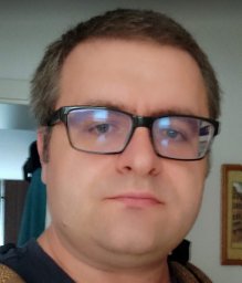 Koray Tascilar合作论文数Istanbul University, Trakya University Medical Faculty143
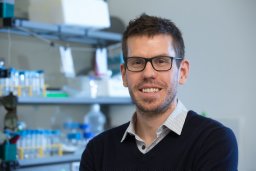 Gerhard Krönke合作论文数Department of Internal Medicine 3, University of Erlangen-Nuremberg130Beyer Christian合作论文数114
 Bernhard Manger合作论文数112Bergmann Christina合作论文数Department of Internal Medicine III and Institute for Clinical Immunology, University of Erlangen-Nuremberg111
 Johannes Knitza合作论文数Arbeitsgemeinschaft Junge Rheumatol Deutsch Gesel101Stephanie Finzel合作论文数98
 Luis Munoz合作论文数Friedrich-Alexander-University Erlangen-Nürnberg;Universitätsklinikum Erlangen98
 Josef Smolen合作论文数Universitätsklinik für Innere Medizin III, Medizinische Universität Wien97Melanie Hagen合作论文数82A. Kleyer合作论文数79Zaiss Mario M合作论文数73
 Philip J. Mease合作论文数Division of Rheumatology Clinical Research, Swedish Medical Center;University of Washington71Filippo Fagni合作论文数Dept Internal Med 3, Friedrich Alexander Univ FAU Erlangen Nuremberg66Pawel Zerr合作论文数66
 Michael Sticherling合作论文数64Klaus Engelke合作论文数64Raimondo Maria Gabriella合作论文数Division of Rheumatology and Clinical Immunology, Humanitas Clinical and Research Center61D'Agostino Maria-Antonietta合作论文数58Distler Alfiya合作论文数58
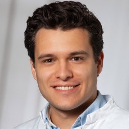 Ricardo Grieshaber-Bouyer合作论文数Brigham & Womens Hosp, Div Rheumatol Immunol & Allergy, 75 Francis St, Boston, MA 02115 USA55
 Judith Haschka合作论文数55Andréas Mackensen合作论文数53
 Reinhard E. Voll合作论文数Department of Rheumatology and Clinical Immunology, Medical Center, University of Freiburg;Faculty of Medicine, University of Freiburg53
 Arthur Kavanaugh合作论文数Center for Innovative Therapy, Division of Rheumatology, Allergy and Immunology, University of California, San Diego;School of Medicine, University of California, San Diego52
 Iain McInnes合作论文数School of Infection & Immunity, University of Glasgow;College of Medical, Veterinary & Life Scinces, University of Glasgow52
 Anna-Maria Liphardt合作论文数Institute of Aerospace Medicine, German Aerospace Center (DLR e.V.)52
 Katrin Palumbo-Zerr合作论文数50Simon Rauber合作论文数Friedrich Alexander Univ Erlangen Nurnberg FAU, Dept Internal Med Rheumatol & Immunol 3, Erlangen, Germany49Silvia Hayer合作论文数Department of Internal Medicine III, University of Vienna49
 Jule Taubmann合作论文数47Michael Aigner合作论文数47D. Simon合作论文数45Kurt Redlich合作论文数45
 Michal Tomcik合作论文数43
 Dafna Gladman合作论文数Institute of Medical Science, Temerty Faculty of Medicine, University of Toronto;UHN-Toronto Western Hospital;Toronto Western Research Institute43Soare Alina合作论文数43




