中国目前有950万痴呆人口,占世界痴呆人口的20%.以三甲医院专科医生为主体的医疗体系远远不能满足痴呆患者诊治需求,迫切需要我国的医疗体系进行相应的改革和完善.专科医生服务模式需要改革,要对基层全科医生就痴呆疾病诊治能力进行培训,加强社区全科医生对此疾病的诊治能力.本文将针对社区全科医生建立痴呆疾病诊治能力的培训模式及内容进行初步探讨.
自1982年经颅多普勒超声(TCD)在临床应用以来,该项技术在脑血管病诸多方面得到了广泛的应用。2004年,美国神经病学学会组织相关的专家组,撰写了这篇经颅多普勒超声治疗及技术评估的报告。这是第一篇依据循证医学概念全面论述TCD临床应用价值的报告。在这篇报告中,协作组复习了TCD及其相关技术经颅彩色编码超声(TCCS)的诊断价值,以寻求明确下述问题,并提供证据:(1)TCD是否为临床提供了有用的信息;(2)应用这种信息能否优化临床决策,从而改善患者的预后;(3)TCD是否优于其他诊断性检查。
欧洲卒中组织(ESO)于2008年5月更新了欧洲卒中促进会(EUSI)2000和2003版的缺血性卒中和短暂性脑缺血发作的治疗指南.2008版指南包含了2003年以来的新进展.本文编译了2008版指南中的主要建议.
<正>编者按本刊第1期主要报道了TCD在颅内、外狭窄性病变及侧支循环、脑血管储备功能评价、诊断右向左心脏分流及在颅内动脉支架成形术中栓子监测中的应用。另外,还介绍了最新发布的TCD的操作标准。
继续医学教育对临床学医生来说是非常重要的,是医学院校教育的有效补充。本文着重分析了开展继续医学教育的必要性,并列举了北京市三级甲等医院神经内科开展的继续医学教育的必要性、内容及类型,发现北京市神经内科继续医学教育的类型呈现多元化,资源丰富。强调临床医生在开展继续医学教育的过程中,应加强自我学习意识、合理选择课程,并应关注个人管理能力及外语应用能力的提高.
经颅多普勒超声(TCD)作为一种常规检查手段在临床上的应用越来越广泛,但各个医院的检查程序和诊断标准并不统一.因此,国际专家组织结合基础理论研究及临床经验,开始规范TCD的操作方法,统一诊断标准,确定使用范围.第一部分介绍脑血管疾病的频谱TCD检查.颞窗常用于观察大脑中动脉(MCA),大脑前动脉(ACA)、大脑后动脉(PCA)、颈内动脉C1段(ICA)及前交通动脉(AComA)、后交通动脉(PComA)的侧支循环;眼窗用于观察眼动脉(OA)和ICA虹吸部;枕窗用于观察椎动脉(VA)和基底动脉(BA).尽管Willis环的构成存在显著的个体差异,但完整的诊断性TCD检查还是应该评价双侧的脑动脉,包括:大脑中动脉M2段(深度范围30~40 mm),M1段(深度范围40~65 mm)[大脑中动脉M1段中点的深度位于50 mm处(范围45~55 mm),平均长度约16 mm(范围5~24 mm)],大脑前动脉A1段(深度范围60~75 mm),颈内动脉C1段(60~70 mm),大脑后动脉P1~P2段(平均深度63 mm,范围55~75 mm),前交通动脉(70~80 mm),后交通动脉(58~65 mm),眼动脉(40~50 mm),颈内动脉虹吸部(55~65 mm),椎动脉(40~75 mm),以及基底动脉近端(75~80 mm)、中段(80~90 mm)、远段(90~110 mm).经下颌下窗检测颈段ICA远端(40~60 mm)可以计算VMCR/VICA比值或Lindegaard指数,用于评价蛛网膜下腔出血后血管痉挛的程度.诊断性TCD检查的目的是探查上述动脉节段的特征性频谱,确定动脉血液流动方向,计算脑动脉血流速度和搏动指数.对于频谱多普勒和具有M-模的超声装置来说,建立标准化的检查程序,诸如选择探头的位置、角度和深度及血管的区分等,将有助于该项检查的临床应用和推广.
卒中患者除有各种躯体症状外,还常伴发情感障碍,其中以卒中后抑郁(PSD)最多见.抑郁降低了患者参与康复的欲望及能力,也限制了患者参加社会活动.PSD可直接影响卒中患者的神经功能康复及生活质量.对PSD患者进行早期诊断和治疗有利于患者躯体功能恢复,改善预后.
Objective: To find a better way to predict the clinical events caused by carotid vulnerable lesions via comparison study on high-resolution magnetic resonance imaging (MRI) and ultrasonography (US).Methods: Twenty-seven symptomatic stroke patients were recruited, all of which were given both high-resolution MRI and US at bilateral carotids. Respective correlations of high-resolution MRI and US outcomes with clinical events were performed and the lesion numbers identified by high-resolution MRI and US were statistically analysed.Results: Six carotid arteries in six patients were excluded because of uninterpretable high-resolution MRI findings or patients' intolerance. In the remaining 48 carotids analysed, the number of carotid with vulnerable/stable lesion was 17/31 by high-resolution MRI and 25/23 by US, respectively. Contingency coefficient was 0.40 between vulnerable lesion by high-resolution MRI and clinical event (p=0.004), and 0.19 (p=0.221) by US and clinical event, respectively. The difference of detected lesion numbers between high-resolution MRI and US was statistically significant (p=0.039) through matched chi-square test.Conclusion: High-resolution MRI may be a better way than US in predicting the clinical events caused by carotid vulnerable lesions.
2007年11月23~24日,由中国卒中杂志主办的"第二届卒中规范化管理培训项目"在北京召开,来自全国各地的约150名神经科医生参加了本次培训.著名卒中专家王拥军教授以管理学思想为主线,以案例教学为模式,将数十年的临床实践与循证医学相融合,作了"缺血性脑血管病的分层诊断与处理"专题讲座,现将讲座内容摘录如下,以飨广大读者.
Objective To investigate short-term hemodynamic changes in selected patients with middle cerebral artery(MCA) stenting by transcranial Doppler sonography(TCD).Methods Stenting was given to 29 cases (31 MCAs) of patients with moderate and severe MCA stenosis if they had recurred symptoms during the standard antiplatelet therapy.TCD was applied to assess the hemodynamic changes in the stenosis segment and stenotic distal segment before stenting, 1 h and 3 d after stenting. Results Angiography showed that the rate of post-procedure residual stenoses of MCA were less than 20%.Compared with the pre-stentinng peak systolic velocity [PSV, (273±77)cm/s], the post-stenting PSV significantly decreased,which was (162±38) cm/s (P<0.01) at 1 hour and (168±45)cm/s ( P<0.01)at 3 days, respectively.Three cases (3 MCA) experienced recurrent stenosis-like spectra at stenosis segments 3 d after stenting and brain CT showed that 2 out of 3 cases suffered intracranial hemorrhage,which was potentially induced by hyperperfusion; PSV doubled in the stenotic distal segment in at least 28.6% patients.There was no statistic difference between pre- and post-stenting in the contralateral MCA (P>0.05).Conclusions Stenting can dramatically improve the hemodynamic compromise of stenosis segments and their distal segments in selected patients with symptomatic MCA stenosis, however, potential hyperperfusion risk might he taken into consideration after MCA stenting.
Low-dose aspirin aspirin exerts anti-thrombotic action by acetylating and inactivating cyclooxygenase-1,preventing the production of thromboxane A2 in platelets,therefore low-dose aspirin is considered as a preventative of ischemic diseases such as myocardial and cerebral infarction. However,many studies have revealed that aspirin has other beneficial actions in addition to its antiplatelet activity. In this review,we discuss the involvement of inflammation in atherosclerosis and how Low-dose aspirin aspirin exerts its anti-inflammation action in atherosclerotic diseases.
<正>《中国卒中杂志》创刊已2年了,在2年的时间内,在杂志社领导吕伟伟社长的关怀下,在杂志主编王拥军教授的精心策划下,在杂志编委会所有成员的大力支持下,在编辑部工作人
Objective To analyze the potential causes of abnormal hemodynamic changes after stenting in se- lected patients with symptomatic middle cerebral artery(MCA)stenosis.Methods Patients with symptomatic MCA stenosis underwent MCA stenting if they had recurred symptoms of cerebral ischemia while receiving an- tiplatelet therapy.Transcranial Doppler(TCD)examination was performed before and 3 days after stenting.The causes of abnormal hemodynamics were analyzed by TCD spectra,clinical symptoms and neuroimaging.Results Among the 29 patients(31 stenotic MCA),TCD spectra in 27 MCA restored to nearly normal level after stenting. The stenosis-like spectra at stenotic segment appeared in four patients(four MCA)within 3 days after stenting and 2 patients occurred intracranial hemorrhagic changes on CT scans.The potential causes were hyperperfusion syn- drome with 3 patients,and another severe stenosis was found at down branch of MCA in 1 patient.Conclusion The potential causes may be hyperperfusion syndrome or phenomenon and stenosis of other branch of MCA in pa- tients with stenosis removing on angiography but stenosis-like spectra on TCD after stenting.
<正>序言本共识文件由美国心脏病学院基金会(ACCF)临床专家共识文件(CECD)专项工作组发起,心血管造影和介入学会(SCAI)、血管医学和生物学协会(SVMB)、介入放射学会(SIR)以及美国介入与治疗神经放射学会(ASITN)共同完成。本文件旨在提供有关颈动脉支架成形术(CAS)现状的权威观点。
<正>神经科学界万众嘱目的盛会——天坛国际脑血管病会议2007’于2007年6月30日至7月1日在北京国际会议中心隆重举行。本次会议由卫
Objective To identify the efficacy of the motion-mode transcranial Doppler(M-mode TCD) in the evaluation of hemodynamics of deep cerebral vein in patients with cerebral venous thrombosis (CVT). Methods M-mode TCD was performed with a range-gated 2-MHz transducer in 4 normal control subjects and in 4 patients with CVT. Sex and age were matched between two groups. Deep middle cerebral veins(dMCV) were monitored and peak blood flow velocity and pulsating index were recorded. Results In normal control group, the identification rate of dMCV was 75% and mean peak blood flow velocities of bilateral dMCV was 14±2 cm/s; In four patients with CVT, the identification rate dMCV was 87.5%. Elevated mean blood flow velocities (single or bilateral) in all patients were detected on M-mode TCD and mean peak blood flow velocities of bilateral dMCV was 55±25 cm/s. There was statistically significance in mean peak blood flow velocity between two groups(P0.01). Conclusion M-mode TCD provides a reliable, noninvasive, and rapid technique for intracranial deep vein examination. Increased venous blood flow velocity can be used as an indirect index for CVT diagnosis.
在刚刚过去的2006年,有关卒中医疗所取得结果有的令人兴奋,有的令人失望.虽然在卒中危险因素的管理、卒中预防、急性卒中的干预、并发症的管理及康复等众多领域的临床试验有了结果,但是能用于临床的结论却很少.今后,需要继续对危险因素的管理,如强化降脂等进行研究,对颈动脉支架的有效性及安全性、急性缺血性卒中以及颅内血肿扩大的治疗方案等方面积累数据.
此指南由美国心脏协会(AHA)/美国卒中协会(ASA)卒中委员会、高血压研究委员会、护理质量与预后研究跨学科工作组联合制定,是美国神经病学学会指定的神经科医师的教学工具。指南制定的目的为当前急性脑出血的诊断及治疗提供综合性的建议。
Objective To explore the regularities of acquired hepatocerebral degeneration(AHD)by analyz- ing the clinical and neuroimaging characteristics.Methods The data of clinical manifestation,laboratory examina- tion and neuroimaging findings in 9 cases of AHD had been collected.Results 5 cases had definite previous cirrho- sis,while 4 cases had no previous chronic liver disease,which afterward turned out to be hepato-vascular disease, diffuse calcification of liver,pancreas and spleen,and cirrhosis.The main clinical manifestation included mental dis- turbance,cognitive abilities decline,Parkinson-like syndrome,dysarthria,ataxia,and sleep disorder.Hepato-enzy- mology revealed mild or moderate elevation,and all cases had high level of blood ammonia.EEG showed diffuse slow electrical activity of brain,4 of which had triphasic wave.Magnetic resonance imaging of the brain revealed symmetrical high T_1signals in the bilateral globus pallidus,cerebral peduncle,cerebellum and subcortex and T_2high- signal intensities in the bilateral basal ganglia,midbrain,pons,and medulla olivary nucleus.Conclusion AHD is a neurologic syndrome associated with hepatocellular degeneration and hepato-vascular diseases.Hepato-enzymologic changes,elevated level of blood ammonia,EEG consistent with metabolic disorder,combined with typical manifesta- tion of neuroimaging,have pivotal significance in the diagnosis of AHD.
 Yongjun Wang (王拥军)合作论文数Beijng Tiantan Hospital, Capital Medical University27
 Bin Du (杜彬)合作论文数8
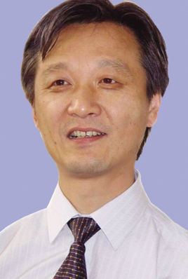 Weijian Jiang (姜卫剑)合作论文数首都医科大学附属北京天坛医院急诊介入科8
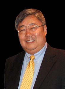 Jianping Dai (戴建平)合作论文数Beijing Tiantan Hospital, Capital Medical University7Haiqiang Qin (秦海强)合作论文数Beijing Tiantan Hospital, Capital Medical Univerity7Jie Long (龙洁)合作论文数5Min Jin合作论文数5SuXiang Wang (王素香)合作论文数5
 Mingwang Zhu (朱明旺)合作论文数Sanbo Brain Hospital, Capital Medical University4
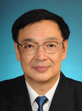 Peiyi Gao (高培毅)合作论文数Beijing Tiantan Hospital, Capital Medical University4JiaXing Huang (黄家星)合作论文数3CunJiang Li (李存江)合作论文数3Jiang Weijian合作论文数3
 Xingquan Zhao (赵性泉)合作论文数Beijing Tiantan Hospital, Capital Medical University3
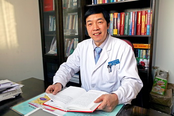 Xinghu Zhang (张星虎)合作论文数Department of Neurology, Beijing Tiantan Hospital,Capital Medical University3
 Baoguo Wang (王保国)合作论文数Sanbo Brian Hospital Capital Medical University3
 Huaguang Zheng (郑华光)合作论文数Beijing Tiantan Hospital, Capital Medical University3
 Wanliang Du合作论文数2Qian Jia合作论文数2
 Yi Ju合作论文数2
 Xiaokun Qi (戚晓昆)合作论文数Chinese PLA General Hospital2Yong Zhou (周永)合作论文数Beijing Tiantan Hospital, Capital Medical University2
 Liping Liu (刘丽萍)合作论文数Beijing Tiantan Hospital, Capital Medical University2
 Xu Yang合作论文数Department of Mathematics, College of Letters and Science, University of California, Santa Barbara2
 Dongning Wei (魏东宁)合作论文数The 8th Medical Center of Chinese PLA General Hospital2H. Q. Zhao合作论文数1Yan Wang合作论文数1Kangxiang Ji合作论文数1Liao Xiaoling合作论文数1Mingchen Ding (丁铭臣)合作论文数1guirong hu (胡桂荣)合作论文数1Fengli Che合作论文数1J Long (龙洁)合作论文数1Yong-jun Wang (王拥军)合作论文数1Xiping Gong合作论文数1hanqing gao (高晗清)合作论文数1Yuelong Xu (许跃龙)合作论文数1Xiaotong Xu合作论文数1huirong zhao (赵惠荣)合作论文数1Xueying Yu合作论文数1Heng Zhou合作论文数1Jian-zhong FU (付建忠)合作论文数1GangZhi Wei (魏岗之)合作论文数1YaQiu Bai (白亚秋)合作论文数1ZhangWei Wu (吴章薇)合作论文数1YiZhen Wang (王毅珍)合作论文数1ChunLu Li (李春禄)合作论文数1Tao Cui (崔韬)合作论文数1




