PATIENTS:This report describes the digital workflow for the design and fabrication of a telescopic prosthesis that incorporates a polyetherketoneketone (PEKK) primary crown and PEKK framework to restore multiple teeth in the mandible of a 56-year-old female patient. The process includes intraoral scanning, computer-aided design, digital milling of the telescopic crown and PEKK framework, and 3D printing of the casts. Good retention, mastication, aesthetics, and biological outcomes are observed in the four-year follow-up. DISCUSSION:Two clinical outcomes are observed. (1) A PEKK telescopic prosthesis can be fabricated using digital workflows, which demonstrates an initial retention value sufficient for intraoral function. (2) No significant wear or decline in the retention force is observed over a four-year follow-up period. CONCLUSIONS:The PEKK telescopic prosthesis facilitated favorable outcomes when restoring extensive mandibular tooth loss. We suggest that PEKK can be effectively used in the fabrication of telescopic prostheses supported by natural teeth.
This clinical report presents the use of an oral surgical robotic system (Yakebot Oral Surgical Robotic System; Yake Wisdom (Beijing) Technology Co, Ltd) for autonomous post space preparation and the immediate cementation of a custom ceramic post-and-core. Preoperative planning was conducted utilizing DentalNavi Oral Surgical Plan Software program, which facilitated the concurrent design and fabrication of the 3-dimensionally (3D) printed ceramic (3Y-TZP; Zrcoop) post-and-core. Postoperative cone beam computed tomography (CBCT) analysis demonstrated the high accuracy of the post space preparation, with an average angular deviation of 0.6 degrees, global coronal deviations of 0.21 mm, and global apical deviations of 0.33 mm. Following tooth preparation and intraoral scanning, a zirconia crown (4Y-TZP; Wieland) was designed, fabricated, and cemented onto the tooth (RelyX Ultimate; 3M ESPE). The patient reported satisfaction with the esthetic outcome and the reduced number of visits required. This autonomous system enabled accurate post space preparation and immediate cementation of the custom ceramic post-and-core, ensuring an excellent fit by guiding the drill's location, orientation, and depth according to the preoperative design. The robotic procedure enhanced operational safety during space preparation and ensures an optimal fit with the custom post-and-core, thereby streamlining the digital workflow.
Introduction and aims Conventional removable partial denture (RPD) clasp fabrication with wrought wire depends on operator skill, limiting efficiency and reproducibility. This study compared robotically and manually bent clasps for fabrication efficiency, adaptation, reproducibility, and initial retentive force. Methods Two groups of clasps (n = 10 per group) were bent using either a robotic clasp-bending machine or an experienced technician. Bending time was recorded. Clasp adaptation and reproducibility were evaluated using scanning and 3D registration software. Initial retentive force was measured using a specialized clasp retention testing system for (1) dentures incorporating robot-bent clasps with 3D-printed bases and (2) dentures incorporating manually bent clasps with conventionally processed acrylic resin bases. Data were analysed using independent-samples t tests or Mann–Whitney U tests, as appropriate, with α = 0.05. Results Robotic clasp fabrication was significantly faster (median, 69.9 seconds) than manual bending (median, 336.9 seconds; P < .001). Robotically bent clasps demonstrated superior adaptation, with smaller clasp–abutment gaps at the clasp body (83.1 µm vs 121.5 µm; P < .001) and reduced variability. At the mid-clasp arm, disto-axial angle of the clasp body, and the middle and lingual aspects of the proximal groove, robotically bent clasps also showed closer adaptation than manually bent clasps (P < .05). Initial retentive force did not differ significantly between groups. Conclusions Robot-assisted clasp bending significantly improved efficiency, clasp adaptation, and reproducibility compared with manual bending, while preserving comparable initial retentive force between groups. Clinical relevance This digital approach offers a practical option for routine RPD fabrication, facilitating more consistent clasp quality and potentially reducing clinical and laboratory workload.
IntroductionPolymethyl methacrylate (PMMA)—a widely applied dental resin-based material—contributes to oral PMMA microplastics (PMMA-MPs) exposure through masticating. PMMA-MPs may facilitate pathogen adhesion, posing substantial risks to oral health. Dental caries represents the most prevalent chronic infectious oral disease, characterized by progressive lesions that may induce pain, tooth structure loss, and compromised masticatory efficiency. Streptococcus mutans have been widely identified as the primary etiological agents responsible for caries pathogenesis. This study aimed to investigate the effects of PMMA-MPs on oral microbiota closely associated with dental caries.MethodsThe impacts of PMMA-MPs were assessed using a standardized murine oral exposure model, followed by the quantification of PMMA-MPs-associated shifts in oral microbiota using high-throughput 16S rRNA gene sequencing. Compared with the control group, PMMA-MPs significantly altered the diversity of oral microbial communities in mice, with a notable increase in the relative abundance of Streptococcus. Based on the 16S sequencing results, S. mutans was selected for subsequent in vitro experiments.ResultsPMMA-MPs markedly enhanced the growth, biofilm formation, and virulence factor synthesis of S. mutans. Transcriptomic analysis revealed that PMMA-MPs may promote biofilm formation through pathways including ABC transporters, quorum sensing, and purine metabolism. Additionally, PMMA-MPs exposure enhanced bacterial antibiotic tolerance.DiscussionOverall, our results revealed that PMMA-MPs can alter the composition of the oral microbial community, while enhancing both the virulence factors and antibiotic tolerance of S. mutans biofilms.
Additive manufacturing of Ti-6Al-4V porous dental implants has become a prominent advancement in the field of prosthetic dentistry, providing enhanced osseointegration and biomechanical long-term stability. To further enhance the biomechanical properties, this study designed different porous structures with axial gradient porosity, investigated their stress distribution, and permeability behavior. The implant–bone mechanical interaction was studied using finite element analysis (FEA). To observe the actual mechanical performance and biological characteristics of implants, the experimental analysis was performed on the laser powder bed fusion LPBF-fabricated specimens together with cytocompatibility tests. The FEA results showed that a Gyroid structure with axial gradient porosity of 40–80
STATEMENT OF PROBLEM:Digitally fabricated occlusal devices represent a novel approach to treating temporomandibular disorders (TMDs), yet they have not been adequately evaluated. PURPOSE:The purpose of this pilot clinical study was to develop a manufacturing process for a digitally fabricated occlusal device, assess its therapeutic effectiveness for various types of TMDs, and examine factors influencing treatment outcomes. MATERIAL AND METHODS:The records of patients with TMDs treated with a digitally fabricated occlusal device from May 2023 to October 2024 were retrospectively analyzed. They were divided into 3 groups based on the diagnosis: anterior disc displacement with reduction (ADDwR), anterior disc displacement without reduction (ADDwoR), or unilateral anterior disc displacement with reduction and unilateral anterior disc displacement without reduction (ADDwR +ADDwoR). Visual analog scale (VAS) scores for pain, diet, mandibular function (MF), and quality of life (QoL) and maximum interincisal opening (MIO) were compared before and at least 8 weeks after treatment. A nonparametric test was used to analyze changes in VAS scores and MIO before and after treatment (α=.05). Effects of sex, age, horizontal or vertical overlap, wearing time, and thickness of the device on the treatment were analyzed using binary logistic regression. RESULTS:Forty-one patients with a mean ±standard deviation age of 27.95 ±11.04 years and a mean ±standard deviation follow-up of 12.51 ±14.10 weeks were evaluated. Pain, diet, MF, and QoL were significantly improved after treatment with a digitally fabricated occlusal device (P<.05), with effective rates of 80.6%, 73.5%, 74.4%, and 66.7%, respectively. Wearing time and thickness of the device affected the treatment (P<.05), whereas sex, age, and horizontal or vertical overlap did not affect the treatment (P>.05). CONCLUSIONS:Digitally fabricated occlusal devices were found to be effective treatment options for TMDs. Wearing a digitally fabricated occlusal device for 24 hours a day led to improved therapeutic outcomes. A 1- to 2-mm-thick device was found to be more effective than 2- to 3- or ≥3-mm options.
A digital workflow for fabricating a polyetherketoneketone (PEKK) periodontal splint is described. The antibacterial properties of PEKK and the precision and efficiency of digital technology led to the provision of a splint with no adverse effects on oral hygiene or periodontal maintenance during a 2-year follow-up.
Digital technologies have become an integral part of complete denture restoration. With advancement in computer-aided design and computer-aided manufacturing (CAD/CAM), tools such as intraoral scanning, facial scanning, 3D printing, and numerical control machining are reshaping the workflow of complete denture restoration. Unlike conventional methods that rely heavily on clinical experience and manual techniques, digital technologies offer greater precision, predictability, and efficacy. They also streamline the process by reducing the number of patient visits and improving overall comfort. Despite these improvements, the clinical application of digital complete denture restoration still faces challenges that require further standardization. The major issues include appropriate case selection, establishing consistent digital workflows, and evaluating long-term outcomes. To address these challenges and provide clinical guidance for practitioners, this expert consensus outlines the principles, advantages, and limitations of digital complete denture technology. The aim of this review was to offer practical recommendations on indications, clinical procedures and precautions, evaluation metrics, and outcome assessment to support digital restoration of complete denture in clinical practice.
A digital workflow is introduced for the same-day delivery of transitional removable partial dentures using a robotic clasp-bending machine. A resin printer creates artificial teeth and denture bases while the robotic machine automatically fabricates clasps from the design data. Clinicians assemble the components in the clinic, delivering the denture during the same visit. This approach significantly enhances clinical efficiency and reduces the waiting time for patients requiring a transitional removable partial denture.
The oral cavity, the gateway to the digestive system, represents a critical entrance for micro- and nanoplastics (MNPs) to enter the human body. Few studies have assessed the long-term accumulation of MNPs in the oral cavity and their potential harm to resident cells. This study investigated the presence of MNPs in human dental calculus and evaluated the cytotoxic and inflammatory effects of polyethylene (PE) on human gingival fibroblasts (HGFs). Twenty-six types of microplastics were identified in human dental calculus, with polyamide (PA, 41.4 %), PE (32.7 %), and polyurethane (PU, 7 %) emerging as the predominant components. In vitro experiments revealed that PE-MNPs significantly reduced HGFs' viability, increased apoptosis rates, and impaired cell migration in a dose-dependent manner. Furthermore, PE-MNPs exposure activated the nuclear factor kappa-light-chain-enhancer of activated B cells signaling pathway and upregulated mRNA expression of pro-inflammatory cytokines (interleukin [IL]-1β and IL-6), as evidenced by elevated phosphorylation of NF-κB. This study revealed that MNPs persistently accumulate in the oral cavity, potentially driving chronic inflammatory activation in gingival fibroblasts and compromising tissue repair mechanisms.
STATEMENT OF PROBLEM:The use of intraoral scanners (IOSs) correlates with clinical outcome and patient satisfaction. While the accuracy of IOSs has been well evaluated, studies on the effect of scanning duration on data accuracy are limited. PURPOSE:The purpose of this in vitro study was to investigate the relationship between different scanning durations and the accuracy of the scanned data. MATERIAL AND METHODS:Two experienced operators used the same intraoral scanner (TRIOS 3; 3Shape A/S) to scan a gypsum cast, but with 5 different scanning durations (30 seconds, 60 seconds, 90 seconds, 120 seconds, and 180 seconds), and the trueness of the scanned data was assessed. Ten scans for each duration group were performed, and all the acquired data were evaluated for precision analysis. In addition, each scanned complete arch cast was divided into anterior and posterior regions at the canine teeth, and the 3-way ANOVA test was used to assess the scanning trueness and precision of the scanned anterior and posterior dental arch. RESULTS:The intraoral scanning results between the 2 operators were highly consistent. The data of the 30-second group showed the lowest trueness and precision (P<.001), whereas no significant difference was found among the other groups (P>.05). The trueness and precision of the scanning data in the posterior region was inferior to that in the anterior region (P<.001). CONCLUSIONS:The duration time of the intraoral scanning (ranging from 60 seconds to 180 seconds) did not influence the accuracy of the acquired data, while excessively rapid scanning adversely affected accuracy.
Aim or purpose: To develop a designing software of digital oral positioning stent for radiotherapy of head and neck, and to evaluate its clinical effect. Materials and methods: 30 patients with nasopharyngeal cancer who received oral examination before radiotherapy in the prosthodontics department from July 1, 2021 to December 31, 2021, were randomly selected and divided into three groups according to the patients' wishes,10 per group: one group without radiotherapy oral positioning stents, one group with traditional radiotherapy positioning stents, and another group with digital radiotherapy positioning stents. The manufacturing time and comfort of the two positioning stents were evaluated, and the radiation doses of the radiotherapy target areas and surrounding healthy tissues were statistically analyzed at the end of radiotherapy. Results: The manufacturing time of digital oral positioning stents(209±7.38 min)was much less than that made of traditional manufacturing method(490±10.33 min), p<0.01.The comfort of patients' wearing digital stents(First wearing:4.9±2.21, At the end of radiotherapy:4±1.56) was better than that of traditional ones(First wearing:7.2±2.10, At the end of radiotherapy:6±2.28), p<0.05. Conclusions: Oral positioning stents for nasopharyngeal carcinoma radiotherapy could greatly reduce the exposure dose of tongue and madible of patients. Digital oral positioning stents designed and manufactured by independently developed software had higher production efficiency than the traditional method, and patients’ comfort to wear was better.
Objective The addition of reinforcement bars is a commonly used method to improve the fabrication trueness of selective laser melting removable partial denture alloy frameworks. However, the effects of different reinforcement bar designs on the trueness of the entire framework remain unclear. This study investigated the trueness of removable partial denture frameworks of pure titanium fabricated by selective laser melting under different reinforcement bar settings. Method A virtual framework was designed based on the Kennedy Class I partially edentulous model using computer-aided design software. Frameworks with different reinforcement bar settings (Ti-A without reinforcement bar, Ti-B with a single horizontal bar joining the lingual bar, Ti-C with two more bars at the anterior region, Ti-D with another horizontal bar at the anterior region, and Ti-E with one more bar at the posterior region, n = 5) were printed using pure titanium powder using a direct metal laser melting machine. The fabricated frameworks were scanned, and their fabrication trueness was compared with the designed virtual framework using one-way ANOVA. Results The overall mean discrepancies for Ti-A, Ti-B, Ti-C, Ti-D, and Ti-E were 0.111, 0.047, 0.073, 0.068, and 0.047 mm, respectively. For the group of Ti-A set with no reinforcement bars, larger discrepancies were observed compared with the other four groups (P < .05). Groups Ti-B and Ti-E showed better trueness of the RPI clasps, rests, and distal ends (P < .05). Conclusions Adding reinforcement bars improved the printing trueness of the pure titanium frameworks, and different settings resulted in various degrees of improvement. Setting a single reinforcement bar to join the lingual bar or an additional reinforcement bar at the distal end significantly enhanced the printing trueness of the RPI clasps, rests, and distal ends.
Objectives: To investigate the influence of the 3D printed micro-structured surfaces on the bond strength of zirconia to resin cement. Methods: Zirconia specimens were divided into five groups based on manufacturing technique and surface preparation: (1) milled zirconia (M group); (2) milled zirconia airborne abraded (MA group); (3) printed zirconia (M group); (4) printed zirconia airborne abraded (PA group); and (5) printed zirconia with micro-structured surface (PM group). The surface morphology, cross-sectional morphology, and elemental composition were observed using a scanning electron microscope (SEM). Surface roughness was measured using a laser scanning confocal microscope (SLCM). Shear bond strength (SBS) was measured using a universal testing machine after bonding resin cement (n = 10). The failure modes of the bonded fracture interfaces were observed and counted using a stereomicroscope and a SEM. In addition, boundary dimensional accuracy (n = 10) and micro-structural dimensional accuracy (n = 20) of printed zirconia specimens with micro-structured surfaces were measured using digital calipers and Fiji software. The crystalline phase changes before and after surface treatment were investigated using X-ray diffractometry. Data was analysed using one-way ANOVA and Tukey HSD post-hoc tests (alpha = 0.05). Result: The surface micro-structures of the PM group had regular morphology and no obvious defects. The surface roughness results showed that the PM group had higher Sa (42.21 +/- 1.38 um) and Ra (21.25 +/- 1.80 um) values than the other four groups (p < 0.001). The SBS test showed that the bond strength of the PM group reached 11.23 +/- 0.66 MPa, which was 55.97% (p < 0.001) higher than that of the P group (7.20 +/- 1.14 MPa). The boundary dimensional accuracy of the PM group was proficient (diameter: 99.63 +/- 0.31%, thickness: 98.05 +/- 1.12%), and the actual fabrication dimensions of the hexagonal micro-structures reached 77.45%-80.01% of the original design. The micro-structured surface did not affect the crystalline phase of zirconia. Conclusions: The current study illustrates that 3D-printed microstructured surfaces effectively improve the bond strength of zirconia to resin cements. Clinical significance: With the advantage of 3D printing, this study provides a new idea for improving the bonding properties of zirconia.
The mechanical properties and permeability properties of artificial bone implants have high-level requirements. A method for the design of trabecular-like porous structure (TLPS) with mixed porosity is proposed based on the study of the mechanical and permeability characteristics of natural bone. With this technique, the morphology and density of internal porous structures can be adjusted, depending on the implantation requirements, to meet the mechanical and permeability requirements of natural bone. The design parameters mainly include the seed points, topology optimization coefficient, load value, irregularity, and scaling factor. Characteristic parameters primarily include porosity and pore size distribution. Statistical methods are used to analyze the relationship between design parameters and characteristic parameters for precise TLPS design and thereby provide a theoretical basis and guidance. TLPS scaffolds were prepared by selective laser melting technology. First, TLPS under different design parameters were analyzed using the finite element method and permeability simulation. The results were then verified by quasistatic compression and cell experiments. The scaling factor and topology optimization coefficient were found to largely affect the mechanical and permeability properties of the TLPS. The corresponding compressive strength reached 270–580 MPa; the elastic modulus ranged between 6.43 and 9.716 GPa, and permeability was 0.6 × 10⁻⁹–21 × 10−9; these results were better than the mechanical properties and permeability of natural bone. Thus, TLPS can effectively improve the success rate of bone implantation, which provides an effective theory and application basis for bone implantation.
BACKGROUND:The integrity and stability of collagen are crucial for the dentin structure and bonding strength at dentin-resin interface. Natural plant-derived polypehenols have been used as collagen crosslinkers.OBJECTIVE:The aims of the study were to develop novel chitosan oleuropein nanoparticles (CS-OL-NPs), and to investigate the CS-OL-NPs treated dentin's the resistance to enzymatic degradation and mechanic property.METHODS:CS-OL-NPs were developed using the ionotropic gelation method. Release and biocompatibility of the CS-OL-NPs were tested. Twenty demineralized dentin collage specimens were randomized into four interventions groups: A, Deionized Water (DW); B, 5% glutaraldehyde solution (GA); C, 1 mg/ml chitosan (CS); and D, 100 mg/L CS-OL-NPs. After 1-min interventions, dentin matrix were evaluated by the micro-Raman spectroscopy for the modulus of elasticity test. Collagen degradation was assessed using hydroxyproline (HYP) assay.RESULTS:CS-OL-NPs were spherical core-shape with a size of 161.29 ± 8.19 nm and Zeta potential of 19.53 ± 0.26 mV. After a burst release of oleuropein in the initial 6 h, there was a long-lasting steady slow release. CS-OL-NPs showed a good biocompatibility for the hPDLSCs. The modulus of elasticity in the crosslinked groups were significantly higher than that in the control group (P< 0.05 for all). The specimens treated with CS-OL-NP showed a greater modulus of elasticity than those treated with GA and CS (P< 0.05 for both). The release of HYP in the crosslinked group was significantly lower than that in the non-crosslinked groups (P< 0.05 for all).CONCLUSION:CS-OL-NPs enhanced the dentin mechanical property and resistance to biodegradation, with biocompatibility and potential for clinical application.
The digital fabrication of a maxillary obturator with a 3D-printed polyetheretherketone (PEEK) framework is described. Digital oral data were scanned for the computer-aided design (CAD) of the framework and the 3D printing of a preliminary resin cast. The framework was accurately printed from a PEEK filament material. A secondary impression was made to fabricate the definitive cast. The PEEK framework exhibited precise fit, excellent retention, and reduced weight compared with a typical metal framework.
In the process of artificial metal bone implantation, the mismatch of elastic modulus and bad permeability behavior lead to obvious stress shielding and poor bone tissue ingrowth. For these problems, this paper proposed a new design method of the variable porosity porous structure based on the stress line and Voronoi diagram. In the design process, the principal stress line was determined by mechanical finite element analysis, which controlled the seeds distribution of the 3D Voronoi diagram, making local porosity con-forms to the stress distribution. In addition, the designed samples were fabricated through laser powder bed fusion (L-PBF) technology with TC4 powders. The mechanical charac-teristics were evaluated by static mechanical simulation with ABAQUS and compression tests, and the permeability characteristics were analyzed by fluid simulation with COMSOL and in vitro and vivo test. The research results showed that the designed structure obtained the relatively low elastic modulus and higher mechanical strength, the designed structure has closer permeability characteristics to the reconstructed natural bone, and was conducive to cell proliferation. So the porous structure based on the stress line and Voronoi diagram might have good application potential in the bionic design of bone implants.(c) 2023 The Author(s). Published by Elsevier B.V. This is an open access article under the CC BY-NC-ND license (http://creativecommons.org/licenses/by-nc-nd/4.0/).
With the increasing demand for bone repair, the bionic bone scaffolds have become a research hotspot. A sub-regional design method of the bionic bone scaffolds, using macrostructural topology, is proposed in this paper, aiming to provide a functionally enhanced region division method for the gradient design. The macrostructural topology was carried out by the bi-directional evolutionary structural optimization (BESO), dividing the predefined design domain into sub-region A and sub-region B. Subsequently, a combined probability sphere model and a distance-to-scale coefficient mapping model are established to implement the graded porosification based on the Voronoi tessellation. This approach takes geometric and mechanical continuity into fully account and assures a reasonable distribution of characteristic parameters, yielding to improve the mechanical strength under specific stress conditions. Finally, the scaffolds were fabricated by the laser powder bed fusion (LPBF) process using the Ti-6Al-4V powder. The results of compression tests are satisfactory, showing that the as-built specimens implement sub-regional functionality. The apparent elastic modulus and the ultimate strength range, respectively, between 1.50 GPa and 7.12 GPa (for the first module) and between 38.55 MPa and 268.03 MPa (for the second module), which conform to the required level of natural bone, providing a possibility for clinical application.
 Yimin Zhao (赵铱民)合作论文数The Third Affiliated Hospital, Air Force Medical University51
 Shizhu Bai合作论文数22Yan Dong合作论文数16Zhihong Feng合作论文数14Yingjie Yi合作论文数12
 Xi Chen (陈晞)合作论文数12
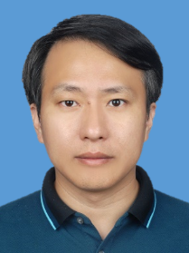 Lida Shen (沈理达)合作论文数College of Mechanical & Electrical Engineering, Nanjing University of Aeronatics and Astronautics11Jiali Meng合作论文数10Jihua Chen (陈吉华)合作论文数Air Force Medical University10
 Shaofeng Zhang (张少锋)合作论文数中国人民解放军第四军医大学9Yunpeng Bi合作论文数9Min Tian (田敏)合作论文数8
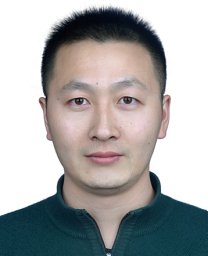 Deqiao Xie (谢德巧)合作论文数7Bo Wang合作论文数7Ya-qi ZHENG (郑亚琪)合作论文数7FangFang Sun (孙方方)合作论文数7DaPeng Zhang (张大鹏)合作论文数7
 Zongjun Tian (田宗军)合作论文数College of Mechanical & Electrical Engineering, Nanjing University of Aeronautics and Astronautics;School of Mechanical Engineering, Jiangsu Ocean University7
 Wang Chang Jiang合作论文数School of Engineering and Informatics, University of Sussex7
 Lin Wang合作论文数Department of Chemical Engineering, Monash University7
 Youwen Yang (杨友文)合作论文数School of Materials Science And Engineering, Hefei University of Technology7
 Chen Jiao合作论文数6
 Chao Long (龙超)合作论文数School of Environment, Nanjing University6Jiaqi Zhang合作论文数5Ling Ding合作论文数5
 Huixin Liang合作论文数5Zhongshan Wang合作论文数5Xinghua Feng (封兴华)合作论文数5Qi Xi合作论文数4Wen Zhao (赵雯)合作论文数4Bing Zhou (周冰)合作论文数4Ming Fang (方明)合作论文数4YuanYuan Li (李媛媛)合作论文数4Bei Li合作论文数4
 Jianhua Wei (魏建华)合作论文数Air Force Medical University4Shu-ming WANG (王书明)合作论文数4Li Zhang合作论文数3Jiasen Gu合作论文数3Yangdong He合作论文数3Kun Ji合作论文数3Guan-yang SUN (孙冠阳)合作论文数3Xiao-lan YE (叶晓兰)合作论文数3Biao Kang (康彪)合作论文数3JianLong Jing (景建龙)合作论文数3BaoLin Liu (刘宝林)合作论文数3
 Dehua Li (李德华)合作论文数College of Physics and Electronic Engineering, Sichuan Normal University3
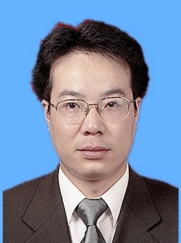 Jianfeng Zhao (赵剑峰)合作论文数College of Mechanical & Electrical Engineering, Nanjing University of Aeronautics and Astronautics3Liang Kong (孔亮)合作论文数Department of Maxillofacial Surgery, Oral Hospital, Fourth Military Medical University3




