Background:The management of advanced thyroid malignancies, particularly poorly differentiated thyroid carcinoma (PDTC), presents significant diagnostic and therapeutic challenges, especially when there is unusual bone involvement. While thyroid cancers typically do not exhibit local invasion to surrounding tissues until advanced stages, this case underscores the necessity for prompt diagnosis and a multidisciplinary approach to treatment to improve patient outcomes. The rarity of sternal infiltration by thyroid cancer and the complexities involved in surgical interventions highlight the need for awareness among healthcare professionals regarding potential diagnoses and treatment pathways. Case Description:We present the case of a 52-year-old male who presented with a painful, progressive anterior chest mass that was diagnosed as bone-infiltrating PDTC. Initial evaluations, including imaging and biopsies, revealed extensive infiltration of the sternum and surrounding structures originating from the thyroid gland. Following an interdisciplinary tumour board discussion, the patient underwent an en bloc resection of the thyroid gland along with affected chest structures and reconstruction with human acellular dermis. Postoperatively, he received radioactive iodine therapy. Despite initial management, follow-up imaging at six months indicated the emergence of bone metastases, prompting the initiation of external beam radiation therapy and recommendations for systemic treatment with a tyrosine kinase inhibitor. Conclusions:This case illustrates the diagnostic complexities and aggressive treatment required for advanced thyroid malignancies with rare bone involvement. Highlighting the importance of a timely and collaborative approach, it emphasizes that surgical intervention, while challenging, can still offer potential for local control in carefully selected patients. However, the case also serves as a reminder of the necessity for ongoing surveillance and the readiness to adapt treatment strategies in response to emerging metastatic disease, contributing to our understanding of management protocols for similar cases in clinical practice.
BACKGROUND:Esophageal adenocarcinoma (EAC) represents one of the most increasing malignancies in Western countries. The disease is multifactorial, involving modifiable risk factors and genetic susceptibility variants. These variants can be aggregated to a polygenic risk score (PRS) that reflects individual genetic risk. Investigation of the effects of lifestyle factors, PRS, and co-medication on EAC age at onset (AAO) is critical for shaping prevention strategies. METHODS:A detailed questionnaire was used to assess pre-diagnostic exposure to lifestyle factors and clinical information from a large German EAC cohort. Linear regression analysis was performed to identify factors associated with EAC AAO in 1742 EAC patients. PRS was available for 1190 patients. Subgroup analyses were conducted to estimate the effects of the analyzed factors on AAO according to age group (early vs. late onset), sex, and prior diagnosis of Barrett's esophagus (BE). RESULTS:Earlier AAO was significantly associated with gastroesophageal reflux (GER), smoking and a higher PRS, whereas later AAO was associated with physical activity and higher consumption of fish and fruits. Among co-medication, combined use of proton pump inhibitors (PPIs) and acetylsalicylic acid (ASA) showed the most significant effect on AAO, whereas the use of PPIs and ASA alone showed weaker effects. DISCUSSION:This study represents the largest questionnaire-based analysis to date investigating factors influencing EAC development. Our findings show that the combined use of PPIs and ASA, both cost-effective medications, is associated with delayed EAC onset. In addition, lifestyle and genetics contribute to EAC AAO.
BackgroundSince the first data of the BILCAP trial was presented in 2017, capecitabine became standard adjuvant treatment for intrahepatic cholangiocarcinoma (ICC). For ICC, the efficacy of capecitabine on survival outcomes remains unresolved as the BILCAP trial included all entities of biliary tract cancer. The aim of this study was to evaluate the influence of adjuvant therapy with capecitabine on long-term outcome in a large German single-center cohort.MethodsAll patients who underwent surgical resection of ICC between January 2008 and December 2023 were divided into a pre-BILCAP (2008–2017) and a post-BILCAP group (2018–2023). For further homogenization both groups underwent a propensity score matching (PSM) in a 1:1 fashion. Survival analysis was conducted using Kaplan Meier model.ResultsIn total 334 patients were included, 254 (76%) underwent resection while 80 (24%) were irresectable. After PSM two comparable groups of 75 patients were generated. Median overall survival was 20.5 months for the pre- and 29.1 months for the post-BILCAP group (p = 0.351). Time to recurrence (TTR, median 10.8 versus 21.3 months, p = 0.019) and recurrence-free survival (RFS, median 8.6 and 10.7 months, p = 0.029) were significantly better for the post-BILCAP group.ConclusionAlthough there was a trend to longer survival, we could not demonstrate a significant effect of adjuvant therapy with capecitabine on an intention to treat basis after resection of intrahepatic cholangiocarcinoma. Nevertheless, a significant influence on time to recurrence and recurrence-free survival could be demonstrated. Further observation is necessary to detect the potential benefit of decreased RFS/TTR in the future.
Background/Objectives: Salvage hepatectomy with a prolonged anhepatic phase represents a potential life-saving strategy in patients with toxic liver syndrome (TLS) following orthotopic liver transplantation (OLT). Available evidence is limited to small case series. Methods: We retrospectively analyzed all patients undergoing OLT between 2014 and 2024 at a German transplant center. Among 497 patients, 33 required retransplantation, including four patients who underwent salvage hepatectomy due to refractory TLS. Clinical trajectories were analyzed descriptively. Additionally, a structured narrative review of the literature was performed. Results: Median age was 63 years (range 62-67), and median anhepatic time was 38.4 h (range 14.39-71.55). No patient died during the anhepatic phase. One patient died during retransplantation, and another during the postoperative course. Two patients survived long-term without neurological impairment. A literature review identified 30 relevant studies with reported mortality rates exceeding 50%. Conclusions: Salvage hepatectomy with subsequent retransplantation may serve as a life-saving bridge in selected patients with TLS. However, outcomes remain heterogeneous, and evidence is limited. This study provides detailed insights into perioperative physiological trajectories and clinical decision-making, which are insufficiently described in the current literature.
Background To investigate imaging-based prognostic factors in patients who underwent thermal ablation for colorectal liver metastases (CRLM), with a focus on sarcopenia-related body composition parameters and L1-bone-density in comparison to tumor burden score (TBS).Patients and methods A retrospective analysis was conducted on patients who received thermal ablation for CRLM at our tertiary care center between 2009 and 2023. CT-derived body composition metrics included the psoas muscle volume index (PMVI), the psoas muscle index (PMI), and L1-bone-density. PMVI was automatically extracted using the open-source deep learning tool TotalSegmentator. Comparisons between 1-year survivors and non-survivors were performed using unpaired t-tests.Results A total of 88 patients were included, most had previously undergone hepatic resection (n = 72, 82%). Among sarcopenia-related imaging markers, PMVI showed a significant association with 1-year survival (p = 0.048), with higher PMVI values observed in survivors (mean 113.3 cm3/m3) compared to non-survivors (mean 101.3 cm3/m3). No significant differences were observed for L1-density (p = 0.925) or PMI (p = 0.137). Similarly, the TBS was not significantly associated with 1-year survival (p = 0.182).Conclusions In our cohort of patients treated with thermal ablation for CRLM, PMVI showed significant association with 1-year survival, which was not observed for conventional tumor burden score or other sarcopenia-related imaging parameters.
Accurate estimation of the functional liver remnant is essential for planning safe liver resections. Unintended vessel transections and subsequent perfusion loss may compromise the functional liver remnant and increase the post-operative risk. We present an approach that models portal and hepatic veins as directed graphs to simulate blood flow and predict downstream perfusion loss by cut vessels. Quantitative metrics, including perfused functional liver remnant and spatial mismatch to the planned resection zone, are automatically computed and visualized. We demonstrated on 22 patients and 31 resections zones that in most cases non-perfused regions extended beyond the planned resection zone, indicating potential risk areas for risk stratification. In conclusion, graph-based perfusion modeling provides quantitative and visual feedback to support pre-operative planning of complex liver resections. This approach may aid in identifying high-risk territories and optimizing resection strategies, demonstrating promising value for surgical decision support.
Abstract Purpose The aim of this study was to evaluate the visibility of colorectal liver metastases (CRLM) using photon-counting detector computed tomography (PCD-CT) and to determine the optimal virtual monoenergetic image (VMI) and iodine map reconstructions for improved contrast detection between metastases and surrounding liver parenchyma. Materials and methods A total of 117 patients with 227 CRLM (up to three measurements per patient) who underwent abdominal PCD-CT for staging between 09/2022 and 08/2024 were retrospectively included. VMI were reconstructed at energy levels between 40 and 90 keV (in 10 keV increments), and scanner-generated iodine maps were additionally analysed. To quantify contrast between CRLM and liver parenchyma, the parenchyma-to-lesion ratio (PLR) was calculated for each VMI and iodine map. The contrast-to-noise ratio (CNR) was determined based on attenuation values of the metastases and the bilateral musculus erector spinae, as well as its standard deviation. For the iodine map, lesion and parenchyma iodine concentrations were used analogously. Subjective assessment of metastases visibility on the three best VMIs in PLR and CNR (40–60 keV) and iodine maps were independently performed by three radiologists. Results Lesion and liver attenuation decreased steadily with higher keV levels. Iodine maps showed markedly higher iodine concentration in liver parenchyma than in metastases. The PLR was highest on the iodine map (3.29 ± 2.01), followed by 40 keV (2.19 ± 0.73). Regarding CNR, the 40 keV VMI showed the highest value (1.49 ± 1.70), followed by the iodine map (1.09 ± 0.99). CNR values decreased further at higher energies and significantly reduced at 70–90 keV. Paired superiority testing confirmed 40 keV as the best-performing VMI, showing significantly higher CNR than the iodine map, whereas PLR remained superior on the iodine map. Subjective ratings indicated that the 50 keV VMI provided the best visibility of CRLM. The iodine map consistently received lower subjective ratings across all criteria. Conclusion Both iodine maps and low-keV VMIs, particularly at 40 keV, demonstrated high PLR and CNR values, contributing to improved depiction of CRLM in PCD-CT. The complementary use of these reconstructions may enhance lesion detection and overall diagnostic confidence.
In this study, we address the problem of automatic liver resection planning for major surgical procedures, including hemi-hepatectomy and extended hemi-hepatectomy, using deep learning. Motivated by clinical practice, where Couinaud liver segments are routinely used to describe tumor location and guide surgical decision-making, we investigate whether incorporating this anatomical information can improve model performance and clinical relevance. We propose a point cloud-based geometric deep learning approach based on a modified RandLA-Net architecture to predict liver resection zones. The model was trained and evaluated on 70 hemi-hepatectomy cases from Johannes Gutenberg University, Mainz, Germany (internal dataset). Two composite loss functions were evaluated: cross-entropy (CE) combined with intersection over union (IoU) and CE combined with Dice loss. For each loss function, models were trained with and without Couinaud segment information. Generalizability was assessed on an external dataset of 30 hemi-hepatectomy cases from the colorectal liver metastases (CRLM) cohort. Both loss functions achieved comparable performance across the evaluated datasets, with CE + IoU consistently outperforming CE + Dice. On the internal test set, incorporating Couinaud segment information increased the IoUmean from 0.787 to 0.804 and the F1-score from 0.864 to 0.870. A Wilcoxon signed-rank test on 15 paired cases confirmed a statistically significant improvement in IoUmean (p = 0.030), with 80
Surgical management of gastric submucosal tumors in anatomically challenging sites such as the gastroesophageal junction (GEJ), fundus, or proximal lesser curvature remains technically demanding. The da Vinci single-port (SP) robotic platform may provide a minimally invasive, organ-preserving alternative by enabling stable endoluminal access and precise dissection. We conducted a case series of five patients who underwent SP robot-assisted transgastric resections for circumscribed gastric pathologies between May and August 2025. Patient demographics, tumor characteristics, operative details, pathology, and short-term outcomes were analyzed. Two patients presented with gastric metastases from malignant melanoma, one with a neuroendocrine tumor (NET), one with a gastrointestinal stromal tumor (GIST), and one with a leiomyoma. All lesions were located in anatomically challenging areas and ranged in size from < 1 cm to 5.5 cm. Four patients underwent submucosal dissection, and one required full-thickness resection. Operative times ranged from 66 to 134 min. Median console time was 38 min. No bleeding, conversion, tumor rupture, or spillage occurred. Patients were discharged between postoperative day (POD)-3 and POD-7, with no reoperations, morbidity, or 30-day mortality. Histopathology confirmed R0 resection of one GIST and both melanoma metastases; the NET G2 had positive margins but very low proliferative activity (Ki-67 < 0.1
BackgroundThe composite endpoint for liver surgery (CELS) was developed to predict surgery-related mortality and to improve the design of clinical trials in liver surgery. However, external validation beyond the initial development cohort remains limited. This study aimed to assess the validity and reliability of CELS in a distinct patient population undergoing liver resection for intrahepatic cholangiocarcinoma.MethodsThe primary objective was to assess the association between CELS and surgery-related mortality. Secondary objectives included 30-day mortality, length of hospital stay (LOS), overall survival, and recurrence-free survival. Predictive performance was evaluated using sensitivity, specificity, accuracy, and receiver operating characteristic curve analyses.ResultsA total of 227 patients were included in the analysis (CELS-positive: n = 87; CELS-negative: n = 140). A total of 58 minor, 62 major, and 107 extended resections were performed. The 90-day mortality rate (surgery-related mortality) was 8.9% in the overall cohort and 9.6% in the validation cohort. The CELS-positive group was more frequently affected by surgery-related mortality compared to the CELS-negative group (20.7% vs. 3.6%, p < 0.001). CELS demonstrated a sensitivity of 81.8%, a specificity of 66.3%, and an overall accuracy of 67.8% in predicting surgery-related death. The discriminatory ability of CELS was moderate, with an area under the receiver operating characteristic (ROC) curve of 0.74.ConclusionsCELS demonstrated moderate predictive ability for surgery-related mortality following liver resection for intrahepatic cholangiocarcinoma.
Despite recent advances in surgical techniques and perioperative management, liver surgery is still associated with perioperative morbidity and mortality. This study aims to investigate the spectrum, risk factors and management of postoperative complications. All patients who underwent liver resection between 01/2008 until 12/2023 were identified from a prospective institutional database. The data was analyzed regarding postoperative morbidity and mortality rates. We identified 3 177 liver resections including 1 345 (42.3
BACKGROUND & AIMS:Curative-intent surgery in patients with perihilar cholangiocarcinoma (pCCA) is associated with substantial surgical risks and high early-recurrence rates. This study aimed to develop an ABC system for preoperative staging of patients with pCCA. METHODS:This retrospective international multicenter cohort study included patients with resected pCCA across 27 participating centers from 9 countries (2006-2022). The prognostic performance of the ABC system for overall survival (OS) and recurrence was assessed using multivariable (cause-specific) Cox regression. RESULTS:Among 1307 included patients (median age, 66 [IQR, 57 to 73] years), independent prognostic factors for OS were: tumor size ≥25 mm (adjusted hazard ratio [aHR], 1.32 [95% CI, 1.13 to 1.55]; P=0.0003), CA19-9 ≥500 U/mL (aHR, 1.49 [1.22 to 1.81]; P<0.0001), and WHO performance status ≥1 (aHR, 1.35 [1.12-1.63]; P=0.002). The ABC score for OS consisted of 1 point for each independent prognostic factor (0-3 points); the ABC score for recurrence did not include WHO PS ≥1 (0-2 points). Compared to the ABC-0 group, the highest ABC risk group had a 3.4 times higher 90-day mortality risk (21 vs 6%; P=0.005), a 3.3 times higher 6-month recurrence rate (18 vs 5%; P<0.0001), a 3.0 times shorter median OS (13 vs 39 months; P=0.0001), and a 3.6 times lower 5-year OS rate (11 vs 35%; P=0.018). The prognostic value of the ABC score for OS remained similar in a sensitivity analysis including only patients with a resection post-2015 (Pinteraction=0.90). CONCLUSIONS:Following the ABC system, we should be reluctant to offer resection to patients with an ABC score of 3 with a tumor size ≥25 mm, CA19-9 ≥500 U/mL, and a WHO PS ≥1.
Introduction: Postoperative pulmonary complications, particularly pneumonia, remain frequent after esophagectomy and contribute significantly to morbidity. One-lung ventilation (OLV) is a potential modifiable risk factor, but its impact in minimally invasive (MIE) and robot-assisted Ivor Lewis esophagectomy (RAMIE) within European populations is not well defined. Methods: 619 patients undergoing MIE or RAMIE were analyzed. OLV duration was extracted from operative records. Postoperative pneumonia incidence, overall survival, and perioperative outcomes were assessed. ASA classification and other risk factors were considered. Results: The overall incidence of postoperative pneumonia was 18.6%, with no significant difference between MIE (20.4%) and RAMIE (18.2%). Prolonged OLV duration increased pneumonia risk by 4% per 10 min. Female sex and higher ASA classification were also significant risk factors. Likely reflecting early diagnosis and advanced perioperative management, pneumonia did not affect overall survival, which remained comparable between MIE and RAMIE. Conclusions: Prolonged OLV during MIE and RAMIE increases the risk of postoperative pneumonia without significantly affecting overall survival, reflecting effective complication management. OLV duration may serve as a practical intraoperative indicator to guide risk stratification and optimize postoperative care in minimally invasive and robot-assisted Ivor Lewis esophagectomy.
Vascular endothelial cells (ECs) play a pivotal role in the pathogenesis of a variety of disorders. The characterization of ECs during these processes is limited to the availability of purified cells. We developed a technique for isolation of human macrovascular ECs from small pieces of blood vessels and investigated whether ECs could be used in an assay system capable of measuring the immunoreactivity in vitro. ECs were isolated 0-3 days after collection of small pieces of blood vessels using Collagenase H digestion and immunomagnetic cell separation. The isolated cells were characterized by immunohistochemical staining of CD31 and human leucocyte antigen HLA-I/-II. Interferon-g (IFN-g) stimulated ECs were used in co-culture with allogeneic peripheral blood mononuclear cells (PBMCs). The activation of allogeneic T cells was investigated using an interleukin-2 (IL-2)-specific ELISA. In 85 % of all preparations, the cultivation of ECs was successful. The purity was determined by immunohistochemistry to be nearly 95 %. Cultivated ECs did not express HLA-II and showed only little expression of HLA-I molecules. The expression of HLA-I/-II could be induced by incubation with IFN-g. In co-culture experiments of ECs with allogeneic PBMCs, a T cell immune response could be detected by measuring IL-2. This strategy of isolating human ECs permits to generate ECs from very small quantities of starting material up to three days after vessel collection. ECs might be used in the context of solid organ transplantation for measuring the immune reactivity of the organ recipient but also for a wide variety of applications regarding pathophysiological disorders associated with ECs.
Background Systemic chemotherapy is considered the standard treatment for recurrent perihilar cholangiocarcinoma, whereas evidence regarding repeated surgical resection remains scarce. Methods We performed a retrospective analysis of our prospectively collected institutional database of all patients who underwent resection for recurrent perihilar cholangiocarcinoma. Statistical analysis included Kaplan-Meier analysis for survival data. Results Between 2008 and 2025, 9 patients underwent repeat resection for recurrent perihilar cholangiocarcinoma. Median time between first and second resection was 40 months (range: 21-85 months). Site of recurrence was locoregional/at the hepatic hilum (n = 3), distant intrahepatic (n = 4) and extrahepatic (n = 2). The residual tumor status was R0 in 7 patients and R1 in 2 patients. There was no 90-day mortality. Eight of nine patients developed a second tumor relapse after a median of 10 months (range 3-130 months) after repeat resection. In two patients, re-repeat resection was performed for a second recurrence in the liver. Median OS after repeat resection was 27 months with a 1-, 3-, and 5-year survival of 89%, 39%, 23% compared to 13 months and a 1-, 3-, and 5-year-survival of 54%, 12% and 0% after non-surgical management (P = 0.037). Two patients are alive without evidence of disease 50 and 194 months after first repeat resection. Conclusions In carefully selected patients with recurrent perihilar cholangiocarcinoma, repeat resection is a technically challenging but feasible and safe treatment option which can potentially offer mid- and even long-term survival.
In perihilar cholangiocarcinoma major liver resections are often required to attain oncological radicality, but these approaches are associated with a high perioperative morbidity and mortality. The aim of this study was to assess the outcome of minor resections compared to standard major liver resections. A single-institutional database was queried for all patients undergoing resection for perihilar cholangiocarcinoma between 2008 and 2024. Minor resections were defined as resections of the hilar bifurcation including up to three liver segments, whereas major resections exceeded 3 segments. The primary study endpoint was safety and feasibility of resection represented by 30-day mortality. Secondary endpoints included survival outcomes. The primary outcome was evaluated using logistic regression. Survival outcomes were analysed using Kaplan–Meier method and Cox regression. Additionally, a propensity score matching was conducted, grouping variables by age and Bismuth-Classification. Two hundred sixteen patients underwent resection, 182 in the major resection and 34 in the minor resection group. Patients in the minor resection group were older (71.4 vs. 66.0 years, p = 0.007) and had more comorbidities (Charlson Comorbidity Index: 5.3 vs. 4.7, p = 0.03). After propensity score matching demographic data were balanced between groups. Minor resection was consistently associated with a trend toward lower 30-day mortality rate (2.9
Background: Calcitonin, a tumor marker primarily used to diagnose medullary thyroid carcinoma (MTC), can also be elevated in other conditions, complicating diagnosis. This study aims to provide a clinical evaluation of the real-world consequences of unexplained calcitonin elevation. Methods: We conducted a retrospective cohort study of patients with elevated basal calcitonin levels who presented at the Department of General, Visceral, and Transplantation Surgery, University Medical Center Mainz, between January 2015 and March 2025. Additionally, we reviewed electronic health records from 2007 onward for patients with ICD codes indicating calcitonin hypersecretion. Patients with confirmed MTC or genetic syndromes were excluded. Results: Of 345 patients with elevated calcitonin levels, 167 (48%) met the inclusion criteria, and 29 additional patients with calcitonin hypersecretion were identified via ICD, resulting in 167 patients analyzed. More than half of the patients were female (52%), had an average age of 53.9 years and a high prevalence of goiter (86%). Calcitonin levels were slightly elevated (<20 pg/mL) in 81% of cases and were above 50 pg/mL in only 10 patients. Surgery was performed in 77% of patients, mainly to exclude malignancy. Postoperatively, calcitonin normalized in 86% of patients but remained elevated in eight patients. Two of these patients were found to have false-positive results due to assay interference. Follow-up data were incomplete for a substantial proportion of patients, with a median follow-up of 4.6 months. The mortality rate was 4%, with causes unrelated to calcitonin levels. Conclusions: Elevated basal calcitonin levels, especially slightly elevated levels (<20 pg/mL), are common in clinical practice and often do not appear to be related to malignant disease, so careful investigation is required. Persistently elevated calcitonin levels justify further examinations, especially if other explanations can be ruled out. Only a few patients attend follow-up appointments, which makes patient follow-up challenging.
 Ines Gockel合作论文数Department of Operative Medicine, University Hospital of Leipzig;New Westminster College143
 Peter Galle合作论文数Medical Center, Johannes Gutenberg University Mainz130
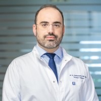 Georgios C. Sotiropoulos合作论文数Athens Medical Grοup;Barts Health NHS Trust112
 Massimo Malago合作论文数Liver Center, University of California at San Diego Medical Center110
 W. Kneist合作论文数Klinik und Poliklinik für Allgemein- und Abdominalchirurgie, Johannes-Gutenberg-Universität Mainz106
 Peter Grimminger合作论文数Department of General, Visceral and Transplantation Surgery, Universitätsmedizin der Johannes Gutenberg-Universität Mainz91
 Mittler Jens合作论文数86Fabian Bartsch合作论文数85
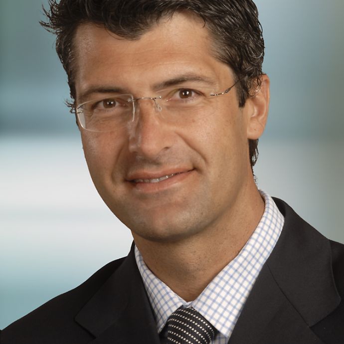 Silvio Nadalin合作论文数Universitätsklinik für Allgemeine, Viszeral- und Transplantationschirurgie, Universitätsklinikum Tübingen79C.E. Broelsch合作论文数74Baumgart Janine合作论文数68Maria Hoppe-Lotichius合作论文数62Evangelos Tagkalos合作论文数Universitätsmedizin der Johannes Gutenberg-Universität Mainz56
 Arndt Weinmann合作论文数55Markus Paschold合作论文数54Arnold Radtke合作论文数53
 Dw Kauff合作论文数52
 Tom Zimmermann合作论文数51
 Tobias Huber合作论文数50George Sgourakis合作论文数42Christoph Broelsch合作论文数Department for General, Visceral and Transplantation Surgery, Essen University Hospital42
 Jens Marquardt合作论文数University of Lübeck;University Hospital Schleswig-Holstein38
 Ernesto Molmenti合作论文数36
 Markus Moehler合作论文数Universitätsmedizin der Johannes Gutenberg-Universität Mainz35A. Paul合作论文数34
 Roman Kloeckner合作论文数Department of Diagnostic and Interventional Radiology, Johannes Gutenberg-University34
 Beate K. Straub合作论文数Institut für Pathologie, Universitätsmedizin Mainz34
 Thomas J. Musholt合作论文数Johannes Gutenberg-Universität Mainz33Florentine Huettl合作论文数Johannes Gutenberg Univ Mainz, Univ Med, Dept Gen Visceral & Transplant Surg, Mainz, Germany32
 Andrea Frilling合作论文数Department of Surgery & Cancer, Faculty of Medicine, Imperial College London32E P Molmenti合作论文数31
 Carl C. Schimanski合作论文数Johannes Gutenberg University of Mainz31Edin Hadzijusufoviç合作论文数30
 Florian Lordick合作论文数University Cancer Center Leipzig, University of Leipzig;Department of Medicine II, Leipzig University Medical Center;Comprehensive Cancer Center Central Germany30
 Johannes Schumacher合作论文数Institut für Humangenetik, Philipps-Universität Marburg;Institute for Human Genetics, University of Bonn;National Institute of Health in Bethesda30
 Darko Castven合作论文数28
 Michael Vieth合作论文数Department of Pathology, Medizinisches Versorgungszentrum, Klinikum Bayreuth GmbH;Institut für Pathologie, Klinikum Bayreuth GmbH28
 Stefan Heinrich合作论文数Technische Universitat Kaiserslautern28
 Wolf Bechstein合作论文数27Wilfried Roth合作论文数27
 Jakob R. Izbicki合作论文数Department of General, Visceral & Thoracic Surgery, University Medical Center Hamburg-Eppendorf25
 Peter Schirmacher合作论文数Translational Spatial Profiling Center, Universität Heidelberg;Pathologisches Institut, Universität Heidelberg;Institute of Pathology, Universität Heidelberg25
 Karl J. Oldhafer合作论文数Klinik für Allgemein- und Viszeralchirurgie , Asklepios Klinik Barmbek25J. Pratschke合作论文数24
 Pierre-Alain Clavien合作论文数Privatklinik Bethanien;Wyss Zurich Translational Center;Hôpital Paul-Brousse;Department of Surgery, University Hospital Zurich;King’s College London24
 Markus Nöthen合作论文数Institut für Humangenetik, Universitätsklinikum Bonn;Institute of Human Genetics, University of Bonn24
 Felix Berlth合作论文数23
 Ioannis P Fouzas合作论文数23




