BACKGROUND:Pancreatic cancer incidence is increasing worldwide while survival remains poor. The incidence varies significantly across different regions. The aim of the present study was to analyse incidence, mortality and survival of patients diagnosed with pancreatic cancer in the canton of Zurich, Switzerland. METHODS:Population-based cancer registry data and cause of death statistics of the canton of Zurich from 1981 to 2021 were analysed. Age-standardised incidence and mortality rates per 100'000 person-years and net survival were calculated. Joinpoint regression analysis was conducted to identify significant changes in time trends while net survival was estimated using the Pohar-Perme method. RESULTS:Age-standardised incidence rate of pancreatic adenocarcinomas (PAC) remained stable between 1981 and 2000 and then increased from 2000 to 2021, while pancreatic neuroendocrine tumours (PNET) incidence increased throughout and pancreatic cancers non histologically or cytologically confirmed (PC Non-HC) decreased. Overall age-standardised mortality rate remained stable. Five-year net survival was moderate for PNET (54.8%), but poor for PAC (4.8%) and PC Non-HC (1.2%). Among patients with PAC, survival declined with increasing age, earlier diagnosis year and advanced stage. CONCLUSIONS:Over the past 40 years, PNET incidence slowly increased. In contrast, PAC incidence significantly increased within the last 20 years. The underlying causes of the observed increase in PAC incidence might be attributable to special risk factors, e.g. obesity, but other risk factors should also be considered. Mortality rates of pancreatic cancer were stable and net survival was poor.
Birt-Hogg-Dubé (BHD) syndrome is an autosomal dominant disorder caused by germline inactivation of the folliculin gene (FLCN). Approximately 25
Radiologists should be familiar with the World Health Organization 2022 renal cell tumor classification and recognize that although most of the rare renal tumors in these categories show gradual enhancement similar to that of papillary renal cell carcinomas, they vary based on patient age, growth pattern, CT attenuation, enhancement degree, homogeneity, T2 signal intensity, and existence of metastases.
The International Society of Urological Pathology Florence Multidisciplinary Consensus Conference establishes a contemporary framework for the definition and standardized reporting of genitourinary cancer precursor lesions. These consensus recommendations provide evidence-based guidance for diagnostic practice, inform clinical management, and define priorities for future research in genitourinary premalignancy.
PURPOSE:Melanoma treatment has advanced with targeted therapies and immunotherapies, but challenges like resistance and response variability remain. Traditional clinical trials and diagnostics struggle to address the molecular heterogeneity of cancer, limiting their effectiveness in overcoming these challenges. The prospective, multicentric observational Tumor Profiler (TuPro) precision oncology project demonstrated the feasibility and the potential of multi-omics-guided therapy selection to account for individual tumor complexity and improve clinical outcomes, particularly in later treatment lines. However, the safety of this multi-omics-guided approach has not yet been established. MATERIALS AND METHODS:We compared the TuPro melanoma cohort (n = 98), which received multi-omics-guided selection of adjuvant, standard of care and beyond standard-of-care therapies in patients with advanced melanoma, to a matched, retrospective synchronous cohort that did not undergo multi-omics tumor profiling. Adverse events, hospitalizations, and adverse event-related costs were analyzed to assess safety and clinical impact. RESULTS:TuPro-guided therapy selection enabled more treatments beyond standard of care (SOC) without increasing high-grade adverse events, hospitalizations, or adverse event-related costs. However, we observed a higher incidence and earlier onset of predominantly low-grade adverse events, primarily within the SOC setting, with shorter durations and higher recovery rates. CONCLUSION:These findings support the clinical feasibility and safety of multi-omics-guided therapies, demonstrating their potential to expand treatment options and efficacy without necessarily increasing toxicity. Prospective studies are needed to validate these results, and further research should focus on leveraging multi-omics data to predict and mitigate adverse events.
Phenotypic composition of tumor cells from PT and distant metastasis. A, Left, heatmap showing the median expression level for each tumor cell cluster after flowSOM clustering. Markers used for clustering are on the x-axis, and flowSOM clusters are on the y-axis. For clustering, all cells were pooled across samples (n = 87 patients, 559,953 cells), and the cell counts in each cluster are shown (bar plot, gray). Euclidean distance with Ward-D2 linkage was used for the hierarchical clustering of rows. Right, box plot shows the per patient proportion of tumor cell phenotypic clusters out of all tumor cells for PT (left) and metastatic (right) samples (n = 76 patients with paired samples). B, Stacked bar plot of all tumor cell phenotypic clusters as a proportion of all tumor cells in PT and metastatic samples for paired patient samples. Rows were hierarchically clustered into 14 patient groups using Euclidean distance of tumor phenotype composition in metastases with Ward-D2 linkage. C, Tumor cell phenotypic cluster enrichment in PT or metastasis determined with differential abundance testing using paired design. The bar plot shows the log2-fold abundance changes (n = 76 patients). D, Tumor cell phenotypic cluster enrichment in each metastatic patient group compared with average metastasis composition by differential abundance testing. Circles display significantly enriched (red) or decreased (blue; FDR < 0.05; multiple testing correction with the Benjamin–Hochberg method) results, with size indicating the log-transformed fold change.
Compared with cancers of other organs, precursor lesions of renal cell neoplasms are rarely discussed. However, there are specific scenarios where cysts or microscopic solid lesions are thought to precede tumor formation. In 2024, the International Society of Urological Pathology (ISUP) held a consensus meeting on precursor lesions of urologic neoplasms in Florence, Italy. This report details the findings of Working Group 3-Precursor Lesions of the Kidney. Papillary adenoma is likely the best-established precursor lesion in the kidney, thought to be an incipient form of papillary renal cell carcinoma (RCC) with shared morphology, immunohistochemistry, and genetics. Likewise, in patients with VHL disease, the kidney often contains multiple small nodules of clear cells and/or cysts lined by one or more layers of clear cells, likely representing early tumor or precursor lesions. Interestingly, a precursor counterpart for clear cell RCC in the sporadic setting is not well established. In other scenarios, cysts are considered potential precursors of neoplasia, such as those in acquired cystic kidney disease (ACKD) and possibly in some hereditary renal tumor syndromes. The consensus panel proposes the following terms "cyst with epithelial proliferation" for tufted/hyperplastic/cribriform cyst lining in ACKD without solid tumor, and "papillary hyperplasia" for tufting of cyst lining in autosomal dominant polycystic kidney. Terms such as "tumorlet," "microtumor," or "incipient tumor" are acceptable for incidental unencapsulated microscopic lesions in hereditary syndrome patients. There is insufficient evidence for the diagnosis of "tubular dysplasia" as a precursor to RCC at the present time.
PURPOSE:Repeated evidence demonstrates limited reproducibility and accuracy of the visual quantification (VQ) of the tumor cell content (TCC) by clinical pathologists for downstream molecular testing. Artificial intelligence (AI)-based digital quantification (DQ) of TCC represents a promising alternative, yet real-world evidence from routine molecular diagnostic workflows remain limited. In this study, we evaluated the analytical performance and practical aspects of analytical validation process of an AI-based DQ tool in routine molecular diagnostics. MATERIAL AND METHODS:The clinical-grade AIM-TumorCellularity (AIM-TC; PathAI©) workflow was tested in molecular diagnostics for samples analyzed by comprehensive genomic profiling (FoundationOne®CDx (F1CDx), Foundation medicine Inc.). The cohort included 300 non-paired resection, biopsy, and cytology/cell block) specimens from primary and metastatic breast (n = 66), lung (n = 117), colorectal (n = 40), pancreatic (n = 38), and prostate (n = 39) cancers, reflecting real-world diagnostic sample heterogeneity of a tertiary care center. We compared TCC estimates generated by pathologists' VQ, AI-based DQ, and molecular quantification (MQ) by bioinformatic deconvolution. RESULTS:Agreement was lowest between VQ and MQ (Spearman Rs = 0.38) and between VQ and DQ (Rs = 0.44), while DQ showed stronger concordance with MQ (Rs = 0.63). Single-cell validation against expert ground truth demonstrated high performance of DQ in tumor cell detection, with sensitivity of 0.98.5, specificity of 0.99, and accuracy of 0.99, based on 27,958 annotated cells across 60 regions of interest comparable to microscopic high-power fields. Analysis of pre-analytical and analytical factors identified specimen type and cautery/crush artifacts as the main pre-analytical contributors to DQ-VQ discrepancies, while overall variations in specimen cellularity was the dominant analytical factor. CONCLUSION:In summary, this study provides the first comprehensive real-world evaluation of AI-based TCC quantification in routine molecular pathology workflow, highlighting its robustness, accuracy, and the critical role of pre-analytical standardization, as well as pathologists` oversight for successful clinical implementation.
Clear cell renal cell carcinoma exhibits striking intra-tumoral heterogeneity at morphological and genetic levels, complicating treatment and contributing to disease progression. CcRCCs with rhabdoid differentiation are highly aggressive tumors characterized by distinct histopathologies. However, the relationship between morphology, underlying molecular alterations, and tumor behavior remains largely unclear. Here, we present Deep Visual Multi-Omics, an approach integrating digital pathology, morphology-guided single-cell isolation, and ultra-sensitive multi-omics profiling to link cell morphologies to their molecular underpinnings. Across five tumors, we profiled 40,000 AI-classified and expert-curated cells. We identified progressive molecular dysregulation across cells with increasing histopathological grade coexisting within heterogeneous tumors as well as distinct molecular alterations associated with aggressive rhabdoid ccRCC cells, including signatures consistent with enhanced FOXM1-driven proliferation, altered cell-matrix interactions, and a putative immunomodulatory phenotype. Notably, rhabdoid cells exhibited elevated expression of IFN-beta, PD-L1, CD38, ITGB2, and integrin signaling, suggesting that they themselves may act as a source of signals influencing the local immune microenvironment. Besides providing new insights into the biology of ccRCC and highlighting avenues for future translational studies, this illustrates the potential of Deep Visual Multi-omics to dissect cancer heterogeneity and characterize high-risk cell populations. Intra-tumor heterogeneity drives tumor progression and therapy resistance. In ccRCC for example, distinct histopathologies coexist and are associated with patient outcome, yet their molecular basis is poorly understood. Deep Visual Multi-Omics links cell morphology to the underlying molecular state. Intra-tumor heterogeneity drives tumor progression and therapy resistance. In ccRCC for example, distinct histopathologies coexist and are associated with patient outcome, yet their molecular basis is poorly understood. Deep Visual Multi-Omics links cell morphology to the underlying molecular state.
A decade into the formal adoption of the World Health Organization (WHO)/International Society of Urological Pathology (ISUP) grading system for clear cell renal cell carcinoma (CCRCC) and papillary renal cell carcinoma (PRCC), newer grading concepts, innovative approaches and some issues have emerged. A comprehensive review of the literature on the WHO/ISUP grading system and other grading approaches for CCRCC and PRCC was conducted. Updates and issues are presented on the following: (1) validation studies on WHO/ISUP grading; (2) grade elements including heterogeneity, grades 1 versus 2, types of multinucleated tumour cells, 'grade spectrum' of spindle cells, impact of sarcomatoid change in grade 4 RCC, percent (%) high grade, % sarcomatoid change and rhabdoid change versus sarcomatoid change; (3) grade applications including observer agreement, needle biopsy grade accuracy (correlation with nephrectomy grade) and grading of multifocal RCCs; (4) grade in papillary tumours including heterogeneity and on small (≤1.5 cm) neoplasms; and (5) novel grading or risk categories including incorporation of necrosis into grading and pattern or architectural-based grading. Practice guidance for some of the issues is provided when feasible or where data are sufficient. This review highlights the recent updates and controversies in the use of WHO/ISUP grading and on novel approaches to grading for CCRCC and PRCC. This review may serve as a best practice guide in addressing some of the grading issues and will help identify gaps in our understanding and use of grading for CCRCC and PRCC that can inform future research.
Compared with cancers of other organs, precursor lesions of renal cell neoplasms are rarely discussed. However, there are specific scenarios where cysts or microscopic solid lesions are thought to precede tumor formation. In 2024, the International Society of Urological Pathology (ISUP) held a consensus meeting on precursor lesions of urologic neoplasms in Florence, Italy. This report details the findings of Working Group 3—Precursor Lesions of the Kidney. Papillary adenoma is likely the best-established precursor lesion in the kidney, thought to be an incipient form of papillary renal cell carcinoma (RCC) with shared morphology, immunohistochemistry, and genetics. Likewise, in patients with VHL disease, the kidney often contains multiple small nodules of clear cells and/or cysts lined by one or more layers of clear cells, likely representing early tumor or precursor lesions. Interestingly, a precursor counterpart for clear cell RCC in the sporadic setting is not well established. In other scenarios, cysts are considered potential precursors of neoplasia, such as those in acquired cystic kidney disease (ACKD) and possibly in some hereditary renal tumor syndromes. The consensus panel proposes the following terms “cyst with epithelial proliferation” for tufted/hyperplastic/cribriform cyst lining in ACKD without solid tumor, and “papillary hyperplasia” for tufting of cyst lining in autosomal dominant polycystic kidney. Terms such as “tumorlet,” “microtumor,” or “incipient tumor” are acceptable for incidental unencapsulated microscopic lesions in hereditary syndrome patients. There is insufficient evidence for the diagnosis of “tubular dysplasia” as a precursor to RCC at the present time.
TFE3 rearranged renal cell carcinoma (TFE3-tRCC) is a rare subtype of renal cell carcinoma (RCC) harboring gene fusions that trigger oncogenic activation of TFE3 transcription factor. The frequency of this rare tumor is surprisingly high in children and young adults (20% to 75% of childhood RCCs) compared to older adults (ranges between 1% and 5%). Importantly, TFE3-tRCC can show aggressive behavior with rapid progression and little to no response to drugs commonly used for renal cancer treatment. In this project we aim to define gene expression programs of distinct TFE3 fusions to uncover molecular events driven by chimeric TFE3 and delineate their consequences for important cancer phenotypes. By identifying fusion-specific pathway alterations, we seek to propose novel therapeutic strategies tailored to the patients with these fusions. Using a cohort of 31 patients with suspected diagnosis of TFE3-tRCC, we have employed a novel customized RNA-based targeted Next generation Sequencing (NGS) panel to determine putative fusion partners of TFE3. Next, high throughput technologies like short read and long read RNA sequencing and ChIP sequencing were used to characterize the molecular landscape downstream of different fusion events. Finally, to explore the influence of the transcriptional diversity on therapeutic decisions, we aim to perform molecularly informed drug screening in our in vitro models followed by in vivo validation. With our newly developed targeted NGS panel, we have characterized our patient cohort and identified 7 distinct TFE3 fusion partners in 25 cases out of 31. The putative partners are ASPSCR1, LUC7L3, MED15, NonO, PRCC, RBM10, and SFPQ. Certain partners confer unique tumor morphologies, e.g., MED15::TFE3 fusion associates with cystic features. Our transcriptomic analysis shows tumors with same fusion tend to cluster together, driven by shared pathway upregulation (OXPHOS, lysosomal pathway, autophagy, proteasome etc.). Differential upregulation of pathways such as immune response, metabolic pathways, mitophagy, RNA splicing etc. highlight fusion specific identity, informing fusion driven molecular characteristics. Preliminary in vitro chromatin accessibility data suggests the potential involvement of the chimeric TFE3 protein in alternative splicing regulation, which is further consistent with splicing enrichment observed in our transcriptomic analysis. Different fusion partners for TFE3 likely impact the dysregulation of the chimeric protein and drives tumorigenesis. However, our limited understanding of the molecular landscape and clinical consequences of the different TFE3 fusions presents as the major obstacle towards development of targeted therapeutic strategies for this disease. In this scenario of unmet clinical needs, we seek to define the molecular features of TFE3-tRCC, thereby uncovering specific vulnerabilities of tumor cells that prompt the development of new targeted therapeutic strategies. Debleena Basu, Hella Bolck, Dorothea Rutishauser, Peter Leary, Daniela C. Garcia (s), Abdullah Kahraman, Niels Rupp, Chantal Pauli, Holger Moch. Development of a mechanistic understanding and novel therapeutic strategies for TFE3-rearranged renal cell carcinoma [abstract]. In: Proceedings of the AACR Special Conference in Cancer Research: Fusion-Positive Cancer: From Discovery to Therapy; 2026 Jan 13-15; Philadelphia PA. Philadelphia (PA): AACR; Cancer Res 2026;86(1_Suppl):Abstract nr A022.
CUPISCO (ClinicalTrials.gov identifier: NCT03498521) demonstrated longer progression-free survival (PFS) with comprehensive genomic profiling (CGP) and subsequent molecularly guided therapies (MGTs), versus standard platinum-based chemotherapy, in patients with previously untreated, unfavorable cancer of unknown primary (CUP) who reached disease control after induction chemotherapy (three cycles). We report efficacy and safety after >1 year of additional follow-up. Eligible patients were randomly assigned (3:1) to MGT (investigator-chosen after discussion in a molecular tumor board) or three further cycles of chemotherapy. The primary end point was PFS. Secondary end points included overall survival (OS) and safety. At data cutoff (December 6, 2024), 436 patients were randomly assigned (326 to MGT; 110 to chemotherapy). Median follow-up was 37.0 months (range, 0.0-67.8). Updated median PFS was 6.1 months (95% CI, 4.7 to 6.5) with MGT and 4.4 months (95% CI, 4.2 to 6.4) with chemotherapy (hazard ratio [HR], 0.75 [95% CI, 0.59 to 0.95]; P = .017); median OS was 15.2 months (95% CI, 13.9 to 18.4) and 12.8 months (95% CI, 9.8 to 15.4), respectively (HR, 0.79 [95% CI, 0.61 to 1.02]; P = .0689). No new safety signals were identified. These updated results aligned with the primary analysis, demonstrating the benefit of CGP with subsequent MGT and highlighting the importance of incorporating CGP at initial diagnosis to guide treatment decisions for patients with unfavorable CUP.
PBMCs collected before vaccination show low/no response to candidate vaccine peptides. (A) The nomenclature of the N-peptides and D-neopeptides. I or II indicate if the peptides were chosen/designed for HLA class I or -II binding; MUT/RNA/GBM indicates a naturally mutated peptide (MUT), derivation from the over-/highly expressed genes (RNA) or from known glioblastoma (GBM) targets; X indicates the number of the peptide; 0 or 1 indicate if the peptides had been artificially mutated (1) or not (0); (’) indicates that peptides contained the naturally mutated amino acid. (B) PBMCs collected before vaccination were stimulated with 4 μg/mL Tetanus toxoid or 2 μM CEF II peptide pool for 7 days, and proliferation was measured by ³H-thymidine incorporation assay. The proliferation strength is depicted as counts per minute (cpm). In each group, the N-peptides and their corresponding D-neopeptides are included, and the designed mutated positions are highlighted in the red box. The blue dotted line indicates the mean value of the no peptide control. The red dotted line indicates the mean value plus three standard deviations of the no peptide control. Values above the red dotted line were considered positive. (C) PBMCs collected before vaccination were stimulated with 5 μM predicted class I-peptides, including N-peptides and D-neopeptides, for 7 days, and proliferation was measured by ³H-thymidine incorporation assay. 10-15 replicate wells were tested for proliferation, and responses were depicted as cpm. (D) PBMCs collected before vaccination were stimulated with 5 μM predicted class II-peptides, including N-peptides and D-neopeptides, for 7 days, and proliferation was measured by ³H-thymidine incorporation assay. 10-15 replicate wells were tested for proliferation, and responses were depicted as cpm.
Federated learning (FL) allows institutions to collaboratively train deep learning models while maintaining data privacy, a critical aspect in fields like computational pathology (CPATH). However, existing studies focus on performance improvement in simulated environments and overlook practical aspects of FL. In this study, we address this need by transparently sharing the challenges encountered in the real-world application of FL for a clinical CPATH use case. We set up a FL framework consisting of three clients and a central server to jointly train deep learning models for digital immune phenotyping in metastatic melanoma, utilizing the NVIDIA Federated Learning Application Runtime Environment (NVIDIA FLARE) across four separate networks from institutes in four countries. Our findings reveal several key challenges: First, the FL model performs the best across all clients' test sets but does not outperform all local models on their own client test set. Second, long experiment duration due to system and data heterogeneity limited experiment frequency, alleviated by optimizing local client epochs. Third, infrastructure design was hindered by hospital and corporate network restrictions, necessitating an open port for the server, which we resolved by deploying the server on an Amazon Web Services infrastructure within a semi-public network. Lastly, effective experiment management required IT expertise and strong familiarity with NVIDIA FLARE to enable orchestration, code management, parameter configuration, and logging. Our findings provide a practical perspective on implementing FL for CPATH, advocating for greater transparency in future research and the development of best practices and guidelines for implementing FL in real-world healthcare settings.
Rapid recurrence is common in triple-negative breast cancer (TNBC). To better understand drivers of recurrence, we use imaging mass cytometry to characterize the tumor phenotype landscapes of 215 TNBC patients. We observe high intertumor heterogeneity with eleven tumor cell phenotypes, each of which dominates in an individual patient, and identify a tumor cell phenotype with reduced basoluminal lineage fidelity and stem-like traits that is correlated with rapid disease recurrence. Scoring of tumor-CD8+ T cell interactions identifies patients with inflamed tumors and high HLADR expression. We combine these features in multi-omics analyses of 8 cohorts with 3737 patients across all molecular subtypes to propose five prognostic breast cancer subtypes distinguished by tumor cytokeratin expression profiles and CD8+ T cell spatial patterns. This stratification scheme has direct clinical implications: inflamed tumors show good prognosis and high immunotherapy response rates, whereas patients dominated by basoluminal tumor cells have poor prognosis.
While advances in the understanding of tumor biology through multi-omics profiling hold the promise of substantially improving patient outcomes, the cost implications of such strategies remain unclear. We therefore performed a comparative cost analysis of patients treated either within the Tumor Profiler (TuPro) melanoma project or from a control cohort who received treatment after standard next-generation sequencing testing. After adjustment of cohorts through inverse probability of treatment weighting, we found no evidence of statistically significant differences in total costs between the two cohorts (95% confidence interval -10% to +67%). Importantly, treatment costs (95% confidence interval -28% to +41%) were similar between the two cohorts. In conclusion, we found no evidence that treatment recommendations guided by advanced multi-omics profiling led to significantly higher treatment costs in a Swiss context.
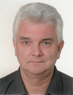 Peter Schraml合作论文数Institute of Pathology and Molecular Pathology, University Hospital Zurich163
 Guido Sauter合作论文数Zentrum für Diagnostik, Universitätsklinikum Hamburg-Eppendorf;Ambulanzzentrum des UKE GmbH, Universitätsklinikum Hamburg-Eppendorf;Fachbereich Pathologie und Gynpathologie, Universitätsklinikum Hamburg-Eppendorf117
 Urs P. Wild合作论文数87
 Alex Soltermann合作论文数Institut für Klinische Pathologie, Medizinische Fakultät, Universität Zürich;Pathologie Länggasse74Michael J. Mihatsch合作论文数Institute for Pathology63Rechsteiner Markus P合作论文数60Zsuzsanna Varga合作论文数59Mj Mihatsch合作论文数58
 Tullio Sulser合作论文数Faculty of Medicine, University of Zurich;Zentrum für Urologie Zürich – Klinik Hirslanden48
 Glen Kristiansen合作论文数Institute of Pathology, University Hospital Bonn47
 Lukas Bubendorf合作论文数Institute for Pathology, University of Basel45
 Thomas Hermanns合作论文数45
 Alexander K Knuth合作论文数Department of Oncology, University Hospital Zurich44TC Gasser合作论文数40
 Arndt Hartmann合作论文数Department of Pathology, Friedrich-Alexander-University of Erlangen-Nürnberg40
 Maries Van Den Broek合作论文数Institute of Experimental Immunology, University of Zürich39
 Reinhard Dummer合作论文数Department of Dermatology, University Hospital Zürich38
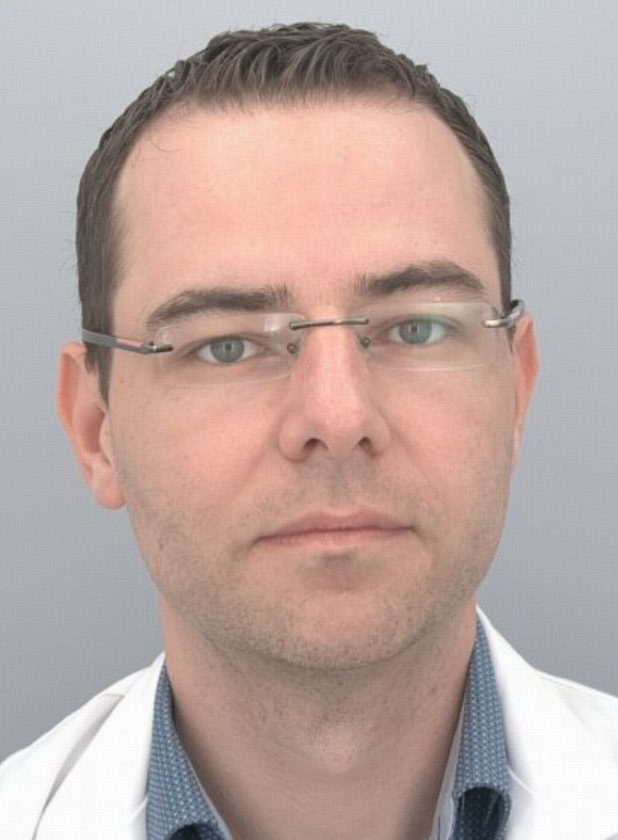 Niels Rupp合作论文数Department of Pathology and Molecular Pathology, University Hospital Zürich37Krek Wilhelm合作论文数36
 Liang Cheng合作论文数Department of Surgery, Warren Alpert Medical School of Brown University;Department of Pathology and Laboratory Medicine, Warren Alpert Medical School of Brown University36
 Mark Rubin合作论文数Department for BioMedical Research, Faculty of Medicine, University of Bern;Bern Center for Precision Medicine, University of Bern35
 Martin Zoche合作论文数Institut für Biologische Informationsverarbeitung, Forschungszentrum Jülich34Jan Richter合作论文数33
 Sven Perner合作论文数University Hospital of Luebeck;Department of Pathology, Leibniz Research Center Borstel32
 Kirsten Mertz合作论文数University Hospital Basel;Basel Research Centre for Child Health31Walter Weder合作论文数30
 Rodolfo Montironi合作论文数Section of Pathological Anatomy, School of Medicine, Polytechnic University of Marche;Department of Biomedical Sciences and Public Health, School of Medicine, Polytechnic University of Marche30
 Michael Weller合作论文数Department of Neurology, University Hospital Zurich29
 Mahul B. Amin合作论文数Labcorp;Keck School of Medicine of the University of Southern California;The University of Tennessee Health Science Center29
 Aurel Perren合作论文数Institute of Tissue Medicine and Pathology, Faculty of Medicine, University of Bern;Institute of Pathology, University of Bern29Florian R Fritzsche合作论文数28
 Alessandra Curioni-Fontecedro合作论文数University of Fribourg;Hospital of Fribourg27
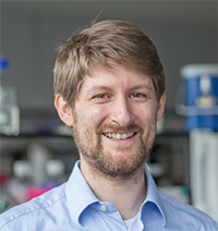 Bernd Bodenmiller合作论文数Department of Quantitative Biomedicine, University of Zurich27Komminoth Paul合作论文数27Antonio Lopez-Beltran合作论文数26Pauli Chantal合作论文数Inst Pathol & Mol Pathol, Univ Hosp Zurich26Srigley J R合作论文数26Urs Wagner合作论文数26
 Luigi Maria Terracciano合作论文数Anatomic Pathology Division, Department of Biomedical Sciences, Humanitas University Hospital25
 Abdullah Kahraman合作论文数25
 Brett Delahunt合作论文数Department of Pathology and Molecular Medicine, Wellington School of Medicine and Health Sciences, University of Otago24Christian Britschgi合作论文数23
 Burkhardt Seifert合作论文数Division of Biostatistics, University of Zurich;Biostatistics and Prevention Institute, University of Zurich23
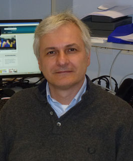 Guido Martignoni合作论文数Department of Pathology, University of Verona;Department of Diagnostics and Public Health, University of Verona23
 Eberli Daniel合作论文数22
 Victor E. Reuter合作论文数Department of Pathology, Memorial Sloan Kettering Cancer Center;Department of Pathology and Laboratory Medicine, Weill Medical College, Cornell University22Verena Tischler合作论文数Univ Zurich Hosp, Inst Surg Pathol, CH-8091 Zurich, Switzerland21Ikenberg Kristian合作论文数21von Teichman Adriana合作论文数21




