Automatic segmentation of 2D echocardiograms is a critical step in diagnosing and assessing heart diseases. Convolutional Neural Networks (CNNs), such as U-Net, have proven effective for medical image segmentation, but they often fall short in capturing long-range dependencies because convolution operations are inherently local. While transformer models capture the global context, they also may not be good in fine-grained localization. This study aims to develop a novel deep learning architecture that addresses these limitations by combining the strengths of CNNs and transformers for improved segmentation accuracy in 2D echocardiograms. We propose a novel encoder-decoder framework enriched with multiple attention mechanisms and hybrid bottleneck combining a Vision Transformer (ViT) and a Multi Receptive Field Block (MRFB). The integration of a ViT enables the modeling of long-range dependencies, while the MRFB improves multi-scale feature extraction and is specifically designed to improve the representation of spatial features and reduce the loss of spatial information. Enhanced by multiple attention mechanisms and deep supervision with Atrous Spatial Pyramid Pooling (ASPP), our model robustly captures both local and global contexts. Trained and validated on the CAMUS dataset, it achieves Dice Similarity Coefficients of 91.11±0.19, 87.60±0.11, and 87.85±0.29 for left ventricle endocardium, epicardium, and left atrium in two-chamber views, and 92.25±0.17, 87.29±0.17, and 91.71±0.10 in four-chamber views. Our model outperforms pure CNN-based (such as UNet), pure transformer-based (such as Swin-unet), and CNN-Transformer hybrid (such as UNETR-2d) architectures, achieving superior segmentation accuracy and robustness. Code is publicly available on the paper’s https://github.com/saeedchamani/CAMUS_Segmentation_Hybrid_Bottleneck repository.
High-Intensity Focused Ultrasound (HIFU) is a non-invasive therapeutic modality that uses high-energy acoustic waves to thermally coagulate tissue at the focal region. The Twinkling Artifact (TA) is a color Doppler artifact caused by the acoustic radiation force and the consequent tissue vibration during Doppler imaging. This paper aims to employ TA for real-time detection and monitoring of HIFU-induced lesions. A dataset gathered in a previous study concerning ex vivo porcine tissue samples was used, in which the real-time backscattered radiofrequency signals were acquired before, during, and after HIFU treatment. To investigate the presence of TA in Doppler images, the amplitude of each pixel is considered in the sequence of frames as time-series or slow-time signals. It is shown that the main frequency of slow-time signals represents the Doppler frequency shift. Doppler images were constructed using the maximum frequency from every 10-sample slow-time signal. By constructing Doppler images, the frequency shifts within tissue during HIFU treatment were visually and analytically assessed. Our frequency analysis of RF data confirmed the occurrence of TA during HIFU exposure. Furthermore, a novel method was developed for lesion formation monitoring, with less than a 5% error rate in depth and width measurements for depicting coagulated tissue dimensions.
Breast cancer is the most common and lethal cancer among women worldwide. Early detection using medical imaging technologies can significantly improve treatment outcomes. Automated breast ultrasound, known as ABUS, offers more advantages compared to traditional mammography and has recently gained considerable attention. However, reviewing hundreds of ABUS slices imposes a high workload on radiologists, increasing review time and potentially leading to diagnostic errors. Consequently, there is a strong need for efficient computer-aided detection, CADe, systems. In recent years, researchers have proposed deep learning-based CADe systems to enhance mass detection accuracy. However, these methods are highly dependent on the number of training samples and often struggle to balance detection accuracy with the false positive rate. To reduce the workload for radiologists and achieve high detection sensitivities with low false positive rates, this study introduces a novel CADe system based on a self-supervised framework that leverages unannotated ABUS datasets to improve detection results. The proposed framework is integrated into an innovative 3-D convolutional neural network called DATTR2U-Net, which employs a multi-task learning approach to simultaneously train inpainting and denoising pretext tasks. A fully convolutional network is then attached to the DATTR2U-Net for the detection task. The proposed method is validated on the TDSCABUS public dataset, demonstrating promising detection results with a recall of 0.7963 and a false positive rate of 5.67 per volume that signifies its potential to improve detection accuracy while reducing workload for radiologists. The code is available at: github.com/Pooryamn/SSL_ABUS.
Increasing the frame rate of ultrasound imaging while keeping image quality is important for following fast movements, especially the heart. There are different modalities for B-mode image recording, including line-by-line scanning with linear, phased, convex array, synthetic aperture imaging (STA), plane waves (PWI), then the combination of plane waves (CPWI), and so on. Researchers have tried to increase the frame rate in each case using different methods. Three approaches for this aim are data acquisition, post-processing, and beamforming. This article reviews these approaches and their solutions for compensating image quality reduction. Ultrafast ultrasound imaging, which provides exceptional temporal resolution (high frame rate), is promising in diagnosing heart diseases due to its ability to capture rapid heart movements. It can record images faster than conventional imaging, usually exceeding 1000 frames per second. This can be achieved through plane wave imaging (PWI). However, high frame rate data acquisition can lead to a decrease in image quality. Transmitting at different angles and then combining plane wave imaging is a popular method to enhance PWI quality but reduces the frame rate by the number of angles. As a result, researchers have aimed to increase the temporal resolution while compensating for the loss of quality.
Ultrasound (US) has become a widely used imaging modality in clinical practice, characterized by its rapidly evolving technology, advantages, and unique challenges, such as a low imaging quality and high variability. There is a need to develop advanced automatic US image analysis methods to enhance its diagnostic accuracy and objectivity. Vision transformers, a recent innovation in machine learning, have demonstrated significant potential in various research fields, including general image analysis and computer vision, due to their capacity to process large datasets and learn complex patterns. Their suitability for automatic US image analysis tasks, such as classification, detection, and segmentation, has been recognized. This review provides an introduction to vision transformers and discusses their applications in specific US image analysis tasks, while also addressing the open challenges and potential future trends in their application in medical US image analysis. Vision transformers have shown promise in enhancing the accuracy and efficiency of ultrasound image analysis and are expected to play an increasingly important role in the diagnosis and treatment of medical conditions using ultrasound imaging as technology progresses.
Background: Noninvasive therapies such as focused ultrasound were developed to be used for cancer therapies, vessel bleeding, and drug delivery. The main purpose of focused ultrasound therapy is to affect regions of interest (ROI) of tissues without any injuries to surrounding tissues. In this regard, an appropriate monitoring method is required to control the treatment. Methods: This study is aimed to develop a noninvasive monitoring technique of focused ultrasound (US) treatment using sparse representation of US radio frequency (RF) echo signals. To this end, reasonable results in temperature change estimation in the tissue under focused US radiation were obtained by utilizing algorithms related to sparse optimization as orthogonal matching pursuit (OMP) and accompanying Shannon’s entropy. Consequently, ex vivo tissue experimental tests yielded two datasets, including low-intensity focused US (LIFU) and high-intensity focused US (HIFU) data. The proposed processing method analyzed the ultrasonic RF echo signal and expressed it as a sparse signal and calculated the entropy of each frame. Results: The results indicated that the suggested approach could noninvasively estimate temperature changes between 37°C and 47°C during LIFU therapy. In addition, it represented temperature changes during HIFU ablation at various powers, ranging from 10 to 130 W. The normalized mean square error of the proposed method is 0.28, approximately 2.15 on previous related methods. Conclusion: These results demonstrated that this novel proposed approach, including the combination of sparsity and Shanoon’s entropy, is more feasible and effective in temperature change estimation than its predecessors.
This research proposes a deep learning method for breast mass segmentation in 2D ultrasound images. The low quality of ultrasound images and indistinct boundaries of masses cause difficulties in the automatic segmentation of breast masses. To solve this problem, we introduce an autoencoder-based model that is equipped with resolution enhancement blocks. The proposed model is composed of U-Net, along with residual blocks and attention blocks. Its objective is to increase the accuracy of breast mass segmentation by enhancing the feature maps and paying more attention to the region of interest, and preventing the loss of features in the encoder part of U-Net, this helps to rebuild an image with better quality in the decoder section. The proposed model achieved an accuracy of 98.84% and a Dice score of 84.72% for image containing benign tumor and 82.14% for image containing malignant tumor. For the IoU metric, the model obtained scores of 85.95% and 82.21% for images containing benign and malignant tumors, respectively. These results showed an improvement in segmentation accuracy compared to the performance of previous models based on U-Net. This highlights the usefulness and advantage of using U-Net and resolution enhancement blocks for breast mass segmentation1.
Cardiovascular diseases (CVD) are today a major cause of death globally that is diagnosed by measurement and quantification of left ventricle (LV) wall motion (WM) abnormality of the heart. The aim of this study was to assess the utility of left ventricular (LV) entropy, a novel measure of disease derived from two-dimensional (2D) echocardiography images that assesses the probability distribution of pixel intensities in the LV. The purpose of this research is to develop the method of LV entropy to predict heart diseases. In this algorithm, a frame is usually chosen as the reference frame to extract the region of interest (ROI) around LV and then it is mapped to all images in a cardiac cycle. Then Shannon Entropy transform was applied to calculate the distribution of pixel intensities across the LV so we obtained entropy curves and compared them. The main idea is to find a motion estimation accuracy. The results obtained by our method are quantitatively evaluated to those obtained by an experienced echocardiographer visually on 22 normal cases and 19 myocardial infarction (MI) cases in apical four-chamber (A4C) view. The entropy of diastole in MI cases was 0.50 (0.29-0.58) while in normal cases was 0.75 (0.64-1.13). The entropy of systole in MI cases was 0.64 (0.26-1.04) while in normal cases was 0.81 (0.63-1.26). The percent change of entropy for diastole and systole between normal and MI cases are 33.3% and 20.2%. The results indicate that the LV entropy curves of MI cases have less changes than normal cases.
Mitral valve (MV) diseases constitute one of the etiologies of cardiovascular mortality and morbidity. MV pathologies need evaluating and classifying via echocardiographic videos. Transformers have significantly advanced video analytics. MV motion is divided by Carpentier functional classification into four types: normal, increased, restricted, and restricted only during systole. This paper introduces CarpNet, a deep transformer network that incorporates video transformers capable of direct MV pathology Carpentier's classification from the parasternal long‐axis (PLA) echocardiographic videos. The network, instead of processing frames independently, analyzes stacks of temporally consecutive frames using multi‐head attention modules to incorporate MV temporal dynamics into the learned model. To that end, different convolutional neural networks (CNNs) are evaluated as the backbone, and the best model is selected using the information of the PLA view. The use of information obtained by our proposed deep transformer network from consecutive echocardiographic frames yielded better results concerning the Carpentier functional classification than information obtained by CNN‐based (single‐frame) models. Using the Inception_Resnet_V2 architecture as the backbone, CarpNet achieved 71% accuracy in the test dataset. Deep learning and transformers in echocardiographic videos can render quick, precise, and stable evaluations of various MV pathologies.
Introduction:Functional neuroimaging has developed a fundamental ground for understanding the physical basis of the brain. Recent studies have extracted invaluable information from the underlying substrate of the brain. However, cognitive deficiency has insufficiently been assessed by researchers in multiple sclerosis (MS). Therefore, extracting the brain network differences among relapsing-remitting MS (RRMS) patients and healthy controls as biomarkers of cognitive task functional magnetic resonance imaging (fMRI) data and evaluating such biomarkers using machine learning were the aims of this study. Methods:In order to activate cognitive functions of the brain, blood-oxygen-level-dependent (BOLD) data were collected throughout the application of a cognitive task. Accordingly, a nonlinear-based brain network was established using kernel mutual information based on the automated anatomical labeling atlas (AAL). Subsequently, a statistical test was carried out to determine the variation in brain network measures between the two groups on binary adjacency matrices. We also found the prominent graph features by merging the Wilcoxon rank-sum test with the Fisher score as a hybrid feature selection method. Results:The results of the classification performance measures showed that the construction of a brain network using a new nonlinear connectivity measure in task-fMRI performs better than the linear connectivity measures in terms of classification. The Wilcoxon rank-sum test also demonstrated a superior result for clinical applications. Conclusion:We believe that non-linear connectivity measures, like KMI, outperform linear connectivity measures, like correlation coefficient in finding the biomarkers of MS disease according to classification performance metrics. Highlights:The performance of some brain regions (the hippocampus, parahippocampus, cuneus, pallidum, and two segments of the cerebellum) is different between healthy and MS people.Non-linear connectivity measures, such as Kernel mutual information, perform better than linear connectivity measures, such as correlation coefficient, in finding the biomarkers of MS disease. Plain Language Summary:Multiple sclerosis (MS) can disrupt the function of the central nervous system. The function of brain network is impaired in these patients. In this study, we evaluated the change in brain network based on a non-linear connectivity measure using cognitive task-based fMRI data between MS patients and healthy controls. We used Kernel mutual information (KMI) and designed a graph network based on the results of connectivity analysis. The the paced auditory serial addition test was used to activate cognitive functions of the brain. The classification was employed for the results using different decision tree -based technique and support vector machine. KMI can be considered a valid measure of connectivity over linear measures, like the correlation coefficient. KMI does not have the drawbacks of mutual information technique. However, further studies should be implemented on brain data of MS patients to draw more definite conclusions.
Low intensity focused ultrasound (LIFU) is a novel approach that could activate drug release and considerably improve the delivery of anticancer drug. LIFU treatment has some features like is able to penetrate deep into the tissue and being non-invasive, as a consequence LIFU displays great capability for controlling the drug release and improving the chemotherapy treatment efficiency. The goal of this study is to research the feasibility of the entropy parameter of RF time series of ultrasound backscattered signals for measuring the changes in temperature induced by a LIFU device. Entropy Imaging is a technique for reconstructing ultrasound images based on the average uncertainty of time-series in a signal. Furthermore, the Shannon Entropy can quantify the uncertainty of a random process and is usually used as a measure for the information content of probability distributions. In this study, we use the Entropy Imaging method for measuring the LIFU-induced temperature changes in the deep region of ex vivo porcine tissue samples. The results obtained show that the changes of entropy parameter of RF time series signal are proportional to temperature changes recorded by a calibrated thermocouple in the temperature range of 37-47°C. In conclusion, in this study we show that Shannon entropy of RF time series signal possesses promising features like succinctly capturing the available information in a system by considering the uncertainty in a given data that can be used, as a new method, to measure temperature changes non-invasively and quantitatively in the deep region of tissue.
A semi-automatic method for the segmentation of the Left Ventricle in echocardiography images is presented. The manual segmentation of the left ventricle in all image sequences takes a lot of time. The proposed method is based on sparse representation and the design of overcomplete dictionaries based on prior knowledge of the intensity variation time curves (IVTC). We used the sparse recovery algorithm of orthogonal matching pursuit (OMP) to find the sparse coefficients of the IVTC signals. We obtained the histogram of non-zero sparse coefficients for all images. The binary images from successive frames were constructed via thresholding. In addition, we defined one image representing all the frames, dividing all the points of the heart into three groups. One group involved the points located inside the cavities in all frames. The second group included the points that belonged to the tissue in all frames. Points that in some frames are located inside the cavities and in some other frames are located inside the tissue. The results on 2D echocardiographic images acquired from both healthy and patient subjects showed good agreement with manual tracing and took a short time for the contour, including the whole left ventricle. According to the cardiology specialist, the value of ejection fraction is correctly calculated, and the error percentages were 0.83 and 2.33 for two healthy data samples. The proposed method can be applied to 3D echocardiography images to obtain the left ventricular volume. This approach also can be used for other types of medical images.
Speckle tracking echocardiography (STE) is widely used to quaantify regional motion and deformation of heart tissues. Before tracking, a segmentation step is first carried out, and only a set of nodes in the segmented model are tracked. However, a random selection of the nodes even after tissue segmentation could lead to an inaccurate estimation. In this paper, a convolutional neural network (CNN)-based method is presented to detect trackable speckle spots that have important properties of the texture for speckle tracking. The proposed CNN was trained and validated on 29500 ultrasound manually labelled image patches extracted from the echocardiography of 65 people. Using the proposed network, in silico experiments for automatic node selection were conducted to investigate the applicability of the proposed method in speckle tracking. The results were statistically highly significant (P<0.001) and demonstrated that the proposed method has the least tracking error among various existing methods.
Graph theory allows us to gain better insight concerning changes in brain functional architecture associated with cognitive impairments in the early stages of multiple sclerosis (MS). In the present study, we employed a machine-learning system based on graph measures from functional networks constructed by cognitive-task-related functional magnetic resonance imaging (fMRI) data. We used a predefined atlas to define the brain regions and Pearson’s correlation to describe the connectivity strength between the regions. Then, several graph metrics were extracted for each subject. After that, the most efficient subsets of features were selected through the Wilcoxon rank-sum test, and the linear support vector machine (SVM) classifier was employed to distinguish between MS and healthy subjects. The node degree, subgraph centrality, K-coreness, and PageRank centralities measured in the left fusiform, hippocampus, and parahippocampal gyri regions demonstrated an accuracy of 85% through the combination of all local measures. Two optimal global measures, modularity and small-worldness index, and individual betweenness centrality feature improved the identification of MS patients with a sensitivity of 81.25%. Our results indicated the potential of the proposed system to identify cognitive changes in early MS for diagnostic purposes.
Plane-wave imaging (PWI) is an interesting technique for increasing frame rate in medical ultrasound imaging, but it suffers from poor image quality. To overcome this problem, the coherent plane-wave compounding (CPWC) approach has been introduced, which provides the possibility of achieving the desired resolution and contrast by combining images obtained from different angles. In this paper, we proposed a new technique based on adaptive compounding of resulted radio frequency (RF) image data at different angles to obtain a high resolution image. We called this method coherent plane wave adaptive compounding (CPWAC). The adaptive method used here is the minimum variance (MV) approach. An important benefit of the CPWAC method is that the calculation and inversion of the covariance matrix is done only once. We applied the proposed method on simulated, experimental, and in vivo datasets of the plane wave imaging challenge in medical ultrasound (PICMUS) data by considering 75 and 25 plane waves (PW). The results showed that the proposed CPWAC improves the lateral full width at half maximum (FWHM) by 35% for both simulated and experimental datasets, even by using 33% of the total angles compared to CPWC, which indicates simultaneous improvement of lateral resolution and frame rate. In addition, for the experimental dataset, this method has improved the {CR, CNR} about {103.8%, 100%} and {30.28%, 28%} in the near-field and far-field, respectively. Therefore, while the proposed method does not add much time overhead compared to the original CPWC, it improves the image quality of the CPWC significantly.
The frame rate improvement is an essential issue in dynamic ultrasonic imaging for better displaying rapid heart movements. In this study, a new technique using the compressive sensing (CS) theory was introduced for the frame rate improvement of two‐dimensional (2D) and three‐dimensional (3D) dynamic ultrasonic imaging. In the suggested procedure, a fewer radio frequency (RF) lines were received. Subsequently, the CS theory by the recommended approach was used to reconstruct the nonacquired RF lines. The suggested process uses the temporal and spatial information in the neighborhood samples of the desired sample in RF frame sequences. We utilized the dictionary learning technique to construct the sparsifying over‐complete dictionary. Here, we used the 2D in vivo carotid artery data and the 3D simulated echocardiography for the quality assessment of the suggested approach. According to the qualitative and quantitative results, reconstructions in the suggested approach are more precise, and the reconstruction errors are much lower than the conventional techniques. The tissues' structure in reconstructed videos by the suggested approach is preserved to be applicable for medical diagnosis goals. The frame rate can be improved by a factor of 10 with suitable quality for medical diagnosis goals. The implementation of the suggested approach does not require any more hardware or alternation in the devices' available hardware. The frame rate can be improved by only implementing effective programming on the ultrasound systems.
The first step in the automatic evaluation of the cardiac prosthetic valve is the recognition of such valves in echocardiographic images. This research surveyed whether a deep convolutional neural network (DCNN) could improve the recognition of prosthetic mitral valve in conventional 2D echocardiographic images. An efficient intervention to decrease the misreading rate of the prosthetic mitral valve is required for non-expert cardiologists. This intervention could serve as a section of a fully-automated analysis chain, alleviate the cardiologist's workload, and improve precision and time management, especially in an emergent situation. Additionally, it might be suitable for pre-labeling large databases of unclassified images. We, therefore, introduce a large publicly-available annotated dataset for the purpose of prosthetic mitral valve recognition. We utilized 2044 comprehensive non-stress transthoracic echocardiographic studies. Totally, 1597 patients had natural mitral valves and 447 patients had prosthetic valves. Each case contained 1 cycle of echocardiographic images from the apical 4-chamber (A4C) and the parasternal long-axis (PLA) views. Thirteen versions of the state-of-the-art models were independently trained, and the ensemble predictions were performed using those versions. For the recognition of prosthetic mitral valves from natural mitral valves, the area under the receiver-operating characteristic curve (AUC) made by the deep learning algorithm was similar to that made by cardiologists (0.99). In this research, EfficientNetB3 architecture in the A4C view and the EfficientNetB4 architecture in the PLA view were the best models among the other pre-trained DCNN models.
To solve the problem of resolution and contrast in plane wave imaging (PWI), coherent plane wave compounding (CPWC) was introduced, in which scanning was performed at different angles, which can achieve the desired image quality by combining the images obtained from PWI at different angles. However, the application of this idea reduces the frame rate in proportion to the number of plane waves (PWs) or angles, so that in this modality, when dealing with some applications such as shear wave imaging (SWI) and strain imaging, there is always a compromise between the frame rate and the image quality. Tensor completion (TC) is a powerful technique to recover missing information of a low-rank tensor from limited observations based on rank minimization. In this article, we present an idea based on TC to make this compromise lighter; in other words, with a smaller number of angles, we can achieve the desired quality of the output image. To evaluate the proposed idea, plane wave imaging challenge in medical ultrasound (PICMUS) datasets was used, which were recorded at 75 different angles. The results of the resolution evaluation showed that using 20% of the coherent PWs and reconstructing other 80% by TC, compared with the situation of using only 20% of the coherent PWs provided a resolution improvement of 14.97% and 17.4% in the simulated and experimental point targets, respectively. Also, the results of the contrast investigation showed that the contrast ratio (CR) improved by 72.6%, 62.9%, and 111.4% in the simulated cyst target data, experimental cyst targets, and in vivo carotid cross section, respectively. The results confirmed that using 20% of the coherent PWs and reconstructing other 80% by TC, the image quality is very close to that obtained by considering all 75 angles, so that the difference in resolution is less than 2% and the difference in contrast to noise ratio (CNR) is less than 5 dB. Therefore, with this idea, it can be said that less compromise is needed; in other words, despite having a higher frame rate, an acceptable quality can be achieved.
One of the most important methods in medical ultrasound imaging is the synthetic transmit aperture (STA). Despite the image quality improvement in the STA, this method suffers from several limitations, including a limited data acquisition rate and an increase in the overall time to form a single frame. Tensor completion (TC) is a powerful technique that uses rank minimization to recover missing information from a low-rank tensor. This paper provides a novel random synthetic transmit aperture (RSTA) method based on using only a randomly selected part (a fraction) of the linear array elements in the transmit mode to increase the data acquisition rate and then applying the tensor completion (TC) to improve the image quality. By the proposed method, as it is not necessary to transmit all elements sequentially, the data acquisition rate is improved and the overall time for creating an image is also significantly reduced. We investigated the proposed idea by using several simulated and experimental phantoms. Results showed that the proposed method could increase the data acquisition rate up to three times with the image quality difference of less than 6% compared to the original STA method.
Cardiac multimodal image fusion can offer an image with various types of information in a single image. Many coronary stenosis, which are anatomically clear, are not functionally significant. The treatment of such kind of stenosis can cause irreversible effects on the patient. Thus, choosing the best treatment planning depend on anatomical and functional information is very beneficial. An algorithm for the fusion of coronary computed tomography angiography (CCTA) as an anatomical and transthoracic echocardiography (TTE) as a functional modality is presented. CCTA and TTE are temporally registered using manifold learning. A pattern search optimization algorithm, using normalized mutual information, is used to find the best match slice to TTE frame from CCTA volume. By employing a free-form deformation, the heart's non-rigid deformations are modeled. The spatiotemporal registered TTE frame is embedded to achieve the fusion result. The accuracy is evaluated on CCTA and TTE data obtained from 10 patients. In temporal registration, mean absolute error of 1.97 $$\, \pm \, $$ 1.23 is resulted from comparing the output frame numbers from the algorithm and from manual assignment by an expert. In spatial registration, the accuracy of the similarity between the best match slice from CCTA volume and TTE frame is resulted in 1.82 $$\, \pm \, $$ 0.024 mm, 6.74 $$\, \pm \, $$ 0.013 mm, and 0.901 $$\, \pm \, $$ 0.0548 due to mean absolute distance, Hausdorff distance, and Dice similarity coefficient, respectively. Without the use of ECG and Optical tracking systems, a semiautomatic framework of spatiotemporal registration and fusion of CCTA volume and TTE frame is presented. The experimental results showed the effectiveness of our proposed method to create complementary information from TTE and CCTA, which may help in the early diagnosis and effective treatment of cardiovascular diseases (CVDs).
 Zahra Alizadeh Sani合作论文数Tehran University of Medical Science48
 Parisa Gifani合作论文数38
 Maryam Shojaeifard合作论文数Rajaie Cardiovascular Medical and Research Center, Iran University of Medical Sciences23
 Jahan Tavakkoli合作论文数Ryerson University16
 Ahmad Shalbaf合作论文数13
 Emad Fatemizadeh合作论文数Biomedical Signal and Image Processing Laboratory, Sharif University of Technology11
 Farzan Haddadi合作论文数11
 Reza Shalbaf合作论文数School of Electrical Engineering, Iran University of Science &Technology;School of Electrical Engineering, Iran University of Science & Technology10
 Maryam Noroozian合作论文数Roozbeh Psychiatric Hospital, Tehran University of Medical Sciences9
 Mohammad Reza Mohammadi合作论文数Psychiatry and Psychology Research Center, Tehran University of Medical Sciences9
 Ali Sheikhani合作论文数Biomedical Engineering, Science and Research Branch, Islamic Azad University9
 Shigeo Ohtsuki合作论文数8
 Parisa Rangraz合作论文数Science & Research branch, Islamic Azad university6
 Mohammad Reza Daliri合作论文数5Leila Mohammadi合作论文数Sci & Res Branch, Islamic Azad Univ5
 Sajjad Afrakhteh合作论文数Sch Elect Engn, Iran Univ Sci & Technol5
 Ehsan Kozegar合作论文数5
 Jalali, M.合作论文数Dept. of Biomed. Eng., Iran Univ. of Sci. & Technol.;c;Dept. of Biomed. Eng., Iran Univ. of Sci. & Technol.5
 Seyedeh Naghmeh Miri Ashtiani合作论文数5
 Seyed Kamaledin Setarehdan合作论文数Faculty of Electrical and Computer Engineering, University of Tehran5
 Mohsen Soryani合作论文数Iran University of Science and Technology5Majid Vafaeezadeh合作论文数4
 Arash Hajjam合作论文数4
 Tao Tan (檀韜)合作论文数Faculty of Applied Sciences, Macao Polytechnic University4Hamed Rakhshan合作论文数4Sani, Z.A.合作论文数3motonao tanaka合作论文数3Masoumeh Salamati合作论文数3
 Hosseinipour Mina C合作论文数UNC Project, Lilongwe, Malawi3
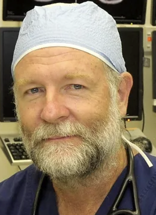 Jamie Sleigh合作论文数University of Auckland3
 Rahimi Amir Abbas合作论文数Molecular Medicine Department, Pasteur Institute of Iran3
 Nasrin Ahmadinejad合作论文数3Ali Hosseinsabet合作论文数3Darvishi Saeed合作论文数Faculty of Biomedical Engineering, Islamic Azad University3Zahra Alizade-Sani合作论文数3
 Logan Voss合作论文数Department of Anesthesia, Waikato Hospital3Pari Golabi合作论文数3
 Fereshteh Yousefirizi合作论文数3Pooya Sobhe Bidari合作论文数2Mohammad Reza Nasiri Avanaki合作论文数2Fatemeh Zareayan Jahromy合作论文数2Talayeh Ghodsizad合作论文数2
 Samrand Sharifi合作论文数Dept Biomed Engn, Islamic Azad Univ2
 Hamed Yeganegi合作论文数School of Life Sciences, Technical University of Munich2
 Farzad Azarmi合作论文数2Nasrin Amini合作论文数Dept Biomed Engn & Med Phys, Shahid Beheshti Univ Med Sci2
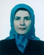 Fariba Bayat合作论文数Cardiovascular Research Center, Shahid Beheshti University of Medical Sciences2
 Mohammad Mahdi Khalilzadeh合作论文数Electr. Eng. Dept., Khajeh Nasir Toosi Univ. of Technol. (KNTU),;c;Electr. Eng. Dept., Khajeh Nasir Toosi Univ. of Technol. (KNTU),2Mehrnam, A.合作论文数2




