Sinusoidal obstruction syndrome (SOS) is a known adverse effect of oxaliplatin, causing significant morbidity and cessation of chemotherapy. Recent studies suggest single-nucleotide polymorphisms of certain genes may predispose carriers to oxaliplatin-induced liver toxicity. This study aims to evaluate candidate SOS-predisposing single-nucleotide polymorphisms by correlating routinely available next-generation sequencing (NGS) data with clinical and histologic evidence of SOS. Seventy-nine colorectal cancer cases with liver metastases and clinical NGS data were identified. A combination of clinical, biochemical, and histologic criteria was used to identify 12 cases with confirmed SOS. In addition, 10 control cases were selected with no clinical or histologic evidence of SOS. Clinical data and SOS-related findings were collected. For each patient, the expanded panel NGS data obtained for clinical management were reanalyzed to assess 12 polymorphisms in ERCC1, ERCC2, ABCB1, GSTP1, DPYD, MTHFR, ABCC2, UGT1A1, GSTT1, and GSTM1 known to be associated with oxaliplatin toxicity, as identified in the PharmGKB database. Statistical analyses, including the Fisher exact test and odds ratios, were performed. The strongest nominal signal was observed in the prevalence of ERCC1 rs11615 (A>G) in SOS and control groups (allelic P = .006; false discovery rate q = 0.072). The odds ratio for developing SOS associated with rs11615 (A>G) was 0.15 (95% CI: 0.04-0.59), indicating a protective effect of ERCC1 rs11615 (A>G). There was a noticeable numerical increase in ascites frequency, levels of aspartate aminotransferase and alanine aminotransferase, and the frequency of neuropathy in the SOS group, although not statistically significant. These findings highlight the use of expanded analysis of routinely obtained NGS panel results at the time of colorectal cancer diagnosis at many medical centers to personalize the chemotherapy regimen. Our data suggest that patients who carry the ERCC1 rs11615 (A>G) variant may be protected from developing oxaliplatin-induced SOS, and those lacking the G allele should be monitored more carefully after oxaliplatin use.
PURPOSE:To evaluate treatment effectiveness of radiation segmentectomy (RS) using yttrium-90 and combined transarterial chemoembolization (TACE) plus thermoablation (TACE-microwave ablation [MWA]) for treatment of hepatocellular carcinoma (HCC) by evaluating tumor necrosis (TN) on liver explant among patients bridged to transplant with HCC measuring up to 5 cm. MATERIALS AND METHODS:A single-institution retrospective review was performed from October 2010 to February 2024 to include consecutive patients who underwent TACE-MWA or RS using glass microspheres for treatment-naive HCC measuring up to 5 cm and then liver transplant. All treatments and liver transplant were performed at the same institution. No crossover treatment was allowed. TACE and ablation were performed sequentially during the same admission. RS was defined as target dose greater than 200 Gy to up to 2 segments, performed in 2 outpatient settings for mapping and administration, respectively. Radiologic response (RR) was evaluated with contrast-enhanced computed tomography (CT) and/or magnetic resonance (MR) imaging prior to transplant. The percentage of TN was determined on liver explant. Complete pathologic necrosis (CPN) was defined as 100% TN. RESULTS:A total of 25 and 28 patients underwent TACE-ablation and RS, respectively, with 40 tumors in each group. The mean segmental dose of the RS group was 284.5 Gy (SD ± 67.1). Tumor sizes were comparable between TACE-ablation and RS groups (mean, 2.1 cm [SD ± 0.9] vs 2.4 cm [SD ± 0.8]; P = .239). No significant difference was observed between 2 groups regarding percentage of TN on pathology (mean, 79.0% [SD ± 29.0] vs 83.6% [SD ± 29.6]; P = .484). The rate of CPN was also similar between 2 groups (mean, 45.0% vs 61.5%; P = .141) between tumors treated with TACE-ablation and RS, respectively. Tumor size of ≤3 cm was independently associated with CPN (odds ratio, 18.3; 95% CI, 2.14-156.48; P = .008) but not treatment modality. Tumors with complete RR demonstrated a higher degree of TN on liver explant than those did not (42.7% vs 26.6%, P = .0071). CONCLUSIONS:For tumor measuring up to 5 cm, TACE-ablation and RS were equivalent in achieving TN among patients bridged to transplant based on data from liver explant.
The incidence of early onset (age < 50 years at diagnosis) appendiceal adenocarcinoma (EOAA) is rising alarmingly. Reported data on etiology, treatment, and outcomes is scarce. In this study, we report clinical outcomes for patients with EOAA. Patients diagnosed with appendiceal adenocarcinoma between 2013 and 24 were stratified on the basis of age at diagnosis as having either EOAA (< 50 years) or average-age onset appendiceal adenocarcinoma (AOAA; ≥ 50 years). Clinicopathological and genomic data were abstracted from electronic medical records. Baseline clinicopathologic features and survival outcomes were compared between patients with EOAA and AOAA. Of 181 eligible patients, 49 (27.1
To determine the degree of tumor necrosis on pathology and clinical outcomes among intrahepatic cholangiocarcinoma (iCCA) patients who were treated with transarterial radioembolization (TARE) and the bridge or downstaged to surgery. A single center retrospective review was performed to include consecutive patients who underwent TARE using glass microspheres and then subsequently underwent surgical resection or transplant. Baseline characteristics, dosimetry, radiologic response, histological tumor necrosis, and overall survival were evaluated. A total of 20 patients (F: M = 14:6; age: 57.3 ± 10.8 years) underwent TARE and then received resection (n = 15) or living-donor liver transplant (n = 5). A total of 23 selective TARE (two segments or less) were delivered, with a mean dose 301.7 ± 177.5, whereas 18 lobar (left or right hepatic artery) doses were delivered, with a mean dose of 150.3 ± 44.1 Gy. Concurrent systemic therapy was administered in 15 patients. The mean time from the initial TARE to surgery was 7.1 ± 9.2 months. On pathology, complete tumor necrosis, > 90
Amiodarone-induced liver injury (AILI) is a known risk of amiodarone therapy, with presentations ranging from asymptomatic aminotransferase elevations to severe or fatal hepatitis and cirrhosis. Due to limited understanding of its histopathologic features, we conducted a retrospective cross-sectional re-analysis of liver biopsy samples from patients on amiodarone from two centers. Of the 48 liver biopsy samples, 42 (87%) exhibited histologic evidence of AILI. All patients showed minimal or mild macrovesicular steatosis. Ballooned hepatocytes were observed in 36 cases (86%), with 25 (69%) displaying a periportal distribution, 8 (22%) centrilobular, and 3 (8%) panacinar in distribution. Mallory-Denk bodies were found in 36 samples (76%)-18 (50%) were numerous and 18 (50%) multiple. Cholestasis was present in 10 patients, 7 (70%) of whom died. In contrast, 10 (31%) of the 32 patients without cholestasis died. This represents a significantly increased mortality risk for patients with AILI and cholestasis (p = 0.03). While AILI shares features with the more generally known metabolic dysfunction-associated steatotic liver disease, our findings indicate that a prominence of periportal distribution of ballooned hepatocytes and Mallory-Denk bodies despite a minimum of macrovesicular steatosis are characteristic of AILI. Furthermore, cholestasis in biopsy samples may suggest a poorer prognosis in patients on amiodarone.
Background. Findings have linked GNAS-activating mutations, frequent in appendiceal adenocarcinoma (AA), with improved overall survival but poor response to chemotherapy. The authors hypothesized that GNAS-activating mutations are associated with differential outcomes in AA treated with chemotherapy. Methods. Patients seen at the authors' center between 2013 and 2023 who received systemic chemotherapy for metastatic/recurrent AA were identified. The primary outcome was disease event-free survival (EFS), defined as time from start of chemotherapy (5-fluorouracil/capecitabine based) to earliest disease event, including death, clinical/radiographic recurrence, or progression. Study outcomes were assessed using Kaplan-Meier estimations and Cox proportional hazards regression. Results. The study included 48 patients. In 18 (37.5 %) of the 48 patients, GNAS-activating mutations were seen. Patients with GNAS mutations were more likely to have lower grades of disease (p = 0.003), with lower proportions of lymphovascular invasion (p = 0.005) and perineural invasion (p = 0.03), but a higher median peritoneal carcinomatosis index (p = 0.03). In the multivariable analysis, GNAS mutations (10.7 months [95 % confidence interval {CI}, 7.1-19.2] vs 20.3 months [95 % CI, 18.6-29.4; adjusted HR {aHR}, 3.75; 95 % CI, 1.84-7.63] p < 0.001) and metachronous metastases (aHR, 5.14; 95 % CI, 2.08-12.69; p < 0.001) were associated with worse EFS. Both CC0-1 resection (aHR, 0.12; 95 % CI, 0.05-0.28; p < 0.001) and CC2-3 resection (aHR, 0.28; 95 % CI, 0.10-0.81; p = 0.02) were associated with prolonged EFS. There was no significant difference in the OS from the date of metastases diagnosis between the GNAS(mt) and GNAS(wt) patients (HR, 0.68; 95 % CI, 0.31-1.47; p = 0.33). Conclusions. With systemic chemotherapy, GNAS-mutated metastatic/recurrent AAs have worse EFS despite less frequent high-risk features. Routine somatic mutation-testing of patients with AA should be considered for prognostication and possibly therapeutic decision-making.
Chimeric antigen receptor T-cell (CAR-T) therapy targeting for B-cell maturation antigen (BCMA) is used to treat patients with multiple myeloma (MM). To investigate gastrointestinal changes associated with CAR-T therapy, we performed a retrospective study. A total of 10 patients with 26 gastrointestinal biopsy specimens who underwent CAR-T therapy for MM were identified. Except for one patient with residual multiple myeloma, all specimens demonstrated markedly reduced or absent plasma cells. The most prominent biopsy findings occurred in the small intestine, primarily the duodenum, and included lamina propria lymphocytic infiltration, villous atrophy, foveolar metaplasia, an absence of plasma cells, and an increase in intraepithelial lymphocytes. There was an average of 7 apoptotic bodies per 10 high-power fields (hpf). The terminal ileum was also notable for increase in apoptotic bodies (average of 9.5 apoptotic bodies/10 hpf), villous atrophy, and lamina propria lymphocytic infiltration. The stomach biopsies overall typically showed mild inflammation with no increase in apoptotic bodies. A few colonic specimens demonstrated active colitis and prominent apoptotic bodies, while the majority of the colonic biopsies did not have significant findings other than melanosis coli, and an absence or reduction of plasma cells. The disproportionately greater injury in the duodenum versus the colon highlights the importance of upper endoscopic evaluation in symptomatic patients after CAR-T therapy. The mechanisms for these patterns of injury and differential anatomic findings are unknown; however, an immune-mediated injury associated with CAR-T therapy is suspected. Our study identifies a unique pattern of intestinal injury in patients with MM who received BCMA-targeted CAR-T therapy; this encompasses histologic findings more profound in the small intestine, which include absence of plasma cells, an increase in apoptotic bodies, lymphocytic infiltration, and villous atrophy.
Only a minority of patients at high likelihood of a gastrointestinal polyposis syndrome (GPS) are appropriately referred for workup. This proof-of-concept study evaluates a GPS screening rubric based exclusively on information in prior pathology reports and intended to facilitate pathologist engagement in GPS screening and referral. We sought to (1) identify patients who would benefit from further GPS workup, (2) assign a probable polyposis syndrome category (adenomatous, hamartomatous, serrated, or mixed), and (3) suggest a specific syndrome, such as familial adenomatous polyposis, whenever possible. We retrospectively tested the rubric against the pathology records of 108 patients (median, 6 reports/patient) with an established clinical diagnosis of GPS (adenomatous (N = 88), hamartomatous (N = 18), and mixed (N = 2) polyposis syndromes). Records were reviewed chronologically (mean, 4.4 min/patient) by a GI pathologist blinded to clinical history. Ninety-five patients (88
Rituximab (RTX) is a monoclonal anti-CD20 antibody widely used to treat B-cell neoplasms and autoimmune conditions. RTX has recently been linked to an enteropathy characterized by diarrhea, malabsorption, and hypogammaglobulinemia, closely resembling common variable immunodeficiency (CVID) enteropathy. We present the first dedicated histopathologic assessment of RTX-associated CVID-like enteropathy. Study inclusion criteria were the presence of diarrhea, weight loss, or other gastrointestinal symptoms in the setting of current/prior RTX use and associated hypogammaglobulinemia. Twenty-two patients (15 male:7 female; mean age at biopsy/resection, 63.4 years) across 9 tertiary medical centers met inclusion criteria and had small bowel (N = 20) and/or colon (N = 17) specimens (biopsies/resections) available for review; 71.4% of specimens dated from ≤5 years of last RTX dose. Cases were systematically evaluated by gastrointestinal pathologists at each institution. Key histologic features in the small bowel included sparse/absent lamina propria plasma cells (N = 10; 50%), intraepithelial lymphocytosis (N = 12; 60%), villous atrophy (N = 11; 55%), increased crypt apoptotic bodies (N = 6; 30%), and active inflammation (N = 5; 25%). Common features in the colon included sparse/absent plasma cells (N = 7; 41.2%), increased crypt apoptotic bodies (N = 7; 41.2%), active inflammation (N = 5; 29.4%), and intraepithelial lymphocytosis (N = 4; 23.5%). Goblet cell loss was appreciated in small bowel and/or colon specimens from 2 patients. Follow-up biopsies (interval, 2 months to 4 years) were available for 7 patients and largely recapitulated the histology of the index specimens, though 1 patient demonstrated improvement in villous blunting and intraepithelial lymphocytosis. In summary, the histologic spectrum of post-RTX CVID-like enteropathy encompasses lamina propria plasma cell depletion, increased crypt apoptotic bodies, small bowel villous atrophy, and goblet cell loss. While the underlying pathophysiology remains uncertain, the clinicopathologic picture may reflect post-RTX B-cell/plasma cell impairment. Although histologic findings may be subtle and variable, pathologists should be aware of this entity and should seek a history of RTX use in patients whose biopsies exhibit these CVID enteropathy-like features.
 Marc Bissonnette合作论文数The University of Chicago Medical Center115
 Shuyuan Xiao (肖书渊)合作论文数The Second Clinical College of Wuhan University84
 Namrata Setia合作论文数Biological Sciences Division, The University of Chicago76
 Lindsay Alpert合作论文数Department of Pathology, The University of Chicago71Urszula Dougherty合作论文数66Alexander B. Taylor合作论文数65
 Loren Joseph合作论文数Department of Pathology, University of Chicago61Reba Mustafi合作论文数56
 Maria Westerhoff合作论文数Department of Pathology, Medical School, University of Michigan-Ann Arbor53Joel Pekow合作论文数Department of Medicine and the Digestive Disease Research Core Center, The University of Chicago50
 Alessandro Fichera合作论文数Corresponding author. 3409 Worth StreetWorth Tower, Suite 600Dallas, TX 75246.47
 Irving Waxman合作论文数Digestive Disease Service Line for Rush University System for Health, Rush University Medical Center;Division of Digestive Diseases & Nutrition, Rush University Medical Center45Pai Rish K合作论文数Cleveland Clin, Dept Anat Pathol, Cleveland, OH 44195 USA43
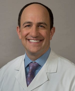 David T. Rubin合作论文数Division of the Biological Sciences, University of Chicago;Digestive Diseases Center, University of Chicago Medicine;MacLean Center for Clinical Medical Ethic;Comprehensive Cancer Center, University of Chicago43
 Maria Tretiakova合作论文数Department of Pathology, University of Chicago43
 Hanlin Wang合作论文数David Geffen School of Medicine, University of California, Los Angeles32
 Yanchun Li合作论文数The University of Chicago31Uzma D. Siddiqui合作论文数31Stephen P. Holloway合作论文数Department of Biochemistry and Applied Molecular Biology, University of Manchester Institute of Science and Technology (UMIST),29
 Amy Noffsinger合作论文数27
 Mitchell C. Posner合作论文数Department of Radiation Oncology, The University of Chicago27
 Michael Millis合作论文数Dept Surg, Univ Chicago26
 Christopher R. Weber合作论文数State University of New York, University of Maine24
 Sharad Khare合作论文数Department of Medicine, University of Chicago23Lei Zhao合作论文数21Sf Kuan合作论文数20Xiaohang Cao合作论文数20
 Mark K Ferguson合作论文数Department of Surgery, University of Chicago20J. Richard Thistlethwaite合作论文数20Helen S. Te合作论文数Biological Sciences Division, The University of Chicago19Haider I. Haider合作论文数18
 Katherine Meckel合作论文数Department of Economics, Division of Social Sciences, University of California, San Diego18Devkumar Mustafi合作论文数18Lindsay Yassan合作论文数Biological Sciences Division, The University of Chicago18Sadiq Farhana合作论文数18
 Joan S. Valentine合作论文数Department of Chemistry and Biochemistry, University of California, Los Angeles18Mary P. Bronner合作论文数18Newell K A合作论文数17
 Vani Konda合作论文数Center for Esophageal Diseases, Baylor Scott & White Health;GI Physiology Lab, Baylor Scott & White Health;Division of Gastroenterology and Hepatology, Baylor University Medical Center17Smruti R. Mohanty合作论文数Centre of Plasma Physics17Fatma Ayaloglu-Butun合作论文数16Andrea D Olivas合作论文数16
 Joel K. Greenson合作论文数Department of Pathology, Medical School, University of Michigan16
 Daniel Catenacci合作论文数Catenacci Consulting Limited16
 Andrew Ross合作论文数Virginia Mason Medical Center;Virginia Mason Franciscan Health16Gang He合作论文数16
 Emmanuelle Waubant合作论文数Department of Neurology, School of Medicine, University of California, San Francisco;Weill Institute for Neurosciences, University of California, San Francisco16




