Accumulation of Cu and its allocation to CuCOX correlates with the progression of ccRCC and drives tumor growth.
Polycyclic aromatic hydrocarbons (PAHs), such as benzo[a]pyrene (B[a]P), are major risk factors for lung cancer and other diseases, acting through the aryl hydrocarbon receptor (AHR). Alveolar macrophages (AMs) help regulate the lung microenvironment by responding to inhaled toxicants and resident microbiota. Although small extracellular vesicles (sEVs, aka exosomes) released by AMs mediate intercellular communication and immune responses, the influence of lung microbiota on sEV biogenesis and the mechanisms underlying sEV dysregulation during PAH exposure remain unknown. Here, we investigated the interplay between AMs, B[a]P, and lung microbiota, focusing on sEV-associated miRNAs (exo-miRNAs). Murine AMs (MH-S) were exposed to varying B[a]P concentrations in the presence or absence of murine lung microbiota with or without an AHR antagonist. sEVs from each condition were characterized and profiled for miRNA. Distinct miRNA signatures emerged: high-dose B[a]P enriched miRNAs linked to cancer progression, whereas lung microbiota alone or with low-dose B[a]P induced tumor-suppressor miRNAs that limit proliferation and metastasis and promote apoptosis, an effect enhanced by AHR antagonism. Lung microbiota appeared to counteract high-dose B[a]P by modulating tumor-suppressive exo-miRNAs. This study demonstrates that lung microbiota-induced exo-miRNAs critically shape AM-derived sEV-miRNA signaling during PAH exposure. The identified exosomal miRNAs could serve as important exposure biomarkers and therapeutic targets for mitigating B[a]P-induced toxicity and cancer development.
Abstract Copper (Cu) is a cofactor of cytochrome c oxidase (CuCOX), indispensable for aerobic mitochondrial respiration. This study reveals that advanced clear cell renal cell carcinomas (ccRCC) accumulate Cu, allocating it to CuCOX. Using a range of orthogonal approaches, including metabolomics, lipidomics, isotope-labeled glucose and glutamine flux analysis, and transcriptomics across tumor samples, cell lines, xenografts, and patient-derived xenograft models, combined with genetic and pharmacologic interventions, we explored the role of Cu in ccRCC. Elevated Cu levels stimulate CuCOX biogenesis, providing bioenergetic and biosynthetic benefits that promote tumor growth. This effect is complemented by glucose-dependent glutathione production, which facilitates detoxification and mitigates Cu–H2O2 toxicity. Single-cell RNA sequencing and spatial transcriptomics reveal increased oxidative metabolism, altered glutathione and Cu metabolism, and diminished hypoxia-inducible transcription factor activity during ccRCC progression. Thus, Cu drives an integrated oncogenic remodeling of bioenergetics, biosynthesis, and redox homeostasis, fueling ccRCC growth, which can be targeted for new therapeutic approaches. Significance: The work establishes a requirement for glucose-dependent coordination between energy production and redox homeostasis, which is fundamental for the survival of cancer cells that accumulate Cu and contributes to tumor growth.
Abstract Oncogenic Kras signaling is a key therapeutic target in human cancers. Ras activation is catalyzed by guanine nucleotide exchange factors (GEFs), of which SOS1 is a major player that transduces receptor tyrosine kinase signaling to Ras. In addition to recently devised oncogenic Kras targeting small molecule inhibitors, disrupting the catalytic interaction between Ras and SOS1 has emerged as an attractive therapeutic strategy in working synergistically with Kras inhibitors and overcoming resistance. Previous studies have demonstrated an active “feed forward” loop of active Kras binding to the allosteric site of SOS1 that further stimulates SOS1 catalytic activation of wild type H/N/Kras, which is required for oncogenic Kras driven tumorigenesis. Here we have developed a rational approach that combines virtual screening with experimental screening to identify small molecule inhibitors that target the allosteric site of SOS1 to suppress oncogenic Kras mediated SOS1-regulated wild-type Ras activities. Unlike SOS1 catalytic site inhibitors which can have significant toxicity due to their ability to inhibit normal Ras signaling function, the lead SOS1 allosteric inhibitors developed in our laboratory binds to the allosteric site of SOS1 and blocks active Ras interaction with SOS1: we found these inhibitors can competitively suppresses SOS1-Ras interaction at the SOS1 allosteric site and dose-dependently inhibits the feed-forward SOS1 GEF activity. Mutagenesis and structure-activity relationship studies mapped the site of action to the SOS1 allosteric site, and preliminarily defined the chemical moieties in the inhibitor essential for the activity. The inhibitor showed dose-dependent and selective efficacy in inhibiting WT Ras activity, downstream signaling activities, and associated cell proliferation in KRAS mutation cells but not KRAS wild-type cells. Compared to the SOS1 catalytic inhibitor, BI3406, our inhibitors showed better selectivity for KRAS mutant pancreatic cancer cells vs. WT KRAS cells. These studies establish a proof of principle for the rational design of small molecule inhibitors targeting an oncogenic Kras specific allosteric site of SOS1 and provide a novel therapeutic concept to target KRAS-driven tumors more effectively. Citation Format: Xin Duan, Chris R Evelyn, Jacek Biesiada, James F Johnson, William L. Seibel, Jaroslaw Meller, Yi Zheng. Rational design of a small-molecule inhibitor targeting the allosteric site of sos1 in oncogenic k-ras pancreatic cancer [abstract]. In: Proceedings of the American Association for Cancer Research Annual Meeting 2024; Part 1 (Regular Abstracts); 2024 Apr 5-10; San Diego, CA. Philadelphia (PA): AACR; Cancer Res 2024;84(6_Suppl):Abstract nr 5912.
ABSTRACT Mitogen-activated protein 3 kinase 1 (MAP3K1) has a plethora of cell type-specific functions not yet fully understood. Herein, we describe a role for MAP3K1 in female reproductive tract (FRT) development. MAP3K1 kinase domain-deficient female mice exhibited an imperforate vagina, labor failure and infertility. These defects corresponded with shunted Müllerian ducts (MDs), the embryonic precursors of FRT, that manifested as a contorted caudal vagina and abrogated vaginal–urogenital sinus fusion in neonates. The MAP3K1 kinase domain is required for optimal activation of the Jun-N-terminal kinase (JNK) and cell polarity in the MD epithelium, and for upregulation of WNT signaling in the mesenchyme surrounding the caudal MD. The MAP3K1-deficient epithelial cells and MD epithelium had reduced expression of WNT7B ligands. Correspondingly, conditioned media derived from MAP3K1-competent, but not -deficient, epithelial cells activated a TCF/Lef-luciferase reporter in fibroblasts. These observations indicate that MAP3K1 regulates MD caudal elongation and FRT development, in part through the induction of paracrine factors in the epithelium that trans-activate WNT signaling in the mesenchyme.
Copper (Cu) is an essential trace element required for mitochondrial respiration. Late-stage clear cell renal cell carcinoma (ccRCC) accumulates Cu and allocates it to mitochondrial cytochrome c oxidase. We show that Cu drives coordinated metabolic remodeling of bioenergy, biosynthesis and redox homeostasis, promoting tumor growth and progression of ccRCC. Specifically, Cu induces TCA cycle-dependent oxidation of glucose and its utilization for glutathione biosynthesis to protect against H 2 O 2 generated during mitochondrial respiration, therefore coordinating bioenergy production with redox protection. scRNA-seq determined that ccRCC progression involves increased expression of subunits of respiratory complexes, genes in glutathione and Cu metabolism, and NRF2 targets, alongside a decrease in HIF activity, a hallmark of ccRCC. Spatial transcriptomics identified that proliferating cancer cells are embedded in clusters of cells with oxidative metabolism supporting effects of metabolic states on ccRCC progression. Our work establishes novel vulnerabilities with potential for therapeutic interventions in ccRCC. Accumulation of copper is associated with progression and relapse of ccRCC and drives tumor growth.Cu accumulation and allocation to cytochrome c oxidase (CuCOX) remodels metabolism coupling energy production and nucleotide biosynthesis with maintenance of redox homeostasis.Cu induces oxidative phosphorylation via alterations in the mitochondrial proteome and lipidome necessary for the formation of the respiratory supercomplexes. Cu stimulates glutathione biosynthesis and glutathione derived specifically from glucose is necessary for survival of Cu Hi cells. Biosynthesis of glucose-derived glutathione requires activity of glutamyl pyruvate transaminase 2, entry of glucose-derived pyruvate to mitochondria via alanine, and the glutamate exporter, SLC25A22. Glutathione derived from glucose maintains redox homeostasis in Cu-treated cells, reducing Cu-H 2 O 2 Fenton-like reaction mediated cell death. Progression of human ccRCC is associated with gene expression signature characterized by induction of ETC/OxPhos/GSH/Cu-related genes and decrease in HIF/glycolytic genes in subpopulations of cancer cells. Enhanced, concordant expression of genes related to ETC/OxPhos, GSH, and Cu characterizes metabolically active subpopulations of ccRCC cells in regions adjacent to proliferative subpopulations of ccRCC cells, implicating oxidative metabolism in supporting tumor growth.
Mitogen-Activated Protein 3 Kinase 1 (MAP3K1) is a dynamic signaling molecule with a plethora of cell-type specific functions, most of which are yet to be understood. Here we describe a role for MAP3K1 in the development of female reproductive tract (FRT). MAP3K1 kinase domain-deficient (Map3k1ΔKD) females exhibit imperforate vagina, labor failure, and infertility. These defects correspond to a shunted Müllerian duct (MD), the principle precursor of the FRT, in embryos, while they manifest as a contorted caudal vagina with abrogated vaginal-urogenital sinus fusion in neonates. In epithelial cells, MAP3K1 acts through JNK and ERK to activate WNT, yet in vivo MAP3K1 is crucial for WNT activity in mesenchyme associated with the caudal MD. Expression of Wnt7b is high in wild type, but low in Map3k1 knockout MD epithelium and MAP3K1-deficient keratinocytes. Correspondingly, conditioned media derived from MAP3K1-competent epithelial cells activate TCF/Lef-luciferase reporter in fibroblasts, suggesting that MAP3K1-induced factors released from epithelial cells trans-activate WNT signaling in fibroblasts. Our results reveal a temporal-spatial and paracrine MAP3K1-WNT crosstalk contributing to MD caudal elongation and FRT development. Highlights MAP3K1 deficient female mice exhibit imperforate vagina and infertility Loss of MAP3K1 kinase activity impedes Müllerian duct (MD) caudal elongation and fusion with urogenital sinus (UGS) in embryogenesis The MAP3K1-MAPK pathway up-regulates WNT signaling in epithelial cells MAP3K1 deficiency down-regulates Wnt7b expression in the MD epithelium and prevents WNT activity in mesenchyme of the caudal MD
Microtubule-associated protein 1 light chain 3 gamma (MAP1LC3C or LC3C) is a member of the microtubule-associated family of proteins that are essential in the formation of autophagosomes and lysosomal degradation of cargo. LC3C has tumor-suppressing activity, and its expression is dependent on kidney cancer tumor suppressors, such as von Hippel-Lindau protein and folliculin. Recently, we demonstrated that LC3C autophagy is regulated by noncanonical upstream regulatory complexes and targets for degradation postdivision midbody rings associated with cancer cell stemness. Here, we show that loss of LC3C leads to peripheral positioning of the lysosomes and lysosomal exocytosis (LE). This process is independent of the autophagic activity of LC3C. Analysis of isogenic cells with low and high LE shows substantial transcriptomic reprogramming with altered expression of zinc (Zn)-related genes and activity of polycomb repressor complex 2, accompanied by a robust decrease in intracellular Zn. In addition, metabolomic analysis revealed alterations in amino acid steady-state levels. Cells with augmented LE show increased tumor initiation properties and form aggressive tumors in xenograft models. Immunocytochemistry identified high levels of lysosomal-associated membrane protein 1 on the plasma membrane of cancer cells in human clear cell renal cell carcinoma and reduced levels of Zn, suggesting that LE occurs in clear cell renal cell carcinoma, potentially contributing to the loss of Zn. These data indicate that the reprogramming of lysosomal localization and Zn metabolism with implication for epigenetic remodeling in a subpopulation of tumor-propagating cancer cells is an important aspect of tumor-suppressing activity of LC3C.
Background The objective was to assess secretion of small extracellular vesicular microRNA (exo-miRNA) in head and neck squamous cell carcinoma (HNSCC) according to human papillomavirus (HPV) status, and determine the translational potential as a liquid biopsy for early detection. Methods This study employed a combination of cell culture and case-control study design using archival pretreatment serum. Small extracellular vesicles (sEV) were isolated from conditioned culture media and human serum samples via differential ultracentrifugation. miRNA-sequencing was performed on each sEV isolate. Results There were clear exo-miRNA profiles that distinguished HNSCC cell lines from nonpathologic oral epithelial control cells. While there was some overlap among profiles across all samples, there were apparent differences in exo-miRNA profiles according to HPV-status. Importantly, differential exo-miRNA profiles were also apparent in serum from early-stage HNSCC cases relative to cancer-free controls. Conclusions Our findings indicate that exo-miRNA are highly dysregulated in HNSCC and support the potential of exo-miRNA as biomarkers for HNSCC.
 Jarek Meller合作论文数31Mario Medvedovic合作论文数Laboratory for Statistical Genomics and Systems Biology, University of Cincinnati College of Medicine31Czyzyk-Krzeska Maria F合作论文数22David R. Plas合作论文数Department of Cancer Biology, University of Pennsylvania20Dina Secic合作论文数17
 Behrouz Shamsaei合作论文数Icahn Sch Med Mt Sinai, LINCS Data Coordinat & Integrat Ctr BD2K, New York, NY 10029 USA17
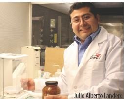 Julio Alberto Landero Figueroa合作论文数Department of Chemistry, University of Guanajuato17Xiang Zhang合作论文数16
 Bhargav Vemuri合作论文数14
 Shuchi Gulati合作论文数Department of Medicine, UC Davis Comprehensive Cancer Center14
 Reisz Julie A合作论文数Dept Biochem & Mol Genet, Univ Colorado Denver14
 Angelo D'Alessandro合作论文数Department of Biochemistry and Molecular Genetics, School of Medicine, University of Colorado14
 Katherine Vest合作论文数Department of Pharmacology, Emory University14
 Patra Krushna C合作论文数Massachusetts General Hospital Cancer Center, Harvard Medical School14
 Caterina Bartolacci合作论文数14
 Cunningham John T合作论文数School of Medicine and Department of Urology, University of California14
 Schmidt Lucas合作论文数14
 Pier Paolo Scaglioni合作论文数14
 James Brugarolas合作论文数UT Southwestern Medical Center13
 Rafal Adamczak合作论文数Department of Informatics
Nicolaus Copernicus University13
Rafal Adamczak合作论文数Department of Informatics
Nicolaus Copernicus University13 Włodzisław Duch合作论文数Department of Informatics, Institute of Engineering and Technology, School of Physics, Astronomy and Informatics, Nicolaus Copernicus University;NeuroCognitive Laboratory, Centre for Modern Interdisciplinary Technologies, Nicolaus Copernicus University11Damaris Kuhnell合作论文数9
 Scott M Langevin合作论文数graduate School of Public Health, University of Pittsburgh9Jaroslaw Meller合作论文数University of Cincinnati College of Medicine9
 Alvaro Puga合作论文数Department of Environmental and Public Health Sciences, University of Cincinnati8
 William Seibel合作论文数Univ Cincinnati, Ctr Drug Discovery, Cincinnati, OH USA7Stanek Jerzy合作论文数7
 Yi Zheng合作论文数Cancer and Blood Diseases Institute, Cincinnati Children's;Department of Pediatrics, University of Cincinnati6Olga Mikhaylova合作论文数5
 Adam Kachel合作论文数5Chris R Evelyn合作论文数5Jiang Wang合作论文数5Megan E Bischoff合作论文数5
 Tomasz Winiarski合作论文数Dept . of Informatics;Nicholaus Copernicus University;Dept . of Informatics, Nicholaus Copernicus University5
 Susan Kasper合作论文数5Birgit Ehmer合作论文数4Wen Niu合作论文数Icahn Sch Med Mt Sinai, LINCS Data Coordinat & Integrat Ctr BD2K, New York, NY 10029 USA4Yan Ren合作论文数BD2K-LINCS DCIC, Department of Environmental Health, University of Cincinnati, Cincinnati, OH 45220, USA.4
 Vinicius Carreira合作论文数College of Medicine, University of Cincinnati4
 Trisha Wise-Draper合作论文数Cincinnati Children's Hospital Medical Center, University of Cincinnati College of Medicine4
 Marcin Blachnik合作论文数Department of Management and Informatics, Silesian University of Technology4Xin Duan合作论文数Institute of Modern Physics, Chinese Academy of Sciences4Emily Kellner合作论文数3
 Collin Wetzel合作论文数3James Reigle合作论文数3Lixia Zhang合作论文数3
 Ricky Yuet-Kin Leung合作论文数Dept Environm Hlth, Univ Cincinnati3
 Katherine Burns合作论文数3
 Chia-I Ko合作论文数3




