BACKGROUND:The incidence of rectal adenocarcinoma is rising in patients < 50 years old compared to those > 50. Nonoperative management and active surveillance have become a standard of care in rectal cancer treatment for those who achieve a complete response to neoadjuvant therapy; however, the use, safety and efficacy of this approach in different age groups has not been well described. OBJECTIVE:Examine oncological outcomes in patients with rectal adenocarcinoma managed by nonoperative management according to patient age. DESIGN:Retrospective cohort study. SETTING:Comprehensive cancer center in New York. PATIENTS:Patients with rectal adenocarcinoma diagnosed between January 2006 and December 2020. MAIN OUTCOME MEASURES:Local regrowth, recurrence-free survival, distant metastasis-free survival, overall survival rates. RESULTS:A cohort of 430 patients with rectal adenocarcinoma who were treated with neoadjuvant therapy and entered watch-and-wait were included. Patients were divided into 3 groups according to age at diagnosis: early-onset (age < 50, n = 98), middle-aged (50-64, n = 163), and late-onset (65+, n = 169). No differences were noted in tumor distance from the anal verge (p = 0.345) between groups. More patients in the early-onset cohort received induction chemotherapy with chemoradiation than late-onset patients, who more often received chemoradiation alone and had clinical stage I disease (p = 0.015). Median follow-up time was 4.0years (IQR 2.6-5.5). The 3-year local regrowth rates in each group were 23.8% (95% CI: 15.2-32.4), 33.0% (95% CI: 25.6-40.4), and 26.1% (95% CI: 19.4-32.8) in the early, middle-aged, and late-onset patients, respectively (p = 0.395). No associated differences in 5-year distant metastasis-free or recurrence-free survival rates were noted between the 3 age groups. The early-onset cohort had an associated superior disease-specific survival from the late-onset cohort. LIMITATIONS:Retrospective nature. CONCLUSIONS:Early-onset rectal cancer patients have at least similar oncological outcomes compared to those in older age groups when treated non-operatively. These data provide reassurance when offering a watch-and-wait approach to patients with early-onset rectal cancer. See Video Abstract.
Appendiceal abnormalities, beyond typical acute appendicitis, are occasionally detected on imaging or endoscopy, leading to surgical evaluation. How often these reflect an underlying neoplasm, however, remains unclear. This study aimed to determine the incidence and associated findings of neoplastic disease in this setting. Patients undergoing diagnostic appendectomy following abnormal findings in endoscopy or imaging at an academic cancer center were reviewed (2011–2021). Patients with preoperative diagnoses of acute appendicitis without a “suspicious” lesion, peritoneal carcinomatosis, or known malignancy were excluded. Radiographic, biochemical and colonoscopic findings were compared between neoplastic and non-neoplastic appendiceal pathology. A total of 132 patients were identified (mean age 64 years, 52
Increasing evidence supports watch-and-wait (WW) surveillance for patients with rectal cancer who show clinical complete response (cCR) at restaging following total neoadjuvant treatment (TNT). However, accurate methods to early detect local regrowth (LR) from follow-up endoscopy images during WW are essential to manage care and prevent distant metastases. Hence, we developed a Siamese Swin Transformer with Dual Cross-Attention (SSDCA) to combine longitudinal endoscopic images at restaging and follow-up and distinguish cCR from LR. SSDCA leverages pretrained Swin Transformers to extract domain agnostic features and enhance robustness to imaging variations. Dual cross attention is implemented to emphasize features from the paired scans without requiring any spatial alignment to predict response. SSDCA as well as Swin-based baselines were trained using image pairs from 135 patients and evaluated on a held-out set of image pairs from 62 patients. SSDCA produced the best balanced accuracy (81.76% ± 0.04), sensitivity (90.07% ± 0.08), and specificity (72.86% ± 0.05). Robustness analysis showed stable performance irrespective of artifacts including blood, stool, telangiectasia, and poor image quality. UMAP clustering of extracted features showed maximal inter-cluster separation (1.45 ± 0.18) and minimal intra-cluster dispersion (1.07 ± 0.19) with SSDCA, confirming discriminative representation learning. All code and trained model checkpoints will be shared through GitHub upon manuscript acceptance.
Clinical trial studies indicate benefit of watch-and-wait (WW) surveillance for patients with rectal cancer showing a complete or near clinical response (CR) directly after treatment (restaging). However, there are no objectively accurate methods to early detect local tumor regrowth (LR) in patients undergoing WW from follow-up exams. Hence, we developed Temporal Rectal Endoscopy Cross-attention (TREX), a longitudinal deep learning approach that combines pairs of images acquired at restaging and follow-up to distinguish CR from LR. TREX uses pretrained Swin Transformers in a siamese setting to extract features from longitudinal images and dual cross-attention to combine the features without spatial co-registration between image pairs. TREX and Swin-based baselines were trained under two settings: (a) detecting LR or CR at the last available follow-up and (b) early detection of LR at 3–6, 6–12, and 12–24 months before clinical confirmation. TREX achieved the highest accuracy in detecting LR with a high sensitivity of 97
Organ preservation is a standard-of-care option for select patients with locally advanced rectal cancer, allowing avoidance of a proctectomy without compromising oncologic outcomes. As compared with (chemo)radiation alone, total neoadjuvant therapy, which delivers both (chemo)radiation and chemotherapy before surgery, increases clinical complete (cCR) and near clinical complete (nCR) response rates and may expand eligibility for organ preservation. Eligibility is determined through a standardized response assessment, which incorporates digital rectal examination, endoscopy, and rectal protocol magnetic resonance imaging, which are performed and interpreted by experienced clinicians. Patients with a cCR/nCR may defer immediate surgery with the view to potential organ preservation, provided they adhere to intensive ‘watch and wait’ surveillance every 3–6 months for 5 years to enable early detection of local regrowth and timely salvage. In carefully selected patients managed within experienced programs, organ preservation achieves oncologic outcomes comparable to immediate surgery and may improve functional and quality-of-life outcomes. Ongoing research focuses on personalizing neoadjuvant therapy to balance response and toxicity and on developing more accurate and feasible approaches for detecting residual disease and regrowth.
Patient-reported outcomes (PROs), initially developed for research, are increasingly recognized as valuable in clinical practice to monitor health status, quality of life, and symptoms from the patient perspective. However, few practical, clinician-friendly PRO tools exist for routine oncology care. We aimed to develop a brief electronic “Colorectal Scorecard” grounded in validated PROM content and evaluate its implementation in a high-volume colorectal surgery practice. A multidisciplinary team co-designed a reduced-item electronic PRO questionnaire covering seven key quality-of-life domains plus a patient-designated priority concern, presented as a one-page color-coded report integrated into the electronic health record (EHR). Included PROM content was drawn from validated instruments (e.g., LARS, CIS, IIEF-5/FSFI domains, ICIQ-UI SF items, EQ-5D-5L/EQ-VAS, EORTC QLQ-CIPN20 items, and a FACT-C work-ability item), with wording simplification guided by patient input. The Scorecard rollout included staff training, patient portal distribution with automated reminders, and in-clinic tablet options. We tracked survey completion rates, drop-off rates, completion times, setting of completion (home portal vs. in-clinic tablet when available), and clinician access/printing when loggable from 2019–2024. Provider and patient feedback were collected through structured informal feedback channels to guide iterative refinements. We compared these data to the prior colorectal PRO report system used from 2013–2019. From 2019 to 2024, approximately 13,400 colorectal patients completed > 19,000 Scorecard surveys as part of routine care. Overall PRO completion more than doubled (from 33
Abstract Background: Radiation therapy in rectal cancer treatment is limited by variable tumor responses among patients and harmful effects on normal tissues. We developed a translational human tumoroid-organoid platform to assess novel tumor-specific strategies for radiation sensitization using matched patient-derived models. Methods: Fifteen rectal cancer-derived tumoroids and three matched normal tissue-derived organoids were established from patients, representing primary tumors, metastases, and recurrences. Four tumoroids were derived from a single patient, encompassing different disease stages: primary tumor and splenic metastasis (pre-progression), and rectal and vaginal recurrences (post-progression). To evaluate tumor-specific radiation sensitizers, both tumoroids and their matched normal organoids were treated with 5-fluorouracil (5-FU), one of four DNA damage repair inhibitors (DDRis; ATMi, DNA-PKi, PARPi, or ATRi), or a DMSO control. Following treatment, samples were irradiated, and cell viability was measured. We assessed intrinsic radiation sensitivity under control conditions and radiosensitizer efficacy using a linear regression model with log-transformed cell viability as the outcome. Whole-exome sequencing characterized the mutation profiles of the tumoroids. Results: Intrinsic radiosensitivity was heterogeneous among tumoroids, with a 10–28% decrease in cell growth per unit increase in radiation dose. Critically, when comparing tumoroids to their matched normal organoids, we observed greater sensitization in tumoroids across all cases and for all DDRis, demonstrating tumor-specific radiosensitization. The most potent radiosensitizer relative to DMSO was DNA-PKi in 7 tumoroids, ATMi in 4 tumoroids, and PARPi in 1 tumoroid. Tumoroids derived after disease progression exhibited increased resistance to radiation and a diminished degree of sensitization with DDRi treatment compared to pre-progression tumoroids. Despite variations in radiation sensitivity and DDRi responses, the genetic profiles of the tumoroids remained largely unchanged. Conclusion: We developed a translational ex vivo tumoroid-organoid platform using matched patient-derived models to test tumor-specific radiation sensitizers in rectal cancer. This platform allowed us to determine tumor-specific sensitization by directly comparing tumoroids with matched normal organoids, demonstrating greater sensitization in tumoroids. Our findings highlight the potential of this platform to uncover precise, tumor-selective treatment options, improve patient responses, reduce toxicity, and address resistant tumors in patients with disease progression. Citation Format: Wini Zambare, Chao Wu, Hannah Kalvin, Hanchen Huang, Sara Yoder, Michael Del Latto, Maria Kierstead, Satoru Meguro, Xi Steven Chen, Mithat Gonen, J. Joshua Smith, Paul B. Romesser. Development of a translational tumoroid-organoid platform revealing tumor-specific radiosensitization in rectal cancer using matched patient-derived models. [abstract]. In: Proceedings of the AACR Special Conference in Cancer Research: Translating Targeted Therapies in Combination with Radiotherapy; 2025 Jan 26-29; San Diego, CA. Philadelphia (PA): AACR; Clin Cancer Res 2025;31(2_Suppl):Abstract nr A012.
Reprogramming of aged donor tissue cells into induced pluripotent stem cells (A-iPSC) preserved the epigenetic memory of aged-donor tissue, defined as genomic instability and poor tissue differentiation in our previous study. The unbalanced expression of RNA exosome subunits affects the RNA degradation complex function and is associated with geriatric diseases including premature aging and cancer progression. We hypothesized that the age-dependent progressive subtle dysregulation of EXOSC2 (exosome component 2) causes the aging traits (abnormal cell cycle and poor tissue differentiation). We used embryonic stem cells as a tool to study EXOSC2 function as the aging trait epigenetic memory determined in A-iPSC because these aging traits could not be studied in senesced aged cells or immortalized cancer cells. We found that the regulatory subunit of PP2A phosphatase, PPP2R5E, is a key target of EXOSC2 and this regulation is preserved in stem cells and cancer.
Abdominal adhesions are a globally disruptive problem to patients and healthcare systems, with limited preventative strategies. Multiple discovery prophylactics have been evaluated previously for adhesions prevention with inadequate transfer to patient care. Clinical translation is fundamentally restricted by the ability of a discovery prophylactic to simultaneously navigate 3 key components of adhesions formation throughout the entire abdomen: the innate immune system, the coagulation system, and the local peritoneal cell populations. Furthermore, challenging handling characteristics and product restrictions have decreased the utilization of clinically available prophylactics by surgeons. The success of future adhesions prevention strategies must also be anchored in clinically valid animal modeling with attention towards future regulatory approval. The purpose of the present roadmap article is to provide a state-of-the-art review of adhesions pathophysiology, hydrogel development, animal modeling, and regulatory science, from which a framework for future developmental strategies may be outlined.
Endoscopic images are used at various stages of rectal cancer treatment starting from cancer screening and diagnosis, during treatment to assess response and toxicity from treatments such as colitis, and at follow-up to detect new tumor or local regrowth. However, subjective assessment is highly variable and can underestimate the degree of response in some patients, subjecting them to unnecessary surgery, or overestimating response that places patients at risk of disease spread. Advances in deep learning have shown the ability to produce consistent and objective response assessments for endoscopic images. However, methods for detecting cancers, regrowth, and monitoring response during the entire course of patient treatment and follow-up are lacking. This is because automated diagnosis and rectal cancer response assessment require methods that are robust to inherent imaging illumination variations and confounding conditions (blood, scope, blurring) present in endoscopy images as well as changes to the normal lumen and tumor during treatment. Hence, a hierarchical shifted window (Swin) transformer was trained to distinguish rectal cancer from normal lumen using endoscopy images. Swin, as well as two convolutional (ResNet-50, WideResNet-50), and the vision transformer architectures, were trained and evaluated on follow-up longitudinal images to detect LR on in-distribution (ID) private datasets as well as on out-of-distribution (OOD) public colonoscopy datasets to detect pre/non-cancerous polyps. Color shifts were applied using optimal transport to simulate distribution shifts. Swin and ResNet models were similarly accurate in the ID dataset. Swin was more accurate than other methods (follow-up: 0.84, OOD: 0.83), even when subject to color shifts (follow-up: 0.83, OOD: 0.87), indicating the capability to provide robust performance for longitudinal cancer assessment.
Background The treatment of locally advanced rectal cancer now includes “watch-and-wait” (WW) management for patients who exhibit a clinical complete response (cCR) to total neoadjuvant therapy (TNT). We assessed patients’ knowledge and preferences regarding WW with the goal of incorporating patient input into clinical trial design. Methods Rectal cancer patients in advocacy groups (COLONTOWN/Rectalburgh and Fight CRC) were surveyed regarding perceptions about treatment strategies, tolerable risks of treatment failure, and preferences on clinical trial design. Surveys were initially distributed in 2019, with the shift toward WW prompting re-survey of select questions in 2023. Results All respondents preferred enrollment in trials that incorporated patient input. Most respondents (76%) preferred treatment that incorporated chemotherapy escalation over radiation escalation. Furthermore, when presented with a hypothetical patient who developed a cCR after chemoradiation (without the option for WW), 58% of patients chose surgical treatment (with 100% risk of permanent ostomy and 80% chance of cure) over omission of surgery (with <80% chance of cure). In contrast, when the hypothetical patient developed a cCR after TNT (with an option for WW), 82% of patients opted for selective WW over surgery. Conclusions This work successfully integrated patient advocacy groups as a resource for obtaining patient input. Surveys revealed patients had a strong preference for chemotherapy escalation, for selective WW when offered, and for incorporation of input into future clinical trial design. These findings describe how well-informed patients approach complex medical decision-making and serve as a starting point for future studies seeking to incorporate patient voices.
Purpose: The CivaSheet is a novel radiation delivery device consisting of unidirectionally shielded Pd-103 sources embedded in a bioabsorbable polymer sheet. This facilitates intraoperative placement of permanent brachytherapy seeds in a surgical tumor bed, while minimizing dose to overlying structures. No clinical trial data have yet been published for this device. We prospectively investigated its feasibility as an alternative to standard intraoperative radiation therapy (IORT). Methods and Materials: This was a prospective study in patients with abdomopelvic malignancies undergoing surgery with an indication for IORT. Prior external beam radiation was allowed. The primary endpoint was feasibility, defined as technically satisfactory placement of CivaSheet in ≥7 of 10 patients. Secondary endpoints were adverse events, local control, and implant stability. Results: Ten patients were enrolled and underwent surgery. Eight patients successfully underwent CivaSheet implantation. The 2 feasibility exclusions were technically unfavorable based on intraoperative assessment of tumor bed configuration. All implanted patients were treated to the pelvic sidewall or presacral space with a median prescription dose of 100 Gy. Median follow-up from the date of implant was 25 months. One patient had a marginal recurrence 3 months after implant, and 1 patient recurred at implant site after 13 months. One and 2-year estimates of local control were 86% and 69%, respectively. No device-related grade 4-5 adverse events were observed. Postimplant stability was deemed satisfactory in all patients. Conclusions: This is the first published clinical trial of a novel unidirectional brachytherapy device. CivaSheet proved to be a feasible technique to deliver intraoperative radiation to patients with high-risk pelvic malignancies after surgical resection, and warrants further study and clinical consideration, particularly in settings where other IORT options are not available.
Abstract Background Precision radiation strategies that expand the therapeutic window by selectively sensitizing tumors and sparing normal tissues are needed. We developed a matched tumoroid organoid preclinical platform to identify and characterize personalized radiosensitization strategies. Methods We established 15 rectal cancer derived tumoroids and 3 matched normal rectal epithelial organoids. Whole exome sequencing characterized mutation profiles and phylogenetic relationships. Tumoroids were treated with one of four DNA damage repair inhibitors (DDRi; ATMi, DNAPKi, PARPi, or ATRi), 5fluorouracil, or a DMSO control, followed by escalating doses of radiation. Cell viability was measured, and intrinsic radiosensitivity as well as radiosensitizer efficacy were characterized using linear regression models. Four tumoroids were derived from one patient at distinct stages and disease sites, including preprogression tumoroids (primary tumor, splenic metastasis) and postprogression tumoroids (rectal recurrence and vaginal recurrence). Results Tumoroid radiosensitivity demonstrated variability, paralleling the spectrum of clinical responses seen in rectal cancer. Genomic analyses revealed two distinct mutational signatures (SBS14 and SBS17b) associated with radioresistance. Radiation sensitization by DDRis was highly heterogeneous, depending on both specific tumoroid and inhibitor choice. In the subset of four tumoroids derived pre and postprogression, post progression tumoroids demonstrated greater radioresistance and diminished DDRi induced radiosensitization. Phylogenetic analysis revealed increased clonal and subclonal complexity in these radiation and DDRi resistant tumoroids. Lastly, comparing matched patient derived tumoroids and normal organoids established that tumoroids were generally more radiosensitive and exhibited enhanced DDRi mediated radiosensitization compared to normal organoids. However, the optimal DDRi for maximizing therapeutic index varied among tumoroids. Conclusion To our knowledge, this is the first ex vivo study to systematically quantify intrinsic radiation sensitivity and DDRi sensitization in a comprehensive, patient specific tumoroid platform. Our findings underscore the utility of a patient matched tumoroid organoid model as a platform to quantify and predict personalized responses to radiation and DDR inhibitors. Moreover, by comparing tumoroids from a single patient across different disease stages, our model reveals how tumors adapt to therapeutic pressure and develop increased radioresistance, offering a valuable potential framework for guiding precision radiotherapy. Moreover, the data show that DDRi efficacy is largely tumor selective and not solely predicted by mutational profiles, highlighting opportunities to refine treatment for radioresistant tumors, minimize injury to normal tissues, and adapt therapy over disease progression. ### Competing Interest Statement P.B.R. received research funding (2019) and served as a consultant for EMD Serono (2018-2024), receives research funding from XRAD Therapeutics (2022-present), is a consultant for Faeth Therapeutics (2022-present), is a consultant for Natera (2022-present), and is a volunteer on the advisory board for the HPV Alliance and Anal Cancer Foundation non-profit organizations. J.J.S. received travel support for fellow education from Intuitive Surgical (August 2015). He also served as a clinical advisor for Guardant Health (March 2019) and as a clinical advisor for Foundation Medicine (April 2022). He served as a consultant and speaker for Johnson and Johnson (May 2022). He served as a clinical advisor and consultant for GlaxoSmithKline (2023-24). ### Funding Statement This work was supported in part by a National Institutes of Health/National Cancer Institute, NIH/NCI) Memorial Sloan Kettering Cancer Center (MSK) support grant (P30CA008748). Dr. Wini Zambare was supported by NCI Surgical Oncology T32 research training grant (T32CA9501-34). Dr. Smith is supported by an NIH/NCI grant (R37CA248289), the Department of Surgery and Colorectal Service at MSK (J. Drebin/J. Garcia-Aguilar), the Wasserman Colorectal Research Fund, and generous donations by Corinne Berezuk, Michael Stieber, and Patrick A. Gerschel (Dr. Paty). Dr. Romesser is supported by an NIH/NCI grant (K08CA255574), EMD Serono research grant, and an MSK Imaging and Radiation Sciences Program (IMRAS) grant, and a Geoffrey Beene Cancer Research Center grant. Finally, this research was also supported by the Sylvester Comprehensive Cancer Center at the University of Miami Miller School of Medicine which receives funding from the National Cancer Institute award P30CA240139 (Dr. Chen). ### Author Declarations I confirm all relevant ethical guidelines have been followed, and any necessary IRB and/or ethics committee approvals have been obtained. Yes The details of the IRB/oversight body that provided approval or exemption for the research described are given below: The Memorial Sloan Kettering Cancer Center gave ethical approval for this work under the existing protocol 16-1071. I confirm that all necessary patient/participant consent has been obtained and the appropriate institutional forms have been archived, and that any patient/participant/sample identifiers included were not known to anyone (e.g., hospital staff, patients or participants themselves) outside the research group so cannot be used to identify individuals. Yes I understand that all clinical trials and any other prospective interventional studies must be registered with an ICMJE-approved registry, such as ClinicalTrials.gov. I confirm that any such study reported in the manuscript has been registered and the trial registration ID is provided (note: if posting a prospective study registered retrospectively, please provide a statement in the trial ID field explaining why the study was not registered in advance). Yes I have followed all appropriate research reporting guidelines, such as any relevant EQUATOR Network research reporting checklist(s) and other pertinent material, if applicable. Yes All data produced in the present study are available upon reasonable request to the authors.
Background: Colorectal cancer (CRC) presents significant challenges in chemotherapy response prediction due to its molecular heterogeneity. Current methods often fail to account for the complexity and variability inherent in individual tumors. Methods: We developed a novel approach using matched CRC tumor and organoid gene expression data. We applied Consensus Weighted Gene Co-expression Network Analysis (WGCNA) across three datasets: CRC tumors, matched organoids, and an independent organoid dataset with IC50 drug response values, to identify key gene modules and hub genes linked to chemotherapy response, particularly 5-fluorouracil (5-FU). Findings: Our integrative analysis identified significant gene modules and hub genes associated with CRC chemotherapy response. The predictive model built from these findings demonstrated superior accuracy over traditional methods when tested on independent datasets. The matched tumor-organoid data approach proved effective in capturing relevant biomarkers, enhancing prediction reliability. Interpretation: This study provides a robust framework for improving CRC chemotherapy response predictions by leveraging matched tumor and organoid gene expression data. Our approach addresses the limitations of previous methods, offering a promising strategy for personalized treatment planning in CRC. Future research should aim to validate these findings and explore the integration of more comprehensive drug response data. Funding: This research was supported by US National Cancer Institute grant R37CA248289, and Sylvester Comprehensive Cancer Center. which receives funding from the National Cancer Institute award P30CA240139. This work was supported by National Institutes of Health (NIH) under the following grants: T32CA009501-31A1 and R37CA248289. This work was also supported by the MSK P30CA008748 grant.
136 Background: National guidelines recommend surgery when pathology analysis of a colorectal polyp reveals high-risk features: positive margin/piecemeal resection (PM/PR), lymphovascular/perineural invasion (LVI/PNI), poor differentiation (PD), or tumor budding (TB). However, some patients choose to avoid surgery, with its morbidity and quality-of-life implications, despite the risk of residual disease. Methods: This single-center cohort study determined the rates of luminal or lymph node disease and distant metastasis in patients who underwent surgery and patients who underwent observation following removal of a malignant colorectal polyp with high-risk features between 2015 and 2022. Results: Of 336 patients who underwent a polypectomy for a malignant polyp in the colon (n = 226) or rectum (n = 110), 312 had a PM/PR, 96 had LVI/PNI, 47 had PD, and 45 had TB; 208 patients (62%) underwent surgery, and 128 (38%) underwent observation. Patients with a malignant polyp in the rectum were more likely to undergo observation than patients with a malignant polyp in the colon (79 [72%] vs. 49 [22%] patients; P < 0.01). In the surgery group, 19 patients (9%) had residual luminal disease, 39 patients (19%) had lymph node disease, and 5 patients (2%) had distant metastasis. In the observation group, disease was identified in 14 patients (11%): regrowth in the lumen, 7 patients (9%); regrowth in lymph nodes, 5 patients (6%); distant metastasis, 1 patient (1%); regrowth with distant metastasis, 1 patient (1%). The 12 cases of regrowth were salvaged: resection, 4 patients; local excision, 2 patients; chemoradiotherapy, 6 patients. Conclusions: The risk of residual disease following removal of a malignant colorectal polyp with high-risk features is considerable. Patients concerned about the morbidity of surgery and postsurgery quality of life should be counseled about this risk, as well as the availability of effective salvage options. High-risk features (HRF) Surgery(n = 208) Observation (n = 128) Luminal/LN disease at surgery(n = 51; 25%) Luminal/LN regrowth during observation(n = 12; 9.4%) Distant metastasis(n = 7; 2.1%) PM/PR (n = 312; 93%) 193 119 49 (25%) 12 (10%) 7 (2.2%) LVI/PNI (n = 96; 29%) 56 40 16 (29%) 3 (7.5%) 3 (3.1%) PD (n = 47; 14%) 33 14 11 (33%) 0 1 (2.1%) TB (n = 45; 13%) 20 25 5 (25%) 1 (4.0%) 1 (2.2%) 1 HRF (n = 215; 64%) 141 74 31 (22%) 8 (11%) 4 (1.9%) 2 HRF (n = 78; 23%) 39 39 11 (28%) 4 (10%) 1 (1.3%) 3 HRF (n = 35; 10%) 25 10 8 (32%) 0 2 (5.7%) 4 HRF (n = 6; 1.8%) 2 4 1 (50%) 0 0
159 Background: Advancing genetic and computational technologies have increased the complexity and quantity of data available in the field of rectal cancer research. As such, there is a need for a centralized source of data to aid and organize current research efforts. To this end, our team has created a unique online biorepository of rectal cancer tumoroids which may serve as a resource for a community of researchers. Methods: The rectal cancer tumoroid biorepository was created in cBioPortal in 2015. Organoids were derived from patient tumor samples (tumoroids). These samples represented multiple treatment timepoints and included primary, recurrent, and metastatic tumors. These tumoroids were further analyzed by various assays such as MSK-IMPACT and IC50 (to fluorouracil, FOLFOX, and FOLFIRI) to obtain patient-specific genomic and chemosensitivity data. A tumoroid profile was then created in cBioPortal for each sample. Clinical information was collected by chart review and annotated into the tumoroid profiles using the unique features of cBioPortal. These included interactive filters and graphical depictions of patient demographics, clinical and pathologic staging, treatment course, clinical response, overall survival, and disease-free survival. Results were organized in an intuitive visual display for convenient analysis. Novel features such as chemosensitivity plots and an interactive patient timeline were incorporated as well. Results: In total, 401 rectal cancer tumoroids have been derived from 177 unique patients. Approximately 32% of the tumoroids were from patients 50 years or younger, and 63% of patients have a treatment naïve sample in our repository. Ten percent of tumoroids were derived from T1-T2 tumors, 56% from T3 tumors, and 17% from T4 tumors. Additionally, 14% of tumoroids were derived from primary tumor recurrences while 6% were derived from metastases. Of the 78% of tumoroids with completely annotated clinical data, 8% were MSI-H, 11% were clinical complete responders, and 5.5% were pathologic complete responders. Lastly, complete IC50 data with clinical correlates has been established for 20% of tumoroids. These data show that tumoroids with a high sensitivity to chemotherapy (FOLFOX) correlated with a 3.3-fold increase in clinical complete response rates in the corresponding patients (p=0.0263). Conclusions: Integrating histopathological, clinicogenomic, and tumoroid response data, this expanding online resource provides a platform to identify valid biomarkers for patient treatment response. This publicly available biorepository built within cBioPortal is intuitive and flexible to accommodate and organize a wide range of data and has the potential to serve as an important resource in rectal cancer research.
210 Background: Colorectal brain metastases (cBM) confer a devastating prognosis with survival of less than 13 months. Identifying genomic signatures to predict and guide treatment for these patients would be a valuable adjunct. This study evaluated genomic and clinicopathological features specific to cBM. Methods: cBM patients from the MSK-MET cohort were evaluated in comparison to other colorectal cancer patients with extracranial metastases (cOM). Clinicopathologic features and genomic alterations were analyzed with MSK-IMPACT, a targeted DNA sequencing panel for solid tumors and matched blood specimens. We considered mutations, copy number alterations, and fusions. Analyses of genomic features were restricted to microsatellite stable (MSS) patients. q-values were computed using Benjamini-Hochberg correction to account for multiple hypothesis testing. Patient record review identified patients with sequenced matched samples from the primary colorectal tumor and the brain metastases, which were then examined for loss and gain of assessed genetic alterations. Results: Of 130 patients with cBM identified from the cohort, 20 samples were from brain metastases, 52 from the primary CRC, and 58 from other metastatic sites. Average time to diagnosis of cBM was 3.5 years after primary colorectal cancer diagnosis and average time to death was 10 months after cBM diagnosis. Compared to the 3,383 cOM patients, cBM patients had significantly higher rates of both MSS CRC primaries (p<0.001, 122/130 cBM were MSS vs. 3,038/3,382 cOM) and primaries that originated in the rectum (p<0.002, with 38/130 cBM primaries from the rectum vs. 695/3,382 cOM rectal primaries). Somatic alterations in the KRAS, BRCA2, CDK8 and ERCC5 genes were significantly more frequent in patients with cBM compared to cOM patients (all p<0.001) (Table 1). Of the 9 patients who had matched primary and metastatic brain tissue sequenced, KRAS alterations were shared between the primary and brain metastasis in 2/9, ERBB3 and ERCC5 in 2/9, p53 in 6/9, and APC in 6/9 patients. In addition, newly acquired private alterations found only in the brain metastasis samples of matched patients included PIK3RI, ARID5B, NOTCH4, CYLD and SMAD4. Conclusions: We identify new genomic and clinical factors in cBM patients, including somatic alterations in potentially clinically actionable targets. To further explore potential clinical utility, these findings require validation in an independent cBM clinicogenomic dataset.[Table: see text]
 Julio Garcia-Aguilar合作论文数Department of Surgeon, Memorial Sloan Kettering Cancer Center50
 Martin R. Weiser合作论文数Department of Surgery, Memorial Sloan Kettering Cancer Center47
 Philip B. Paty合作论文数Memorial Sloan-Kettering Cancer Center46
 Garrett M. Nash合作论文数Memorial Sloan Ketterin Cancer Center34
 Andrea Cercek合作论文数Department of Medicine, Memorial Sloan Kettering Cancer Center32
 Jinru Shia合作论文数Department of Pathology, Memorial Sloan Kettering Cancer Center28
 José Gaston Guillem合作论文数Division of Surgical Oncology, School of Medicine, The University of North Carolina at Chapel Hill25
 Leonard Saltz合作论文数Memorial Sloan Kettering Cancer Center;Weill Medical College, Cornell University25
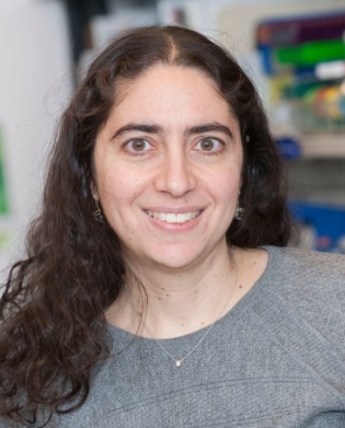 Rona Yaeger合作论文数Memorial Sloan Kettering Cancer Center24
 Emmanouil P Pappou合作论文数Department of Surgery, The Johns Hopkins Hospital22
 Chao Wu合作论文数17
 Paris Tekkis合作论文数Department of Surgery and Cancer, Faculty of Medicine, Imperial College London16Iris H Wei合作论文数15
 Karuna Ganesh合作论文数Internal Medicine - Beth Israel Deaconess Medical Center15
 Mithat Gönen合作论文数Memorial Sloan Kettering Cancer Center14Michael R Marco合作论文数13
 Christopher Crane合作论文数Memorial Sloan Kettering Cancer Center13
 Isaac Wasserman合作论文数Dept Otolaryngol Head & Neck Surg, Massachusetts Eye & Ear Infirm12
 Nancy E. Kemeny合作论文数Division of Solid Tumor Oncology, Department of Medicine, Memorial Sloan Kettering Cancer Center12Maria Widmar合作论文数11
 Lik Hang Lee合作论文数Department of Pathology and Laboratory Medicine, Faculty of Medicine, The University of British Columbia11
 Bryan C Szeglin合作论文数Dept Surg, Colorectal Serv, Mem Sloan Kettering Canc Ctr10
 Jashodeep Datta合作论文数Department of Surgery, University of Pennsylvania Health System10
 Campbell Roxburgh合作论文数School of Cancer Sciences, University of Glasgow;Memorial Sloan Kettering Cancer Center10
 Charles Sawyers合作论文数Memorial Sloan Kettering Cancer Center;Howard Hughes Medical Institute10J. Garcia-Aguilar合作论文数9Arthur Elghouayel合作论文数9
 Afsar Barlas合作论文数9Vakiani Efsevia合作论文数Dept Pathol, Mem Sloan Kettering Canc Ctr9Manova-Todorova Katia合作论文数9Greenhalgh R M合作论文数9
 Alexander Heriot合作论文数Peter MacCallum Cancer Centre;University of Melbourne9
 Alun Davies合作论文数Division of Surgery, Department of Surgery & Cancer, Faculty of Medicine, Imperial College London9Peter Ntiamoah合作论文数8Reiko Nishihara合作论文数8Dhawan Punita合作论文数8
 Andrew Chan合作论文数Clinical and Translational Epidemiology Unit, Massachusetts General Hospital;Mass General Cancer Center, Massachusetts General Hospital8
 Kontovounisios Christos合作论文数Department of Colorectal Surgery, Chelsea and Westminster Hospital8Kosumi Keisuke合作论文数8
 Uppada Srijayaprakash B合作论文数Department of Biomedical Sciences, University of Illinois at Chicago8Jonathan N. Glickman合作论文数8
 Xi Chen (陈曦)合作论文数Department of Systems Biology, School of Life Sciences, Southern University of Science and Technology8
 Shuji Ogino (荻野周史)合作论文数Harvard Medical School, Harvard University;Department of Epidemiology, Harvard T.H. Chan School of Public Health, Harvard University;Brigham and Women's Hospital8Kentaro Inamura合作论文数8
 Neil H. Segal合作论文数Division of Gastrointestinal Oncology, Memorial Sloan Kettering Cancer Center;Department of Medicine, Weill Cornell Medical College8Marios Giannakis合作论文数8
 Eran Sadot合作论文数Ramat Gan8
 Nikolaus Schultz合作论文数Department of Biostatistics and Epidemiology, Memorial Sloan Kettering Cancer Center;Center for Molecular Oncology, Memorial Sloan Kettering Cancer Center7




